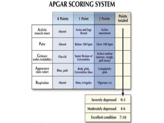
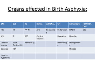
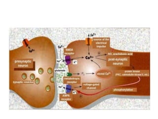
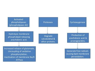
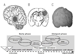
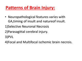
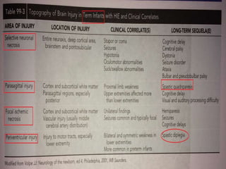
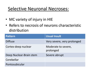
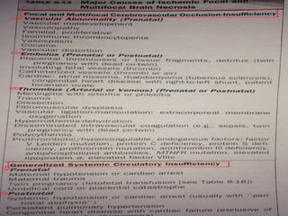
BIRTH ASPHYXIA
HIE-Neurpathology and Pathogenesis
• Perinatal asphyxia is a common neonatal problem and
contributes significantly to neonatal morbidity and
mortality.
• It ranks as the second most important cause of
neonatal death after infections, accounting for around
30 % mortality worldwide.
• In India, between 250,000 to 350,000 infants die each
year due to birth asphyxia, mostly within the first three
days of life.
• In addition, ante-partum and intra-partum asphyxia
contributes to as many as 300,000 to 400,000
stillbirths.
• In our center Birth asphyxia constitutes around 8% of
total admission,and accounts for 17% deaths in our
centre.
Common terminologies:
• Hypoxemia :Decreased arterial concentration of oxygen.
• Hypoxia:Decreased oxygenation to cells or organs.
• Ischemia: Blood flow that is insufficient to organs or tissues to
maintain their normal function.
• Asphyxia:Lack or impaired gas exchange leading to
hypoxia,hypercarbia and acidosis.
• Perinatal Asphyxia: refers to a condition during the first and
second stage of labor in which impaired gas exchange leads to
fetal hypoxemia and hypercarbia
• Perinatal/Neonatal depression: Clinical condition
in immediate post natal period(within 1hour)
with features of depressed mental
status,hypotonia, disturbances in spontaneous
respiration and cardiovascular function .
• Neonatal encephalopathy: Clinical term that
describes abnormal neurobehavioral state
consisting of decreased level of consciousness
with or without brainstem or motor dysfunction.
• Hypoxic- ischemic encephalopathy: Neonatal
encephalopathy with objective data to support a
hypoxic –ischemic mechanism as the underlying
cause for the encephalopathy.
How to define Birth Asphyxia:
• The World Health Organization has defined birth
asphyxia as “failure to initiate and sustain
breathing at birth” and based on Apgar score as
an Apgar score of <7 at 1miniute.
• The National Neonatal Perinatal Database
(NNPD) 2000, used a similar definition for
perinatal asphyxia and defined “moderate
asphyxia as slow gasping breathing or an Apgar
score of 4-6 and severe asphyxia as no breathing
or an Apgar score of 0-3 at one minute of life “
• The National Neonatology Forum of India has
defined asphyxia as “gasping or ineffective
breathing or lack of breathing at one minute of
life”
• The essential criteria for diagnosing perinatal
asphyxia as outlined by ACOG & AAP
1.Prolonged metabolic or mixed acidemia (pH <7 in
cord arterial blood sample)
2. Persistence of an Apgar score of <3 at 5min are
more.
3.Clinical neurologic manifestation as seizures,
hypotonia, coma or HIE in the immediate
neonatal period .
4.Evidence of multi-organ system dysfunction in the
immediate neonatal period
Incidence:
• According to NNPD 2000 data collected from 17
tertiary neonatal intensive care units in India, Apgar
scores<7 at 1 minute (includes moderate and severe
asphyxia) were documented in 9% of all deliveries .
• 2.5% babies continued to have Apgar scores <7 at 5
minutes of age.
• Bag and mask ventilation was used in 4.5% infants and
less than 1% infants needed cardiac compressions and/
or medications for resuscitation at birth.
• Perinatal asphyxia was responsible for 20% of all
neonatal deaths .
• In developed countries perinatal asphyxia accounts for
1-1.5% of live births
Etiology
• Asphyxia can occur in antepartum(30%) or
intrapartum(30%) period as result of impaired gas
exchange across placenta.
• Factors increasing risk of perinatal asphyxia.
1)Impairment of maternal oxygenation
2)Decreased blood flow from mother to placenta.
3)Decreased blood flow from placenta to fetus
4)Impaired gas exchange across placenta or at fetal
tissue level
5)Increased fetal O2 requirement.
Maternal
factors
Placental
&Uterine
factors
Umbilical
cord
Abnormalit-
es
Fetal factors Neonatal
factors
Hyper-
Hypotension
Praevia Prolapse Anemia CCHD
Infection Abruptio Entangleme
nt
Infection PPHN
GDM Infarction Compressio-
-n
Cardiomyop
-athy
Cardiomyopath
-y
Pulmonary
cardiac dis.
Fibrosis Vessel
abnormality
Hydrops Shock
Cocaine use Uterine
rupture
Organs effected in Birth Asphyxia:
CNS CVS RS RENAL ADRENAL GIT METABOLIC HEMATOL-
OGY
HIE MI PPHN ATN Hemorrha
g
Perforation SIADH DIC
ICH TI RDS Cortical
necrosis
Ulceration HypoNA
Cerebral
edema
Poor
Contractiliy
Hemorrhag Hemorrhag
e
Hypoglycemi
a
Seizures <BP HypoCa
Hypo or
hypertonia
Newborn Brain Physiology:
• An overall lower cerebral O2 demand when
compared to adults.
• Areas of active neural development that are
associated with either synapse formation or
activation of enzymes required for ion
homeostasis, with considerably increased
cerebral oxidative metabolism.
• Glucose is the primary source of energy in
cerebral metabolism.
• Capable of utilizing alternative energy
substrates such as ketones, lactate, and free
fatty acids
• Glucose uptake mechanisms are
underdeveloped.
• Absence of energy stores makes the brain
dependent on sustained perfusion.
• Vasoautoregulation in response to increased
cerebral blood pressure or flow is relatively
underdeveloped in the newborn.
• The gradual increase in vascularity of the
developing brain leads to the creation of
watershed areas(i.e., areas not well
vascularized).
• Similarities between processes essential for
brain development and those mediating
cellular injury.
– An increased density of glutamate receptors.
– An increase in glutaminergic synapses in
particular regions of the immature brain.
– Enhanced accumulation of cytosolic calcium
after activation of the glutamate receptor.
• Proportionately more glutamate receptors in
immature rat brain than mature.
HIE-Cellular Mechanisms:
• Pathogenesis of HIE can be divided to 4 steps
1)Decrease in Cerebral energy and membrane
depolarization
2)Increase neurotransmitter release and
neuronal damage
3)Reperfusion
4)Irreversible cell death
Phase II – Phase of increased release
of neurotransmitters and neuronal
damage
• There is sufficient evidence that excitatory
neurotransmitters play a major role in HI-R
injury.
• Excitotoxicity – excessive glutamatergic
activation that leads to cell energy and death
Glutamate & its receptors.
• Glutamate - An excitatory neurotransmitter.
3 subtypes of receptors.
1) N-methyl-D-aspartate (NMDA).
• Plays an important role in normal brain development.
• Expression of receptors changes with maturation.
• Density is higher in regions of active development.
• Different subtypes vary in different regions of the brain
at different gestational ages.
2)Amino-3-hydroxy-5-methyl-4-isoxazole propionic acid
(AMPA).
3) G protein associated metabotropic receptor
• The increase in intracellular calcium sets into
motion an irreversible cascade of events that
leads to cell injury.
• Calcium activates several degradative enzymes
such as phospholipases, proteases, and
endonucleases
• Hypoxic ischemic injury also leads to a change
in iron homeostasis.
• It reduces iron usually maintained in non toxic
state “ferric” to toxic “ferrous” form.
• This reacts with oxygen reactive species to
propagate further injury
Oxidative Stress
• Nitric oxide synthase (NOS)- released during HI-R injury,
acts as a mediator of cell injury.
• NO causes cellular injury by
– Combining with superoxide to form a peroxynitrate
radical that causes lipid peroxidation.
– Generating free radicals by stimulation of COX activity.
– Direct DNA damage.
– Participating in the neurotransmitter response by
reentering the pre synaptic neuron & further increasing
release of glutamate.
Phase 3-Phases of injury during
reperfusion.
• First phase.
– Cerebral energy metabolism restored over 30 mins.
– Resolution of acute cellular hypoxic depolarization &
cell swelling.
• Latent phase.
– Near normal oxidative cerebral metabolism.
– Depressed electroencephalogram and reduced blood
flow.
• Secondary energy failure.
– Inhibition of oxidative phosphorylation.
– Cytotoxic edema leading to delayed seizures.
Phase 4-Cell Death
• Cell death can occur as necrosis or apoptosis
after ischemia.
• A severe insult leads to necrosis, as seen in the
central area of injury.
• A longer duration of less severe injury may
lead to apoptosis, as seen in the penumbra.
Patterns of Brain Injury:
• Neuropathological features varies with
GA,timing of insult and natureof insult.
1)Selective Neuronal Necrosis
2)Parasagittal cerebral injury.
3)PVL
4)Focal and Multifocal ischemic brain necrois.
Selective Neuronal Necroses:
• MC variety of injury in HIE
• Refers to necrosis of neurons characteristic
distribution
Pattern Usual Insult
Diffuse Very severe, very prolonged
Cortex-deep nuclear Moderate to severe,
prolonged
Deep Nuclear-Brain stem Severe abrupt
Cerebellar
Pontosubicular
• Clinical Corelates:stupor or
coma,Seizures,Hypotonia,Oculomotor and
suck/swallow abnormalities.
• Long term Sequelae:Cognitive delay,Cerebral
palsy,Dystonia,Seizure disoder,Ataxia,Bulbar or
pseudo-bulbar palsy.
Parasagittal Cerebral injury:
• Injury is usually B/L & symetrical,superio
medial aspect of cerebral hemisphere.
• Watershed area in term babies
• Posterior aspect of cerebral hemispheres,
especially parieto-occipital region is more
affected.
• MRI study of 173 term infants with HIE,45%
had watershed injury has predominant lesion.
• Clinical corelate:Proximal limb
weakness,Upper limb more affected.
• Long term Sequelae-Spastic
Quadriparesis,Cognitive delay,Visual and
auditory processing difficulty.
Periventricular Leukomalacia:
• Necrosis of white matter in dorsal and lateral
to external angles of lateral ventricles.
• Cystic and Non Cystic PVL
-lesion is observed in PT infants
-infants with post natal survival of more than
few days
-in infants who have IVH,maternal-infant
infection and with cardio pulmonary
disturbances
• Clinical Co relates:B/L symetric weakness in
lower limbs.
• Long term sequelae-Spastic diplegia.
Focal and Multifocal Ischemic
Necrosis:(Arterial Stroke)
• Necroses that occur with distribution of single
major vessel.
• MCA is MC involed artery.
• Clinical corelates:Unilteral findings,seizures
typically focal.
• Long term Sequelae-
Hemiparesis,seizures,cognitive delay.
References
• Volpe JJ:Hypoxic-Ischemic
Encephalopathy:Neuropatholgy and
Pathogenesis,Neurology of Newborn 5th Ed :347-
399,2008.
• Sonia LB,Gonzalez F:CNS Injury and
Neuroprotection,Avery’s Disesease of Newborn 9th
ed:874-880,2012.
• Anne R,Hansen:Perinatal asphyxia and HIE,Manual of
Neonatal Care 7th ed:711-726,2012.
• N Ambalavaan,WA Carlo:Hypoxic-ischemic
Encephalopathy,Nelson textbook of Pediatrics 20th
ed:838-843,2015
Birth asphyxia neurpathology

Birth asphyxia neurpathology

  • 1.
  • 2.
    • Perinatal asphyxiais a common neonatal problem and contributes significantly to neonatal morbidity and mortality. • It ranks as the second most important cause of neonatal death after infections, accounting for around 30 % mortality worldwide. • In India, between 250,000 to 350,000 infants die each year due to birth asphyxia, mostly within the first three days of life. • In addition, ante-partum and intra-partum asphyxia contributes to as many as 300,000 to 400,000 stillbirths. • In our center Birth asphyxia constitutes around 8% of total admission,and accounts for 17% deaths in our centre.
  • 3.
    Common terminologies: • Hypoxemia:Decreased arterial concentration of oxygen. • Hypoxia:Decreased oxygenation to cells or organs. • Ischemia: Blood flow that is insufficient to organs or tissues to maintain their normal function. • Asphyxia:Lack or impaired gas exchange leading to hypoxia,hypercarbia and acidosis. • Perinatal Asphyxia: refers to a condition during the first and second stage of labor in which impaired gas exchange leads to fetal hypoxemia and hypercarbia
  • 4.
    • Perinatal/Neonatal depression:Clinical condition in immediate post natal period(within 1hour) with features of depressed mental status,hypotonia, disturbances in spontaneous respiration and cardiovascular function . • Neonatal encephalopathy: Clinical term that describes abnormal neurobehavioral state consisting of decreased level of consciousness with or without brainstem or motor dysfunction. • Hypoxic- ischemic encephalopathy: Neonatal encephalopathy with objective data to support a hypoxic –ischemic mechanism as the underlying cause for the encephalopathy.
  • 5.
    How to defineBirth Asphyxia: • The World Health Organization has defined birth asphyxia as “failure to initiate and sustain breathing at birth” and based on Apgar score as an Apgar score of <7 at 1miniute. • The National Neonatal Perinatal Database (NNPD) 2000, used a similar definition for perinatal asphyxia and defined “moderate asphyxia as slow gasping breathing or an Apgar score of 4-6 and severe asphyxia as no breathing or an Apgar score of 0-3 at one minute of life “
  • 7.
    • The NationalNeonatology Forum of India has defined asphyxia as “gasping or ineffective breathing or lack of breathing at one minute of life” • The essential criteria for diagnosing perinatal asphyxia as outlined by ACOG & AAP 1.Prolonged metabolic or mixed acidemia (pH <7 in cord arterial blood sample) 2. Persistence of an Apgar score of <3 at 5min are more. 3.Clinical neurologic manifestation as seizures, hypotonia, coma or HIE in the immediate neonatal period . 4.Evidence of multi-organ system dysfunction in the immediate neonatal period
  • 9.
    Incidence: • According toNNPD 2000 data collected from 17 tertiary neonatal intensive care units in India, Apgar scores<7 at 1 minute (includes moderate and severe asphyxia) were documented in 9% of all deliveries . • 2.5% babies continued to have Apgar scores <7 at 5 minutes of age. • Bag and mask ventilation was used in 4.5% infants and less than 1% infants needed cardiac compressions and/ or medications for resuscitation at birth. • Perinatal asphyxia was responsible for 20% of all neonatal deaths . • In developed countries perinatal asphyxia accounts for 1-1.5% of live births
  • 10.
    Etiology • Asphyxia canoccur in antepartum(30%) or intrapartum(30%) period as result of impaired gas exchange across placenta. • Factors increasing risk of perinatal asphyxia. 1)Impairment of maternal oxygenation 2)Decreased blood flow from mother to placenta. 3)Decreased blood flow from placenta to fetus 4)Impaired gas exchange across placenta or at fetal tissue level 5)Increased fetal O2 requirement.
  • 11.
    Maternal factors Placental &Uterine factors Umbilical cord Abnormalit- es Fetal factors Neonatal factors Hyper- Hypotension PraeviaProlapse Anemia CCHD Infection Abruptio Entangleme nt Infection PPHN GDM Infarction Compressio- -n Cardiomyop -athy Cardiomyopath -y Pulmonary cardiac dis. Fibrosis Vessel abnormality Hydrops Shock Cocaine use Uterine rupture
  • 12.
    Organs effected inBirth Asphyxia: CNS CVS RS RENAL ADRENAL GIT METABOLIC HEMATOL- OGY HIE MI PPHN ATN Hemorrha g Perforation SIADH DIC ICH TI RDS Cortical necrosis Ulceration HypoNA Cerebral edema Poor Contractiliy Hemorrhag Hemorrhag e Hypoglycemi a Seizures <BP HypoCa Hypo or hypertonia
  • 13.
    Newborn Brain Physiology: •An overall lower cerebral O2 demand when compared to adults. • Areas of active neural development that are associated with either synapse formation or activation of enzymes required for ion homeostasis, with considerably increased cerebral oxidative metabolism.
  • 14.
    • Glucose isthe primary source of energy in cerebral metabolism. • Capable of utilizing alternative energy substrates such as ketones, lactate, and free fatty acids • Glucose uptake mechanisms are underdeveloped. • Absence of energy stores makes the brain dependent on sustained perfusion.
  • 15.
    • Vasoautoregulation inresponse to increased cerebral blood pressure or flow is relatively underdeveloped in the newborn. • The gradual increase in vascularity of the developing brain leads to the creation of watershed areas(i.e., areas not well vascularized).
  • 16.
    • Similarities betweenprocesses essential for brain development and those mediating cellular injury. – An increased density of glutamate receptors. – An increase in glutaminergic synapses in particular regions of the immature brain. – Enhanced accumulation of cytosolic calcium after activation of the glutamate receptor. • Proportionately more glutamate receptors in immature rat brain than mature.
  • 17.
    HIE-Cellular Mechanisms: • Pathogenesisof HIE can be divided to 4 steps 1)Decrease in Cerebral energy and membrane depolarization 2)Increase neurotransmitter release and neuronal damage 3)Reperfusion 4)Irreversible cell death
  • 19.
    Phase II –Phase of increased release of neurotransmitters and neuronal damage • There is sufficient evidence that excitatory neurotransmitters play a major role in HI-R injury. • Excitotoxicity – excessive glutamatergic activation that leads to cell energy and death
  • 20.
    Glutamate & itsreceptors. • Glutamate - An excitatory neurotransmitter. 3 subtypes of receptors. 1) N-methyl-D-aspartate (NMDA). • Plays an important role in normal brain development. • Expression of receptors changes with maturation. • Density is higher in regions of active development. • Different subtypes vary in different regions of the brain at different gestational ages. 2)Amino-3-hydroxy-5-methyl-4-isoxazole propionic acid (AMPA). 3) G protein associated metabotropic receptor
  • 23.
    • The increasein intracellular calcium sets into motion an irreversible cascade of events that leads to cell injury. • Calcium activates several degradative enzymes such as phospholipases, proteases, and endonucleases
  • 26.
    • Hypoxic ischemicinjury also leads to a change in iron homeostasis. • It reduces iron usually maintained in non toxic state “ferric” to toxic “ferrous” form. • This reacts with oxygen reactive species to propagate further injury
  • 27.
    Oxidative Stress • Nitricoxide synthase (NOS)- released during HI-R injury, acts as a mediator of cell injury. • NO causes cellular injury by – Combining with superoxide to form a peroxynitrate radical that causes lipid peroxidation. – Generating free radicals by stimulation of COX activity. – Direct DNA damage. – Participating in the neurotransmitter response by reentering the pre synaptic neuron & further increasing release of glutamate.
  • 28.
    Phase 3-Phases ofinjury during reperfusion. • First phase. – Cerebral energy metabolism restored over 30 mins. – Resolution of acute cellular hypoxic depolarization & cell swelling. • Latent phase. – Near normal oxidative cerebral metabolism. – Depressed electroencephalogram and reduced blood flow. • Secondary energy failure. – Inhibition of oxidative phosphorylation. – Cytotoxic edema leading to delayed seizures.
  • 31.
    Phase 4-Cell Death •Cell death can occur as necrosis or apoptosis after ischemia. • A severe insult leads to necrosis, as seen in the central area of injury. • A longer duration of less severe injury may lead to apoptosis, as seen in the penumbra.
  • 33.
    Patterns of BrainInjury: • Neuropathological features varies with GA,timing of insult and natureof insult. 1)Selective Neuronal Necrosis 2)Parasagittal cerebral injury. 3)PVL 4)Focal and Multifocal ischemic brain necrois.
  • 35.
    Selective Neuronal Necroses: •MC variety of injury in HIE • Refers to necrosis of neurons characteristic distribution Pattern Usual Insult Diffuse Very severe, very prolonged Cortex-deep nuclear Moderate to severe, prolonged Deep Nuclear-Brain stem Severe abrupt Cerebellar Pontosubicular
  • 36.
    • Clinical Corelates:stuporor coma,Seizures,Hypotonia,Oculomotor and suck/swallow abnormalities. • Long term Sequelae:Cognitive delay,Cerebral palsy,Dystonia,Seizure disoder,Ataxia,Bulbar or pseudo-bulbar palsy.
  • 37.
    Parasagittal Cerebral injury: •Injury is usually B/L & symetrical,superio medial aspect of cerebral hemisphere. • Watershed area in term babies • Posterior aspect of cerebral hemispheres, especially parieto-occipital region is more affected. • MRI study of 173 term infants with HIE,45% had watershed injury has predominant lesion.
  • 38.
    • Clinical corelate:Proximallimb weakness,Upper limb more affected. • Long term Sequelae-Spastic Quadriparesis,Cognitive delay,Visual and auditory processing difficulty.
  • 41.
    Periventricular Leukomalacia: • Necrosisof white matter in dorsal and lateral to external angles of lateral ventricles. • Cystic and Non Cystic PVL -lesion is observed in PT infants -infants with post natal survival of more than few days -in infants who have IVH,maternal-infant infection and with cardio pulmonary disturbances
  • 42.
    • Clinical Corelates:B/L symetric weakness in lower limbs. • Long term sequelae-Spastic diplegia.
  • 43.
    Focal and MultifocalIschemic Necrosis:(Arterial Stroke) • Necroses that occur with distribution of single major vessel. • MCA is MC involed artery.
  • 46.
    • Clinical corelates:Unilteralfindings,seizures typically focal. • Long term Sequelae- Hemiparesis,seizures,cognitive delay.
  • 47.
    References • Volpe JJ:Hypoxic-Ischemic Encephalopathy:Neuropatholgyand Pathogenesis,Neurology of Newborn 5th Ed :347- 399,2008. • Sonia LB,Gonzalez F:CNS Injury and Neuroprotection,Avery’s Disesease of Newborn 9th ed:874-880,2012. • Anne R,Hansen:Perinatal asphyxia and HIE,Manual of Neonatal Care 7th ed:711-726,2012. • N Ambalavaan,WA Carlo:Hypoxic-ischemic Encephalopathy,Nelson textbook of Pediatrics 20th ed:838-843,2015