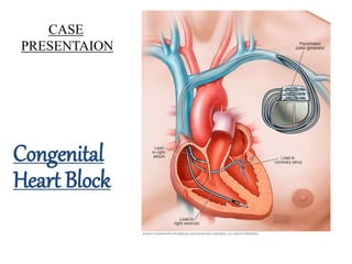
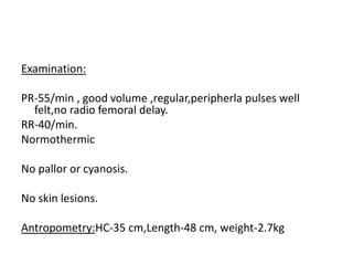
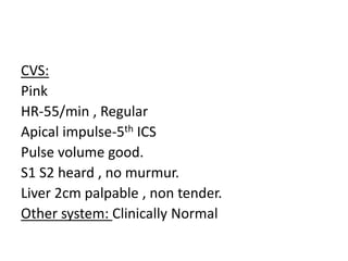
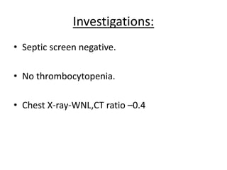
CASE
PRESENTAION
Congenital
Heart Block
• B/O Nirmala , Male child , delivered via LSCS
in Bapuji Hospital on 04/02/17
• Baby cried immediately after birth.
• Shifted to NICU within 1 hour in view of
persistent bradycardia.
Antenatal History:
• Primigravida , concieved spontaneously,ML-1year.
• Had regular ANC and received supplements.
• No h/o joint pain , rash in mother.
• No h/o similar complaints in family members.
Antenatal scan on @24 wk GA:
• Non conduction of every second heart beat.
• Bradycardia
• Atrial bigeminy
Maternal Lupus evolution: ANA Negative
Examination:
PR-55/min , good volume ,regular,peripherla pulses well
felt,no radio femoral delay.
RR-40/min.
Normothermic
No pallor or cyanosis.
No skin lesions.
Antropometry:HC-35 cm,Length-48 cm, weight-2.7kg
CVS:
Pink
HR-55/min , Regular
Apical impulse-5th ICS
Pulse volume good.
S1 S2 heard , no murmur.
Liver 2cm palpable , non tender.
Other system: Clinically Normal
TERM/AGA/S/M/ CONGENITAL HEART
BLOCK IN STRUCTURALLY NORMAL
HEART WITHOUT CCF
Investigations:
• Septic screen negative.
• No thrombocytopenia.
• Chest X-ray-WNL,CT ratio –0.4
2 D Echocardiography:
• Situs solitus/normally
related great vessels.
• Small PDA 3mm L to Rt
Shunt.
• Normal biventricular
function
ECG
• Atrial and ventricular
rate are independent.
• Atrial rate faster than
ventricular rate.
• No PR interval
• F/s/o Complete heart
block.
TERM/AGA/M/CONGENITAL
COMPLETE HEART BLOCK WITH PDA
WITHOUT CCF
CONGENITAL HEART BLOCK
Cardiac conduction system:
• Atrioventricular (AV) block is defined as a delay or
interruption in the transmission of an impulse from
the atria to the ventricles due to an anatomical or
functional impairment in the conduction system.
• Atrioventricular block is considered to be
"congenital" when it occurs spontaneously in a fetus
or young child.
Rhythm Regular
Rate The underlying rate
P Wave Normal
PR Interval Prolonged (>0.20 sec)
QRS Normal (0.06-0.10 sec)
Notes A first-degree AV block is actually a delay rather than a block
First Degree Heart Block
Second degree heart block-Type 1
Rhythm Regular (atrial) and irregular (ventricular)
Rate
Characterized by Atrial rate usually faster than ventricular rate (usually
slow)
P Wave Normal form, but more P waves than QRS complexes
PR Interval Normal or prolonged
QRS Normal or wide
Notes
Second degree Heart block-Type 2
Rhythm Regular (atrial) and irregular (ventricular)
Rate
Characterized by Atrial rate usually faster than ventricular rate (usually
slow)
P Wave Normal form, but more P waves than QRS complexes
PR Interval Normal or prolonged
QRS Normal or wide
Notes
3rd /Complete Degree Heart block
Rhythm Regular, but atrial and ventricular rhythms are independent
Rate Characterized by Atrial rate usually normal and faster than ventricular rate
P Wave Normal shape and size, may appear within QRS complexes
PR Interval Absent: the atria and ventricles beat independently.
QRS Normal.
Notes
Epidemology:
• Incidence 1:20,000 live births.
• Transplacental exposure to maternal autoantibodies related
to SLE or Sjogren's syndrome is responsible for 60 to 90
percent of cases .
• Women withanti-Ro/SSA and/or anti-La/SSB antibodies
incidence 2%.
• Risk in future pregnacies-15%
• 60% diagnosed utero or neonatl,40% of CHB present in
childhood(5-6yrs)
Etiology:
1. Autoimmune antibodies.
2. Structural heart abnormalities due to congenital
heart disease (eg-L-TGA, Endocardial cushion
defects , syndromic ASD)
3. Idiopathic familial congenital CHB
Autoimmune congenital CHB :
• Begins in utero
• Most cases, the block is third degree.
• Damage to developing specialized conduction tissue from
passive transplacental passage of maternal
autoantibodies to Ro/SSA and/or La/SSB intracellular
ribonuclear proteins
• Risk does not correlate directly with the maternal
autoantibody titer.
• Mothers who give birth to a child with autoimmune CHB
have never had symptoms of connective tissue disease.
Pathopysiology:
• Most cases of congenital CHB are immune-mediated and are
characterized pathologically by fibrous tissue that either replaces
the (AV node) and its surrounding tissue.
• Primary damage is by binding of anti-Ro/SSA and/or anti-
La/SSB antibodies to the developing AV node and its surrounding
tissue .
• CHB in patients with congenital heart defects is abnormalities in the
embryologic development of the specialized A-V conduction tissues
that lead to displacement and functional impairment of the AV
node and/or His bundle.
Clinical Features:
• The manifestations of congenital CHB vary according
to the age at presentation, underlying etiology,
ventricular rate of the escape rhythm, and
ventricular function.
• Patients with autoimmune congenital CHB tend to
present earlier .
• In utero presentation — Congenital heart block may
present with fetal bradycardia between 18 and 28
weeks of gestation
Neonatal presentation
• The primary finding in neonates with congenital CHB is a slow
heart rate that is usually less than 100 beats per minute.
• Neonates with congenital CHB otherwise have few specific physical
examination findings, although they may appear pale or diaphoretic
related to the reduction in cardiac output.
• Other clinical clues in the neonate may include:
-Intermittent cannon waves in the neck
-Variable intensity of the first heart sound
-Intermittent gallops and murmurs
-Signs of congestive heart failure .
• As with cases presenting in utero, almost all presenting in the
neonatal period (90 percent in one series) are due to maternal
autoantibodies .
Presentation in later childhood
• Up to 40 percent of patients with congenital CHB
present later in childhood.
• Reasons for the late presentation are , but likely relate
to an intermittent early course or a higher ventricular
rate of the escape rhythm .
• The primary finding in children with congenital CHB is a
slow heart rate with or without bradycardia-related
symptoms, including reduced exercise tolerance and
presyncope or syncope (Stokes-Adams attacks)
• Maternal autoantibody exposure accounts for almost
all cases presenting in utero or the neonatal period,
but for only a few cases occurring at older ages (5
percent in one report) .
Treatment:
In utero treatment —
• Treatment of the fetus with first degree CHB is
primarily expectant,the fetus will tolerate the slow
escape rhythm in the majority of cases.
• Prenatal treatment with glucocorticoids (eg,
oral dexamethasone 4 mg per day,
or betamethasone 3 mg per day) is suggested for
mothers of fetuses with second-degree heart block,
beginning as soon after detection as is feasible and
continuing through the end of pregnancy.
In Utero treatment
• Treatment of third-degree block with glucocorticoids
is controversial, It is generally not advised.
• Glucocorticoids are also considered for signs of a
more global cardiomyopathy.
• Preemptive treatment with hydroxychloroquine (400
mg orally once a day) in pregnant women with anti-
Ro/SSA antibodies who have previously given birth to
a child with cardiac-NL.
Post-natal treatment:
• The principal therapeutic decision involves the need
for, and the potential timing of, permanent
pacemaker insertion.
• Patients with an adequate ventricular heart rate and
no symptoms can usually be followed with serial
observation, while symptomatic patients will require
a permanent pacemaker
• Most patients (approximately 90 percent or greater)
ultimately have a pacemaker inserted , regardless of
when CHB developed
Pacemaker Indications:
1. Symptomatic bradycardia or low cardiac output
2. Wide QRS escape rhythm, complex ventricular
ectopy, or ventricular dysfunction
3. Infants with normal anatomy and a ventricular rate
less than 55 beats per minute
4. Infants with other structural congenital heart
disease and a ventricular rate less than 70 beats per
minute
5. Children beyond the first year of life with an
average heart rate less than 50 beats per minute.
Pacemakers:
• Two types-Epicardial
and transvenous
• Implants for infants and very young
children will usually require epicardial
leads due to small caliber of the
standard insertion veins used for
transvenous devices, as well as the
expectation for thoracic growth that
may eventually place tension on
transvenous leads
• Epicardial leads are also used in
patients of any age with septal defects
that allow intracardiac shunting and
thus increase the risk of
thromboembolic complications .
• A transvenous dual-chamber
pacemaker is preferred at most centers
when there are no significant size
constraints or other contraindication
Prognosis:
• CHB presenting in utero or the neonatal period, which is mostly due to
maternal autoantibodies, is associated with a high early mortality .
• The outcome for patients diagnosed as neonates is better than for those
diagnosed in utero
• Among 175 cases of congenital CHB diagnosed in the fetus, 29 (17
percent) died either in utero or within the first three months of life’
• Offspring born before 34 weeks having a higher mortality rate than those
born later (52 versus 9 percent) .
• Infants with first or second degree heart block at birth can progress to
complete heart block.
• Review of 102 patients who were without symptoms through age 15, 27
(26 percent) had a subsequent syncopal episode, eight of which were
fatal .
• A significant number of patients (5 to 11 percent) develop heart failure
over the long-term, even if a pacemaker is inserted
‘Buyon JP, Hiebert R, Copel J, et al. Autoimmune-associated congenital heart block: demographics, mortality, morbidity and
recurrence rates obtained from a national neonatal lupus registry. J Am Coll Cardiol 1998; 31:1658.
References:
• Buyon JP, Hiebert R, Copel J, et al. Autoimmune-associated congenital
heart block: demographics, mortality, morbidity and recurrence rates
obtained from a national neonatal lupus registry. J Am Coll Cardiol 1998;
31:1658.
• Reid JM, Coleman EN, Doig W. Complete congenital heart block. Report of
35 cases. Br Heart J 1982; 48:236.
• Baruteau AE, Pass RH, Thambo JB, et al. Congenital and childhood
atrioventricular blocks: pathophysiology and contemporary management.
Eur J Pediatr 2016; 175:1235.
• Epstein AE, Dimarco JP, Ellenbogen KA, et al. ACC/AHA/HRS 2008
guidelines for Device-Based Therapy of Cardiac Rhythm Abnormalities:
executive summary. Heart Rhythm 2008; 5:934. Askanase AD, Friedman
DM, Copel J, et al.
• Spectrum and progression of conduction abnormalities in infants born to
mothers with anti-SSA/Ro-SSB/La antibodies. Lupus 2002; 11:145
THANK YOU

Congenital heart block

  • 1.
  • 2.
    • B/O Nirmala, Male child , delivered via LSCS in Bapuji Hospital on 04/02/17 • Baby cried immediately after birth. • Shifted to NICU within 1 hour in view of persistent bradycardia.
  • 3.
    Antenatal History: • Primigravida, concieved spontaneously,ML-1year. • Had regular ANC and received supplements. • No h/o joint pain , rash in mother. • No h/o similar complaints in family members.
  • 4.
    Antenatal scan on@24 wk GA: • Non conduction of every second heart beat. • Bradycardia • Atrial bigeminy Maternal Lupus evolution: ANA Negative
  • 5.
    Examination: PR-55/min , goodvolume ,regular,peripherla pulses well felt,no radio femoral delay. RR-40/min. Normothermic No pallor or cyanosis. No skin lesions. Antropometry:HC-35 cm,Length-48 cm, weight-2.7kg
  • 6.
    CVS: Pink HR-55/min , Regular Apicalimpulse-5th ICS Pulse volume good. S1 S2 heard , no murmur. Liver 2cm palpable , non tender. Other system: Clinically Normal
  • 7.
    TERM/AGA/S/M/ CONGENITAL HEART BLOCKIN STRUCTURALLY NORMAL HEART WITHOUT CCF
  • 8.
    Investigations: • Septic screennegative. • No thrombocytopenia. • Chest X-ray-WNL,CT ratio –0.4
  • 9.
    2 D Echocardiography: •Situs solitus/normally related great vessels. • Small PDA 3mm L to Rt Shunt. • Normal biventricular function
  • 10.
    ECG • Atrial andventricular rate are independent. • Atrial rate faster than ventricular rate. • No PR interval • F/s/o Complete heart block.
  • 11.
  • 12.
  • 13.
  • 14.
    • Atrioventricular (AV)block is defined as a delay or interruption in the transmission of an impulse from the atria to the ventricles due to an anatomical or functional impairment in the conduction system. • Atrioventricular block is considered to be "congenital" when it occurs spontaneously in a fetus or young child.
  • 15.
    Rhythm Regular Rate Theunderlying rate P Wave Normal PR Interval Prolonged (>0.20 sec) QRS Normal (0.06-0.10 sec) Notes A first-degree AV block is actually a delay rather than a block First Degree Heart Block
  • 16.
    Second degree heartblock-Type 1 Rhythm Regular (atrial) and irregular (ventricular) Rate Characterized by Atrial rate usually faster than ventricular rate (usually slow) P Wave Normal form, but more P waves than QRS complexes PR Interval Normal or prolonged QRS Normal or wide Notes
  • 17.
    Second degree Heartblock-Type 2 Rhythm Regular (atrial) and irregular (ventricular) Rate Characterized by Atrial rate usually faster than ventricular rate (usually slow) P Wave Normal form, but more P waves than QRS complexes PR Interval Normal or prolonged QRS Normal or wide Notes
  • 18.
    3rd /Complete DegreeHeart block Rhythm Regular, but atrial and ventricular rhythms are independent Rate Characterized by Atrial rate usually normal and faster than ventricular rate P Wave Normal shape and size, may appear within QRS complexes PR Interval Absent: the atria and ventricles beat independently. QRS Normal. Notes
  • 19.
    Epidemology: • Incidence 1:20,000live births. • Transplacental exposure to maternal autoantibodies related to SLE or Sjogren's syndrome is responsible for 60 to 90 percent of cases . • Women withanti-Ro/SSA and/or anti-La/SSB antibodies incidence 2%. • Risk in future pregnacies-15% • 60% diagnosed utero or neonatl,40% of CHB present in childhood(5-6yrs)
  • 20.
    Etiology: 1. Autoimmune antibodies. 2.Structural heart abnormalities due to congenital heart disease (eg-L-TGA, Endocardial cushion defects , syndromic ASD) 3. Idiopathic familial congenital CHB
  • 21.
    Autoimmune congenital CHB: • Begins in utero • Most cases, the block is third degree. • Damage to developing specialized conduction tissue from passive transplacental passage of maternal autoantibodies to Ro/SSA and/or La/SSB intracellular ribonuclear proteins • Risk does not correlate directly with the maternal autoantibody titer. • Mothers who give birth to a child with autoimmune CHB have never had symptoms of connective tissue disease.
  • 22.
    Pathopysiology: • Most casesof congenital CHB are immune-mediated and are characterized pathologically by fibrous tissue that either replaces the (AV node) and its surrounding tissue. • Primary damage is by binding of anti-Ro/SSA and/or anti- La/SSB antibodies to the developing AV node and its surrounding tissue . • CHB in patients with congenital heart defects is abnormalities in the embryologic development of the specialized A-V conduction tissues that lead to displacement and functional impairment of the AV node and/or His bundle.
  • 23.
    Clinical Features: • Themanifestations of congenital CHB vary according to the age at presentation, underlying etiology, ventricular rate of the escape rhythm, and ventricular function. • Patients with autoimmune congenital CHB tend to present earlier . • In utero presentation — Congenital heart block may present with fetal bradycardia between 18 and 28 weeks of gestation
  • 24.
    Neonatal presentation • Theprimary finding in neonates with congenital CHB is a slow heart rate that is usually less than 100 beats per minute. • Neonates with congenital CHB otherwise have few specific physical examination findings, although they may appear pale or diaphoretic related to the reduction in cardiac output. • Other clinical clues in the neonate may include: -Intermittent cannon waves in the neck -Variable intensity of the first heart sound -Intermittent gallops and murmurs -Signs of congestive heart failure . • As with cases presenting in utero, almost all presenting in the neonatal period (90 percent in one series) are due to maternal autoantibodies .
  • 25.
    Presentation in laterchildhood • Up to 40 percent of patients with congenital CHB present later in childhood. • Reasons for the late presentation are , but likely relate to an intermittent early course or a higher ventricular rate of the escape rhythm . • The primary finding in children with congenital CHB is a slow heart rate with or without bradycardia-related symptoms, including reduced exercise tolerance and presyncope or syncope (Stokes-Adams attacks) • Maternal autoantibody exposure accounts for almost all cases presenting in utero or the neonatal period, but for only a few cases occurring at older ages (5 percent in one report) .
  • 26.
    Treatment: In utero treatment— • Treatment of the fetus with first degree CHB is primarily expectant,the fetus will tolerate the slow escape rhythm in the majority of cases. • Prenatal treatment with glucocorticoids (eg, oral dexamethasone 4 mg per day, or betamethasone 3 mg per day) is suggested for mothers of fetuses with second-degree heart block, beginning as soon after detection as is feasible and continuing through the end of pregnancy.
  • 27.
    In Utero treatment •Treatment of third-degree block with glucocorticoids is controversial, It is generally not advised. • Glucocorticoids are also considered for signs of a more global cardiomyopathy. • Preemptive treatment with hydroxychloroquine (400 mg orally once a day) in pregnant women with anti- Ro/SSA antibodies who have previously given birth to a child with cardiac-NL.
  • 28.
    Post-natal treatment: • Theprincipal therapeutic decision involves the need for, and the potential timing of, permanent pacemaker insertion. • Patients with an adequate ventricular heart rate and no symptoms can usually be followed with serial observation, while symptomatic patients will require a permanent pacemaker • Most patients (approximately 90 percent or greater) ultimately have a pacemaker inserted , regardless of when CHB developed
  • 29.
    Pacemaker Indications: 1. Symptomaticbradycardia or low cardiac output 2. Wide QRS escape rhythm, complex ventricular ectopy, or ventricular dysfunction 3. Infants with normal anatomy and a ventricular rate less than 55 beats per minute 4. Infants with other structural congenital heart disease and a ventricular rate less than 70 beats per minute 5. Children beyond the first year of life with an average heart rate less than 50 beats per minute.
  • 30.
    Pacemakers: • Two types-Epicardial andtransvenous • Implants for infants and very young children will usually require epicardial leads due to small caliber of the standard insertion veins used for transvenous devices, as well as the expectation for thoracic growth that may eventually place tension on transvenous leads • Epicardial leads are also used in patients of any age with septal defects that allow intracardiac shunting and thus increase the risk of thromboembolic complications . • A transvenous dual-chamber pacemaker is preferred at most centers when there are no significant size constraints or other contraindication
  • 31.
    Prognosis: • CHB presentingin utero or the neonatal period, which is mostly due to maternal autoantibodies, is associated with a high early mortality . • The outcome for patients diagnosed as neonates is better than for those diagnosed in utero • Among 175 cases of congenital CHB diagnosed in the fetus, 29 (17 percent) died either in utero or within the first three months of life’ • Offspring born before 34 weeks having a higher mortality rate than those born later (52 versus 9 percent) . • Infants with first or second degree heart block at birth can progress to complete heart block. • Review of 102 patients who were without symptoms through age 15, 27 (26 percent) had a subsequent syncopal episode, eight of which were fatal . • A significant number of patients (5 to 11 percent) develop heart failure over the long-term, even if a pacemaker is inserted ‘Buyon JP, Hiebert R, Copel J, et al. Autoimmune-associated congenital heart block: demographics, mortality, morbidity and recurrence rates obtained from a national neonatal lupus registry. J Am Coll Cardiol 1998; 31:1658.
  • 32.
    References: • Buyon JP,Hiebert R, Copel J, et al. Autoimmune-associated congenital heart block: demographics, mortality, morbidity and recurrence rates obtained from a national neonatal lupus registry. J Am Coll Cardiol 1998; 31:1658. • Reid JM, Coleman EN, Doig W. Complete congenital heart block. Report of 35 cases. Br Heart J 1982; 48:236. • Baruteau AE, Pass RH, Thambo JB, et al. Congenital and childhood atrioventricular blocks: pathophysiology and contemporary management. Eur J Pediatr 2016; 175:1235. • Epstein AE, Dimarco JP, Ellenbogen KA, et al. ACC/AHA/HRS 2008 guidelines for Device-Based Therapy of Cardiac Rhythm Abnormalities: executive summary. Heart Rhythm 2008; 5:934. Askanase AD, Friedman DM, Copel J, et al. • Spectrum and progression of conduction abnormalities in infants born to mothers with anti-SSA/Ro-SSB/La antibodies. Lupus 2002; 11:145
  • 33.