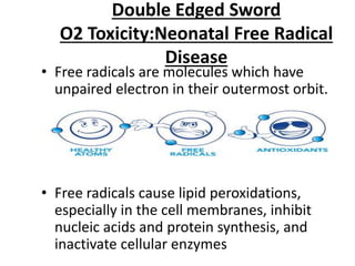
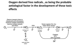
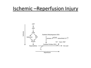
OXYGEN TRANSPORT
AND
OXY-Hb DISSASOCIATION CURVE
• By mass, oxygen is the third-most abundant elemet in
the universe, after hydrogen and helium.
• Oxygen was discovered independently by Carl Wilhelm
Scheele, in 1773 or earlier, and Joseph Priestly in
1774, but Priestley is often given priority because his
work was published first.
• Dr Albert Blodgett -First use of continuous oxygen on
46 year old woman with pneumonia.
• The individual who built on their work and brought
oxygen therapy to a rational and scientific basis was
John Scott Haldane (1860–1936)-‘The therapeutic
administration of oxygen’
Oxygen Transport:
1. Transfer of O2 from Atmosphere to lungs.
2. Diffusion across Alveoli to Blood in lung.
3. Diffusion from Blood to Tissue.
4. Utilization of O2 in tissue(Mitochondria).
1)Oxygen Transport From Atmosphere
to Lungs
ALVEOLAR OXYGEN TENSION : With every breath, the
inspired gas is humidified at 37°C in the upper airway.
• The inspired tension of oxygen (PIO2 ) is therefore
reduced by the added water vapor.
• Water vapor pressure is 47 mm Hg at 37°C.
• In humidified air, in the trachea the normal partial
pressure of O2 at sea level is 149.7 mm Hg:(760-47)0.21
• Alveolar Gas Equation PAO2 =
PiO2 -PACO2/R
-where PIO2 is inspired oxygen tension,
- PACO2 is alveolar CO2 tension (assumed to equal
arterial PCO2)
- R is the respiratory exchange ratio (normally in
the range of 0.8 to 1.0).
The alveolar oxygen tension is approximately
104mm of Hg .
The factors that determine the precise value of
alveolar PO2 are
(1) the PO2 of atmospheric air,
(2) the rate of alveolar ventilation, and
(3) the rate of total body oxygen consumption
Diffusion of Gases Across Respiratory
Membrane:
Exchange of Gases-Henry’s Law:
•When a liquid is exposed to air containing a particular gas,
molecules of the gas will enter the liquid and dissolve in it.
•Henry’s law states that “the amount of gas dissolved in a
liquid will be directly proportional to the partial pressure
of the gas in the liquid-gas interface”.
•As long as the PO2 in the gas phase is higher than the PO2
in the liquid phase, there will be a net diffusion of O2 into
the blood.
•Diffusion equilibrium will be reached only when the PO2 in
the liquid phase is equal to the PO2 in the gas phase.
Factors affecting gas diffusion
1)Partial pressure gradient of the gas across the
alveolarcapillary membrane. (60 mmHg for O2 & 6
mmHg for CO2).
2) Surface area of the alveolar-capillary membrane.
3)Thickness of the alveolar-capillary membrane. (about
0.5 μ).
4)Diffusion coefficient of the gas that depends on:
-Gas solubility. (CO2 is 24 times soluble than O2).
-Molecular weight of the gas. (CO2 M.W. is 1.4 times
greater than O2).
• Net effect: CO2 diffusion is 20 times faster than O2 2)
Rate of gas diffusion =
Diffusion coefficient X Pressure gradient x Surface
area of the membrane /Thickness of the
membrane
The volume of gas transfer across the alveolar-
capillary membrane per unit time is:
Directly proportional to:
- The difference in the partial pressure of gas
between alveoli and capillary blood.
- The surface area of the membrane.
- The solubility of the gas.
Inversely proportional to:
- Thickness of the membrane.
- Molecular weight of the gas.
GAS EXCHANGE
Third Step-Transport in Blood
Physically dissolved O2
• Only 1.5 % of total O2 in
blood.
• Dissolved in plasma and
water of RBC. (because s
olubility of O2 is very lo
w)
• It is about 0.3ml of O2 dis
solved in 100ml arterial
blood (at PO2 100 mmH
g).
Chemically combined O2
• 98.5 % of total O2 in blood.
• Transported in combinatio
n with Hb.
• It is about 19.5 ml of O2 in
100 ml arterial blood.
• Can satisfy tissue needs.
Hemoglobin
•Each hemoglobin molecule is a protein made up
of four subunits bound together.
•Each subunit consists of a molecular group know
as heme and a polypeptide attached to the heme.
•The four polypeptides of a hemoglobin molecule
are,collectively called globin.
•Heme is an ironporphyrin compound that is an es
sential part of the O2binding sites; only the divale
nt form (+2 charge) of iron can bind O2
• Each of the four heme
groups in a hemoglobin molecule contains one at
om of iron (Fe), to which oxygen binds.
• Thus this chain can exist in one of two forms—
deoxyhemoglobin (Hb) and oxyhemoglobin
(HbO2).
Principle of Saturation(SpO2)
monitoring:
In a blood sample containing many Hb molecules, the
fraction of all the Hb in the form of OxyHb is expressedas
the percent Hb saturation
Hb-O2 Disassociation Curve
• The oxygen–hemoglobin dissociation
curve plots the proportion of Hb in its saturated
form on the vertical axis against the prevailing O2 te
nsion on the horizontal axis.
• Important tool for understanding how blood
carries and releases oxygen.
• It is an S-shaped curve that has 2 parts:
- upper flat (plateau) part.
- lower steep part.
• The curve is S shaped because each Hb molecule cont-
ains four subunits.Each binding of O2 to each subunit facili
tates the b-inding of the next on.
Explanation
• The globin units of DeoxyHb are tightly held by electrostatic bonds in
a conformation with a relatively low affinity for oxygen.
• The binding of oxygen to a heme molecule breaks some of these bon
between the globin units, leading to a conformation change such that
the remaining oxygen-binding sites are more exposed.
• Thus, the binding of one O2 molecule to DeoxyHb increases the affini
ty of the remaining sites on the same hemoglobin molecule, and so o
n.
Physiologic significance: -
• Drop of arterial PO2 from 100 to 60 mmHg
little decrease in Hb saturation to 90 % which
will be sufficient to meet the body needs.
• This provides a good margin of safety against
blood PO2 changes in pathological conditions
and in abnormal situations.
• Increase arterial PO2 (by breathing pure O2 )
little increase in % Hb saturation (only 2.5%)
and in total O2 content of blood.
The P50: The PaO2 in the blood at which the
hemoglobin is 50% saturated, typically about
26.6 mmHg for a healthy person.
• Increased P50 indicates a rightward shift of
the standard curve, which means that a larger
partial pressure is necessary to maintain a
50% oxygen saturation. This indicates a
decreased affinity
• Conversely, a lower P50 indicates a leftward
shift and a higher affinity
2-3 DPG
• It interacts with deoxygenated Hb beta subunits
by decreasing their affinity for O2
• It allosterically promotes the release of the
remaining oxygen molecules bound to the
hemoglobin,
• Thus enhancing the ability of RBCs to release
oxygen near tissues that need it most.
• Increased by: exercise, at high altitude, thyroid
hormone, growth hormone and androgens.
• Decreased by: acidosis and in stored blood.
Fetal Hb –O2 Disassociation Curve
• Fetal Hb (HbF) contains 2α and 2γ
polypeptide chains and has no β chain which
is found in adult Hb (HbA).
• So, it cannot combine with 2, 3 DPG that binds
only to β chains.
• So, fetal Hb has a dissociation curve to the left
of that of adult Hb.
• So, its affinity to O2 is high increased O2
uptake by the fetus from the mother.
The Fourth Step Capillary Blood
Within the Cell:
• The blood entering the capillary with a high PO2 begins
to surrender its oxygen because it is surrounded by an
immediate environment of lower PO2
• Initially giving off oxygen dissolved in plasma, and
followed by release of oxygen bound to Hb.
• The principal force driving diffusion is the gradient in
pO2 from blood to the cells
• The oxygen dissociation characteristics of Hb facilitate
the rapid and efficient unloading of oxygen within the
capillary.
• The O2ultimately diffuses from the microcirculation
into the cells and finally into the mitochond
The oxygen cascade describes the process of declining
oxygen tension from atmosphere to mitochondria.
• At sea level:159mmHg
• PIO2 : 140mmHg
• PAO2 : 105 mmHg
• PaO2 : 98 mmHg
• PvO2 : 47mmHg
• Intracellular : < 40 mmHg
• Mitochondria: < 5 mmHg
*Any interference to the delivery of oxygen at any
point in the cascade, significant injury can occur
downstream.
Double Edged Sword
O2 Toxicity:Neonatal Free Radical
Disease
• Free radicals are molecules which have
unpaired electron in their outermost orbit.
• Free radicals cause lipid peroxidations,
especially in the cell membranes, inhibit
nucleic acids and protein synthesis, and
inactivate cellular enzymes
Oxygen derived free radicals , as being the probable
aetiological factor in the development of these toxic
effects.
Ischemic –Reperfusion Injury
THANK

Oxygen transport

  • 1.
  • 2.
    • By mass,oxygen is the third-most abundant elemet in the universe, after hydrogen and helium. • Oxygen was discovered independently by Carl Wilhelm Scheele, in 1773 or earlier, and Joseph Priestly in 1774, but Priestley is often given priority because his work was published first. • Dr Albert Blodgett -First use of continuous oxygen on 46 year old woman with pneumonia. • The individual who built on their work and brought oxygen therapy to a rational and scientific basis was John Scott Haldane (1860–1936)-‘The therapeutic administration of oxygen’
  • 3.
    Oxygen Transport: 1. Transferof O2 from Atmosphere to lungs. 2. Diffusion across Alveoli to Blood in lung. 3. Diffusion from Blood to Tissue. 4. Utilization of O2 in tissue(Mitochondria).
  • 4.
    1)Oxygen Transport FromAtmosphere to Lungs
  • 8.
    ALVEOLAR OXYGEN TENSION: With every breath, the inspired gas is humidified at 37°C in the upper airway. • The inspired tension of oxygen (PIO2 ) is therefore reduced by the added water vapor. • Water vapor pressure is 47 mm Hg at 37°C. • In humidified air, in the trachea the normal partial pressure of O2 at sea level is 149.7 mm Hg:(760-47)0.21
  • 9.
    • Alveolar GasEquation PAO2 = PiO2 -PACO2/R -where PIO2 is inspired oxygen tension, - PACO2 is alveolar CO2 tension (assumed to equal arterial PCO2) - R is the respiratory exchange ratio (normally in the range of 0.8 to 1.0). The alveolar oxygen tension is approximately 104mm of Hg . The factors that determine the precise value of alveolar PO2 are (1) the PO2 of atmospheric air, (2) the rate of alveolar ventilation, and (3) the rate of total body oxygen consumption
  • 11.
    Diffusion of GasesAcross Respiratory Membrane:
  • 12.
    Exchange of Gases-Henry’sLaw: •When a liquid is exposed to air containing a particular gas, molecules of the gas will enter the liquid and dissolve in it. •Henry’s law states that “the amount of gas dissolved in a liquid will be directly proportional to the partial pressure of the gas in the liquid-gas interface”. •As long as the PO2 in the gas phase is higher than the PO2 in the liquid phase, there will be a net diffusion of O2 into the blood. •Diffusion equilibrium will be reached only when the PO2 in the liquid phase is equal to the PO2 in the gas phase.
  • 14.
    Factors affecting gasdiffusion 1)Partial pressure gradient of the gas across the alveolarcapillary membrane. (60 mmHg for O2 & 6 mmHg for CO2). 2) Surface area of the alveolar-capillary membrane. 3)Thickness of the alveolar-capillary membrane. (about 0.5 μ). 4)Diffusion coefficient of the gas that depends on: -Gas solubility. (CO2 is 24 times soluble than O2). -Molecular weight of the gas. (CO2 M.W. is 1.4 times greater than O2). • Net effect: CO2 diffusion is 20 times faster than O2 2)
  • 15.
    Rate of gasdiffusion = Diffusion coefficient X Pressure gradient x Surface area of the membrane /Thickness of the membrane The volume of gas transfer across the alveolar- capillary membrane per unit time is: Directly proportional to: - The difference in the partial pressure of gas between alveoli and capillary blood. - The surface area of the membrane. - The solubility of the gas. Inversely proportional to: - Thickness of the membrane. - Molecular weight of the gas.
  • 16.
  • 17.
  • 18.
    Physically dissolved O2 •Only 1.5 % of total O2 in blood. • Dissolved in plasma and water of RBC. (because s olubility of O2 is very lo w) • It is about 0.3ml of O2 dis solved in 100ml arterial blood (at PO2 100 mmH g). Chemically combined O2 • 98.5 % of total O2 in blood. • Transported in combinatio n with Hb. • It is about 19.5 ml of O2 in 100 ml arterial blood. • Can satisfy tissue needs.
  • 19.
    Hemoglobin •Each hemoglobin moleculeis a protein made up of four subunits bound together. •Each subunit consists of a molecular group know as heme and a polypeptide attached to the heme. •The four polypeptides of a hemoglobin molecule are,collectively called globin. •Heme is an ironporphyrin compound that is an es sential part of the O2binding sites; only the divale nt form (+2 charge) of iron can bind O2
  • 20.
    • Each ofthe four heme groups in a hemoglobin molecule contains one at om of iron (Fe), to which oxygen binds. • Thus this chain can exist in one of two forms— deoxyhemoglobin (Hb) and oxyhemoglobin (HbO2).
  • 21.
    Principle of Saturation(SpO2) monitoring: Ina blood sample containing many Hb molecules, the fraction of all the Hb in the form of OxyHb is expressedas the percent Hb saturation
  • 22.
    Hb-O2 Disassociation Curve •The oxygen–hemoglobin dissociation curve plots the proportion of Hb in its saturated form on the vertical axis against the prevailing O2 te nsion on the horizontal axis. • Important tool for understanding how blood carries and releases oxygen. • It is an S-shaped curve that has 2 parts: - upper flat (plateau) part. - lower steep part.
  • 23.
    • The curveis S shaped because each Hb molecule cont- ains four subunits.Each binding of O2 to each subunit facili tates the b-inding of the next on. Explanation • The globin units of DeoxyHb are tightly held by electrostatic bonds in a conformation with a relatively low affinity for oxygen. • The binding of oxygen to a heme molecule breaks some of these bon between the globin units, leading to a conformation change such that the remaining oxygen-binding sites are more exposed. • Thus, the binding of one O2 molecule to DeoxyHb increases the affini ty of the remaining sites on the same hemoglobin molecule, and so o n.
  • 26.
    Physiologic significance: - •Drop of arterial PO2 from 100 to 60 mmHg little decrease in Hb saturation to 90 % which will be sufficient to meet the body needs. • This provides a good margin of safety against blood PO2 changes in pathological conditions and in abnormal situations. • Increase arterial PO2 (by breathing pure O2 ) little increase in % Hb saturation (only 2.5%) and in total O2 content of blood.
  • 28.
    The P50: ThePaO2 in the blood at which the hemoglobin is 50% saturated, typically about 26.6 mmHg for a healthy person. • Increased P50 indicates a rightward shift of the standard curve, which means that a larger partial pressure is necessary to maintain a 50% oxygen saturation. This indicates a decreased affinity • Conversely, a lower P50 indicates a leftward shift and a higher affinity
  • 32.
    2-3 DPG • Itinteracts with deoxygenated Hb beta subunits by decreasing their affinity for O2 • It allosterically promotes the release of the remaining oxygen molecules bound to the hemoglobin, • Thus enhancing the ability of RBCs to release oxygen near tissues that need it most. • Increased by: exercise, at high altitude, thyroid hormone, growth hormone and androgens. • Decreased by: acidosis and in stored blood.
  • 33.
    Fetal Hb –O2Disassociation Curve • Fetal Hb (HbF) contains 2α and 2γ polypeptide chains and has no β chain which is found in adult Hb (HbA). • So, it cannot combine with 2, 3 DPG that binds only to β chains. • So, fetal Hb has a dissociation curve to the left of that of adult Hb. • So, its affinity to O2 is high increased O2 uptake by the fetus from the mother.
  • 34.
    The Fourth StepCapillary Blood Within the Cell: • The blood entering the capillary with a high PO2 begins to surrender its oxygen because it is surrounded by an immediate environment of lower PO2 • Initially giving off oxygen dissolved in plasma, and followed by release of oxygen bound to Hb. • The principal force driving diffusion is the gradient in pO2 from blood to the cells • The oxygen dissociation characteristics of Hb facilitate the rapid and efficient unloading of oxygen within the capillary. • The O2ultimately diffuses from the microcirculation into the cells and finally into the mitochond
  • 35.
    The oxygen cascadedescribes the process of declining oxygen tension from atmosphere to mitochondria. • At sea level:159mmHg • PIO2 : 140mmHg • PAO2 : 105 mmHg • PaO2 : 98 mmHg • PvO2 : 47mmHg • Intracellular : < 40 mmHg • Mitochondria: < 5 mmHg *Any interference to the delivery of oxygen at any point in the cascade, significant injury can occur downstream.
  • 36.
    Double Edged Sword O2Toxicity:Neonatal Free Radical Disease • Free radicals are molecules which have unpaired electron in their outermost orbit. • Free radicals cause lipid peroxidations, especially in the cell membranes, inhibit nucleic acids and protein synthesis, and inactivate cellular enzymes
  • 37.
    Oxygen derived freeradicals , as being the probable aetiological factor in the development of these toxic effects.
  • 38.
  • 40.