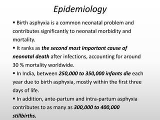
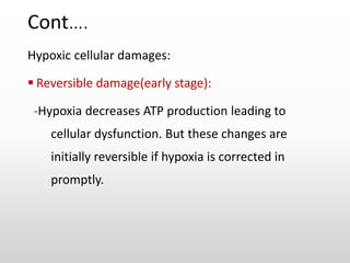
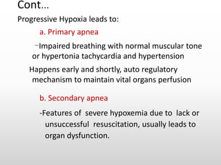
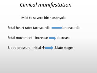
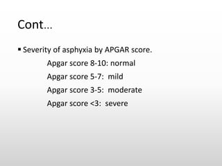
BIRTH ASPHYXIA
Bundala
MD
Objectives
 Introduction
 Epidemiology
 Etiology /risk factors
 Pathophysiology
 Clinical presentations
 Diagnostic criteria
 Management
 Prognosis
 Complications
Introduction
 Failure to initiate spontaneous, sustained breathing
after birth,
OR
 reduction of oxygen delivery and an accumulation
of carbon dioxide owing to cessation of blood
supply to the fetus around the time of birth.
Epidemiology
 Birth asphyxia is a common neonatal problem and
contributes significantly to neonatal morbidity and
mortality.
 It ranks as the second most important cause of
neonatal death after infections, accounting for around
30 % mortality worldwide.
 In India, between 250,000 to 350,000 infants die each
year due to birth asphyxia, mostly within the first three
days of life.
 In addition, ante-partum and intra-partum asphyxia
contributes to as many as 300,000 to 400,000
stillbirths.
Etiology/risk factors
 Pathologically, any factors which interfere with
the circulation between maternal and fetal blood
exchange could result in the happens of perinatal
asphyxia.
 These factors can be maternal factor, delivery
factor and fetal factor.
Cont…
 Maternal factors
Maternal age>35 or <16
Maternal infections
Post term gestation
Multiple gestation
Maternal drug abuse
Poly/oligohydramnios
 Fetal factor:
Multiple birth
Congenital or malformed
Fetus
Abnormal presentation
Meconium aspiration
 Delivery
condition:{Intrapartum}
Abruption of placenta
placenta Preavia
prolapsed cord
premature rupture of
membranes >24hrs
Analgesics, sedation or
drugs depressing the
respiratory centre
Prolonged labour
Pathophysiology
 Due to the presence of those different factors may
cause to placental insufficient which later on cause
impaired fetal gas exchange.
 Impaired fetal gas exchange during in utero and
labor resulting in severe hypoxia before delivery
this may predispose to birth asphyxia,
 Resulting to hypo-ventilation, hyper-capnia, hypo-
perfusion and metabolic acidosis
 When fetal hypoxia occurs, the body responds at
least temporarily by blood flow redistribution to
vital organs such as the brain, heart and adrenal
glands to prevent them from hypoxic damage.
Cont….
Hypoxic cellular damages:
 Reversible damage(early stage):
-Hypoxia decreases ATP production leading to
cellular dysfunction. But these changes are
initially reversible if hypoxia is corrected in
promptly.
Cont…
 Irreversible damage: (late stage)
-Persistent hypoxia leads to irreversible cellular
damage characterized by hypoxemia and marked
loss of Cerebral blood flow autoregulation and
increased celluar damage and cellular energy
dependent pump failure
Cont…
Progressive Hypoxia leads to:
a. Primary apnea
-Impaired breathing with normal muscular tone
or hypertonia tachycardia and hypertension
Happens early and shortly, auto regulatory
mechanism to maintain vital organs perfusion
b. Secondary apnea
-Features of severe hypoxemia due to lack or
unsuccessful resuscitation, usually leads to
organ dysfunction.
Cont…
Other damages:
a. Persistent pulmonary hypertension (PPHN)
b. Hyper/hypoglycemia
c. Hyperbilirubinemia
d. Electrolyte imbalances hyponatremia and
hypocalcemia
Clinical manifestation
Mild to severe birth asphyxia
Fetal heart rate: tachycardia bradycardia
Fetal movement: increase decrease
Blood pressure: Initial ↑ ↓late stages
Diagnostic criteria
 History of fetal distress
 Apgar score <3 at 1 min or <6 at 5 min
 Arterial blood lactate >5mmol/L
 Abnormal neurological state
 Multi-organ involvement
 Mixed acidosis
– Ph <7.2 on arterial cord blood
– Base excess >10
– Pa CO2 > 55 mmHg
Cont…
 Severity of asphyxia by APGAR score.
Apgar score 8-10: normal
Apgar score 5-7: mild
Apgar score 3-5: moderate
Apgar score <3: severe
Management
 1st minute counts (Golden)
Check if the baby is crying and breathing
If baby is not crying or breathing well after
delivery, one need to help baby in the first
minute
 ABCDE resuscitation
• A (air way)
• B (breathing)
• C (circulation)
• D (drug)
• E (evaluation)
Airway
 Open airways by placing the head in the neutral
position
 Clear the secretions from the airway by
suctioning as soon as possible
 If meconium-stained-deep tracheal suctioning
should be performed to removal of all the
meconium to avoid chemical pneumonitis - MAS
Breathing
 Ensure face mask covers nose & mouth connect
to oxygen bag
 Establish respiration of 30-40/min with chest wall
movement
 No response consider advanced airway
management-intubation & mechanic ventilation
Indications for intubation
 When bag and mask is in-effective
 When tracheal suction is required
 When diaphragmentic hernia is present
Circulation
 If heart rate <60/bpm, start external cardiac
compression
 Ratio 3:1 ( 90 compressions to 30 bpm)
Drugs
 In profound bradycardia, give adrenaline(1:10000, 0.1-
0.3ml/kg) by endotracheal tube or umbilical vein
 If no response, intravenous fluid (saline, albumin,
plasma, blood) with 10ml/kg
 In case of acidosis, give 5% sodium bicarbonate (SB)
with 3-5ml/kg
 Apneic and mum was given opiods consider using
naloxone (0.1mg/kg)
Prognosis
 Depends on associated factors, maturity of
the baby, duration and intensity of hypoxia
and acidosis including initiation of
resuscitative measures in the delivery room
 Subsequent competent care and available
facilities also influence the outcome following
birth asphyxia
Cont….
 Apgar score < 5 at 10 minutes : nearly 50 %
death or disability (Leicester)
 No spontaneous respiration after 20 min : 60
% disability in survivors (USA).
 No spontaneous respiration after 30 minutes :
nearly 100 % disability in survivors (Newcastle).
Complications
 Central nervous system
-infarction, intracranial hemorrhage,
cerebral edema, seizure, hypoxic ischemic
encephalopathy
 Cardiovascular
-bradycardia, ventricular hypertrophy,
arrhythmia, hypotension, myocardial
ischemia
Cont…
 Respiratory system
-apnea, respiratory distress syndrome, cyanosis
 KUB
-acute tubular necrosis, bladder paralysis
 Gastrointestinal tract
-necrotizing enterocolitis , stress ulcer
Cont…
 Hematology
-Disseminated intravascular coagulation
 Metabolic
-hypoglycemia, hyperglycemia, hypocalcemia,
hyponatremia
 Integument
-subcutaneous fat necrosis
Hypoxic Ischemic Encephalopathy
 Acquired syndrome of acute brain injury
characterised by neonatal encephalopathy in the
first 3 days of life and evidence of intrapartum
hypoxia in a term baby
 The immature brain is more resistant to hypoxic-
ischemic events as compared to older children
 NE is characterised by an abnormal LOC, abnormal
tone and primitive reflexes in the first week of life.
 Abnormal breathing and seizures may occur
Cont..
 An evolving process initiated by the hypoxic-
ischemic insult leading to decreased blood flow to
the brain followed by the restoration of blood flow
in an injured brain
 This initiates a cascade of pathways which includes
accumulation of extracellular glutamate
 With excessive activation of glutamate receptors,
ca influx and generation of reactive oxygen and
nitrogen species leading to cell death and brain
injuries.
Clinical features
 Apnea
 Bradycardia
 Grunting gasping
breaths
 Cyanosis pallor shock
 Hypotonia / hypertonia
 Hyporesponsiveness
The typical presentation
is that of multi-organ
involvement
 Convulsions
 Coma
 Hypotension
 Shock
 Renal failure
 Hypoglycemia
 Risk of infection
Diagnostic criteria
 A sentinel hypoxic event immediately before
or during labour.
 A sudden, rapid and sustained deterioration
of fetal heart rate.
 Apgar scores of 0-6 for longer than 5
minutes.
 Early evidence of multisystem involvement.
 Early imaging evidence of acute cerebral
abnormality.
Investigations
 Serum electrolytes levels
 Blood sugar levels
 Arterial blood gases
 Renal function tests
 Liver function tests
 Coagulation profile-
PT&PTT
 Cardiac enzymes
 Hb level & FBP
 CXR
 Brain USS
 CT
 MRI
 Echocardigraphy
 EEG
 Retinal and
opthalmological
evaluation
Cont…
Neuro-imaging
 MRI: most app technique and is able to show diff patterns
of injury. If available, it can be done at age 10-14 days
 CT scan: less sensitive than MRI for detecting changes in
the central gray nuclei
 Cranial U/S: not the best in assessing abnormalities in
term infants
 aEEG: has prognostic value and is useful in diagnosing
sub-clinical seizures
Management
 Aim is to treat symptoms and minimize further
organ damage
 Correct standard management is more important
than adding neuro-protective therapy such as
hypothermia
Cont…
 CNS
– Perform a daily neurological assessment and on
admission
– Treat recurrent and persistent
seizures{phenorbabital}
– If mod or severe NE is present at birth, induced
hypothermia may improve the outcome if
commenced asap and not after 6hrs post-del
Cont…
Hypothermia MOA
 Reduces cerebral metabolism prevents oedema
 Decreases energy utilisation
 Reduces cytotoxic amino acid accumulation
 Attenuates secondary neuronal damage
 Inhibits cell death
 Reduces extend of brain damage
– Death or severe disability at 18 months has been
reduced significantly
Cont…
Respiratory System
 Monitor oxygen sats
– If oxygen is needed but respiratory effort is good,
nasal continuous positive airway pressure is
adeqaute
Renal System
 Fluid balance and electrolytes
– Intrinsic renal failure and SIADH commonly occurs
– Initially restrict fluids to 40ml/kg/24 hrs with K
free 10% glucose and 0.45 % n saline
– Monitor urine output, electrolytes, blood glucose
Prognosis And Follow Up
 Neurological examination and aEEG are only
reliable when abnormalities are severe
 Early infant neuro-developmental assessment
can be predictive of the outcome
 Immediate the outcome is Death
 Cerebral palsy, developmental delay and intellectual
disabilities are common in survivors of severe HIE
and multidisciplinary follow-up is required.
Prevention
 Perinatal healthy care
 Prevention of asphyxia
Love is all we need!!

Birth asphyxia

  • 1.
  • 2.
    Objectives  Introduction  Epidemiology Etiology /risk factors  Pathophysiology  Clinical presentations  Diagnostic criteria  Management  Prognosis  Complications
  • 3.
    Introduction  Failure toinitiate spontaneous, sustained breathing after birth, OR  reduction of oxygen delivery and an accumulation of carbon dioxide owing to cessation of blood supply to the fetus around the time of birth.
  • 4.
    Epidemiology  Birth asphyxiais a common neonatal problem and contributes significantly to neonatal morbidity and mortality.  It ranks as the second most important cause of neonatal death after infections, accounting for around 30 % mortality worldwide.  In India, between 250,000 to 350,000 infants die each year due to birth asphyxia, mostly within the first three days of life.  In addition, ante-partum and intra-partum asphyxia contributes to as many as 300,000 to 400,000 stillbirths.
  • 5.
    Etiology/risk factors  Pathologically,any factors which interfere with the circulation between maternal and fetal blood exchange could result in the happens of perinatal asphyxia.  These factors can be maternal factor, delivery factor and fetal factor.
  • 6.
    Cont…  Maternal factors Maternalage>35 or <16 Maternal infections Post term gestation Multiple gestation Maternal drug abuse Poly/oligohydramnios  Fetal factor: Multiple birth Congenital or malformed Fetus Abnormal presentation Meconium aspiration  Delivery condition:{Intrapartum} Abruption of placenta placenta Preavia prolapsed cord premature rupture of membranes >24hrs Analgesics, sedation or drugs depressing the respiratory centre Prolonged labour
  • 7.
    Pathophysiology  Due tothe presence of those different factors may cause to placental insufficient which later on cause impaired fetal gas exchange.  Impaired fetal gas exchange during in utero and labor resulting in severe hypoxia before delivery this may predispose to birth asphyxia,  Resulting to hypo-ventilation, hyper-capnia, hypo- perfusion and metabolic acidosis  When fetal hypoxia occurs, the body responds at least temporarily by blood flow redistribution to vital organs such as the brain, heart and adrenal glands to prevent them from hypoxic damage.
  • 8.
    Cont…. Hypoxic cellular damages: Reversible damage(early stage): -Hypoxia decreases ATP production leading to cellular dysfunction. But these changes are initially reversible if hypoxia is corrected in promptly.
  • 9.
    Cont…  Irreversible damage:(late stage) -Persistent hypoxia leads to irreversible cellular damage characterized by hypoxemia and marked loss of Cerebral blood flow autoregulation and increased celluar damage and cellular energy dependent pump failure
  • 10.
    Cont… Progressive Hypoxia leadsto: a. Primary apnea -Impaired breathing with normal muscular tone or hypertonia tachycardia and hypertension Happens early and shortly, auto regulatory mechanism to maintain vital organs perfusion b. Secondary apnea -Features of severe hypoxemia due to lack or unsuccessful resuscitation, usually leads to organ dysfunction.
  • 11.
    Cont… Other damages: a. Persistentpulmonary hypertension (PPHN) b. Hyper/hypoglycemia c. Hyperbilirubinemia d. Electrolyte imbalances hyponatremia and hypocalcemia
  • 12.
    Clinical manifestation Mild tosevere birth asphyxia Fetal heart rate: tachycardia bradycardia Fetal movement: increase decrease Blood pressure: Initial ↑ ↓late stages
  • 13.
    Diagnostic criteria  Historyof fetal distress  Apgar score <3 at 1 min or <6 at 5 min  Arterial blood lactate >5mmol/L  Abnormal neurological state  Multi-organ involvement  Mixed acidosis – Ph <7.2 on arterial cord blood – Base excess >10 – Pa CO2 > 55 mmHg
  • 14.
    Cont…  Severity ofasphyxia by APGAR score. Apgar score 8-10: normal Apgar score 5-7: mild Apgar score 3-5: moderate Apgar score <3: severe
  • 15.
    Management  1st minutecounts (Golden) Check if the baby is crying and breathing If baby is not crying or breathing well after delivery, one need to help baby in the first minute  ABCDE resuscitation • A (air way) • B (breathing) • C (circulation) • D (drug) • E (evaluation)
  • 16.
    Airway  Open airwaysby placing the head in the neutral position  Clear the secretions from the airway by suctioning as soon as possible  If meconium-stained-deep tracheal suctioning should be performed to removal of all the meconium to avoid chemical pneumonitis - MAS
  • 17.
    Breathing  Ensure facemask covers nose & mouth connect to oxygen bag  Establish respiration of 30-40/min with chest wall movement  No response consider advanced airway management-intubation & mechanic ventilation
  • 18.
    Indications for intubation When bag and mask is in-effective  When tracheal suction is required  When diaphragmentic hernia is present
  • 19.
    Circulation  If heartrate <60/bpm, start external cardiac compression  Ratio 3:1 ( 90 compressions to 30 bpm)
  • 20.
    Drugs  In profoundbradycardia, give adrenaline(1:10000, 0.1- 0.3ml/kg) by endotracheal tube or umbilical vein  If no response, intravenous fluid (saline, albumin, plasma, blood) with 10ml/kg  In case of acidosis, give 5% sodium bicarbonate (SB) with 3-5ml/kg  Apneic and mum was given opiods consider using naloxone (0.1mg/kg)
  • 21.
    Prognosis  Depends onassociated factors, maturity of the baby, duration and intensity of hypoxia and acidosis including initiation of resuscitative measures in the delivery room  Subsequent competent care and available facilities also influence the outcome following birth asphyxia
  • 22.
    Cont….  Apgar score< 5 at 10 minutes : nearly 50 % death or disability (Leicester)  No spontaneous respiration after 20 min : 60 % disability in survivors (USA).  No spontaneous respiration after 30 minutes : nearly 100 % disability in survivors (Newcastle).
  • 23.
    Complications  Central nervoussystem -infarction, intracranial hemorrhage, cerebral edema, seizure, hypoxic ischemic encephalopathy  Cardiovascular -bradycardia, ventricular hypertrophy, arrhythmia, hypotension, myocardial ischemia
  • 24.
    Cont…  Respiratory system -apnea,respiratory distress syndrome, cyanosis  KUB -acute tubular necrosis, bladder paralysis  Gastrointestinal tract -necrotizing enterocolitis , stress ulcer
  • 25.
    Cont…  Hematology -Disseminated intravascularcoagulation  Metabolic -hypoglycemia, hyperglycemia, hypocalcemia, hyponatremia  Integument -subcutaneous fat necrosis
  • 26.
    Hypoxic Ischemic Encephalopathy Acquired syndrome of acute brain injury characterised by neonatal encephalopathy in the first 3 days of life and evidence of intrapartum hypoxia in a term baby  The immature brain is more resistant to hypoxic- ischemic events as compared to older children  NE is characterised by an abnormal LOC, abnormal tone and primitive reflexes in the first week of life.  Abnormal breathing and seizures may occur
  • 27.
    Cont..  An evolvingprocess initiated by the hypoxic- ischemic insult leading to decreased blood flow to the brain followed by the restoration of blood flow in an injured brain  This initiates a cascade of pathways which includes accumulation of extracellular glutamate  With excessive activation of glutamate receptors, ca influx and generation of reactive oxygen and nitrogen species leading to cell death and brain injuries.
  • 28.
    Clinical features  Apnea Bradycardia  Grunting gasping breaths  Cyanosis pallor shock  Hypotonia / hypertonia  Hyporesponsiveness The typical presentation is that of multi-organ involvement  Convulsions  Coma  Hypotension  Shock  Renal failure  Hypoglycemia  Risk of infection
  • 29.
    Diagnostic criteria  Asentinel hypoxic event immediately before or during labour.  A sudden, rapid and sustained deterioration of fetal heart rate.  Apgar scores of 0-6 for longer than 5 minutes.  Early evidence of multisystem involvement.  Early imaging evidence of acute cerebral abnormality.
  • 30.
    Investigations  Serum electrolyteslevels  Blood sugar levels  Arterial blood gases  Renal function tests  Liver function tests  Coagulation profile- PT&PTT  Cardiac enzymes  Hb level & FBP  CXR  Brain USS  CT  MRI  Echocardigraphy  EEG  Retinal and opthalmological evaluation
  • 31.
    Cont… Neuro-imaging  MRI: mostapp technique and is able to show diff patterns of injury. If available, it can be done at age 10-14 days  CT scan: less sensitive than MRI for detecting changes in the central gray nuclei  Cranial U/S: not the best in assessing abnormalities in term infants  aEEG: has prognostic value and is useful in diagnosing sub-clinical seizures
  • 32.
    Management  Aim isto treat symptoms and minimize further organ damage  Correct standard management is more important than adding neuro-protective therapy such as hypothermia
  • 33.
    Cont…  CNS – Performa daily neurological assessment and on admission – Treat recurrent and persistent seizures{phenorbabital} – If mod or severe NE is present at birth, induced hypothermia may improve the outcome if commenced asap and not after 6hrs post-del
  • 34.
    Cont… Hypothermia MOA  Reducescerebral metabolism prevents oedema  Decreases energy utilisation  Reduces cytotoxic amino acid accumulation  Attenuates secondary neuronal damage  Inhibits cell death  Reduces extend of brain damage – Death or severe disability at 18 months has been reduced significantly
  • 35.
    Cont… Respiratory System  Monitoroxygen sats – If oxygen is needed but respiratory effort is good, nasal continuous positive airway pressure is adeqaute Renal System  Fluid balance and electrolytes – Intrinsic renal failure and SIADH commonly occurs – Initially restrict fluids to 40ml/kg/24 hrs with K free 10% glucose and 0.45 % n saline – Monitor urine output, electrolytes, blood glucose
  • 36.
    Prognosis And FollowUp  Neurological examination and aEEG are only reliable when abnormalities are severe  Early infant neuro-developmental assessment can be predictive of the outcome  Immediate the outcome is Death  Cerebral palsy, developmental delay and intellectual disabilities are common in survivors of severe HIE and multidisciplinary follow-up is required.
  • 37.
    Prevention  Perinatal healthycare  Prevention of asphyxia
  • 38.
    Love is allwe need!!