This document discusses birth asphyxia, including its definition, causes, pathophysiology, clinical manifestations, assessment, effects, classification, management, investigations and prognosis. Some key points:
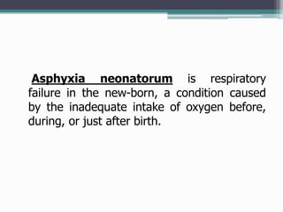
- Birth asphyxia is defined as reduction of oxygen delivery and accumulation of carbon dioxide around birth, leading to respiratory failure in newborns. It is assessed using Apgar scores and fetal monitoring.
- Causes include maternal, delivery and fetal factors that interfere with maternal-fetal circulation such as prematurity, cord problems and placental issues.
- Effects depend on severity and can involve multiple organs, particularly the brain, heart and lungs. Management focuses on stabilizing vital functions and preventing further injury through temperature control