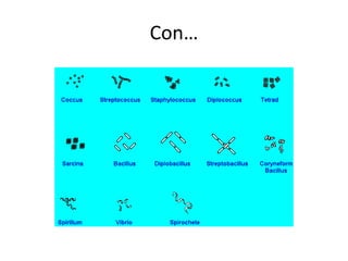
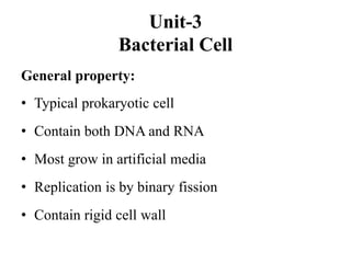
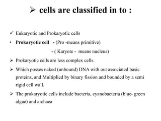
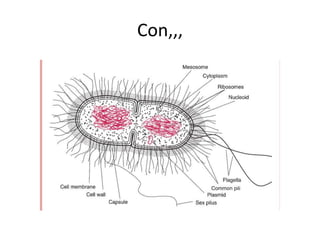
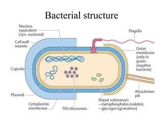
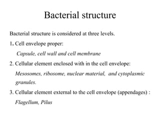
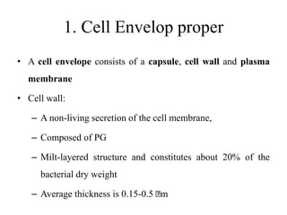
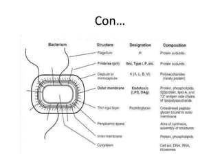
Bacterial of Classification
Bacteria are classified to identify and distinguish one
organism from another and
to group similar organisms by criteria of interest to
microbiologists or other scientists.
Bacteria are classified based on the following :
1. Taxonomy
-is the science of organismal classification
-Lower level taxa -eg. Species
- higher level taxa-eg. kingdome
2. Nomenclature
 Naming of an organism by international rules
 Organisms are named using binomial nomenclature
(except viruses)
 Employ the name of the two level taxa, genes and spps.
 Genus comes before species (E.coli)
 Genus name always capitalized while species is not and
both underlined or italicized. e.g. Salmonella typhi,
Genus name some time use alone but not species.
E.g. Salmonella
Strain→ level below the species
3. Observational and experimental
technique
1. Phenotypic classification of bacteria
 Microscopic and macroscopic morphologies:
is the corner stone for identification
I. Microscopic appearance-depend on shape
e.g. AFS, Gram stain - shape, size and arrangement
e.g. Cocci (sing Coccus)- round (spherical in shape)
Bacilli (sig-Bacillus)- rod in shape
Spirochetes – spiral in shape
Pleomorphic- variable in shape (short rods)
A. Cocci
 Round or oval -measures about 0.5-1.0 mm in diameter
 Single, pairs ,chains or clusters
 Diplococcus(kidney shaped) e.g. Neisseria
 Diplococcus in pairs with pointed ends
e.g. S.pneumonia
 Streptococcus in chains e.g S.pyogens
 Staphylococcus in groups e.g. S.aureus
B. Bacilli
Rod shaped ( stick like)
e.g. - round ends→ Salmonella
- square ends→Bacillus
- club shaped→Corynebacterium
-Comma shaped →Vibrio
C. Spiral
• Spiral shaped bacteria with regular /irregular
distance b/n twisting
Phenotypic Characteristics
Morphologic Characteristics
• This information often can allow identification of an
organism.
• These simple tests can indicate the Gram reaction of the
organism;
 whether it is acid-fast; its motility; the arrangement of
its flagella; the presence of spores, capsules, and
inclusion bodies and, its shape.
Con,,,
Morphology:
- Bacteria vary widely in size, ranging from 0.2 um to
10um long
-There are there basic shapes
1. Spherical or coccoid / cocci- (singular –coccus)
2. Rods or bacilli (singular - bacillus)
3. Spirals or spirilla (Singular - Spirillum
Con…
Generally, Bacteria classification is
based on
1. Morphology
2. Staining
3. Motility
4. Growth
5. Nutritional requirement
6. Biochemical and metabolic activity
7. Pathogenicity
8. Amino acid sequencing of proteins
9. Genetic composition
Unit-3
Bacterial Cell
General property:
• Typical prokaryotic cell
• Contain both DNA and RNA
• Most grow in artificial media
• Replication is by binary fission
• Contain rigid cell wall
 cells are classified in to :
 Eukaryotic and Prokaryotic cells
• Prokaryotic cell - (Pro -means primitive)
- ( Karyote - means nucleus)
 Prokaryotic cells are less complex cells.
 Which posses naked (unbound) DNA with out associated basic
proteins, and Multiplied by binary fission and bounded by a semi
rigid cell wall.
 The prokaryotic cells include bacteria, cyanobacteria (blue- green
algae) and archaea
Con,,,
Eukaryotic cells.
-(Eu- means true,
-(Karyote- means nucleus)
The Eukaryotic cell is more complex, has
membrane bound nucleus, and many organells.
 Eukaryotic cells include protozoa, fungi, algae,
(green, brown and red algae) and plant and animal
cells.
Con,,,
Bacterial structure
Bacterial structure
Bacterial structure is considered at three levels.
1. Cell envelope proper:
Capsule, cell wall and cell membrane
2. Cellular element enclosed with in the cell envelope:
Mesosomes, ribosome, nuclear material, and cytoplasmic
granules.
3. Cellular element external to the cell envelope (appendages) :
Flagellum, Pilus
1. Cell Envelop proper
• A cell envelope consists of a capsule, cell wall and plasma
membrane
• Cell wall:
– A non-living secretion of the cell membrane,
– Composed of PG
– Milt-layered structure and constitutes about 20% of the
bacterial dry weight
– Average thickness is 0.15-0.5 m
Chemical Composition of cell wall
• The major component of cell wall is peptidoglycan (PG)
• The rigidity of the cell wall is due to the presence of this substance
• It consists of a polymer of disaccharides cross-linked by short chains
of amino acids (peptides).
• This molecule is a type of peptidoglycan, which is called murein.
– peptidoglycan layer (murein) is a complex, interwoven network
that surrounds the entire cells and is composed of a single
covalently linked macromolecules.
– Found only in bacterial cell walls
Function of cell wall
• protects the delicate cell protoplast from osmotic lysis.
• Provide rigid support for the cells
• Protects from environments
• Provides staining to bacterial
• Site of action of antibiotic
Cell Wall-less Forms of bacteria
Bacteria with defective cell walls include:
• Protoplasts: bacteria with out cell wall.
• Spheroplast: bacterium with a damaged cell wall.
• Mycoplasma: highly pleomorphic bacteria with out
cell wall
Types of cell wall
I. Gram positive cell wall of bacteria
has two layers (Peptidoglycan (PG) cross linked with
teichoic acid)
– The PG layers is much thicker than Gram negative
bacteria and i.e. 15 – 50 nm thick
The PG layer comprises 50 – 90% of
the cell wall and 20 – 40% of the cell
wall weight
Gram positive cell wall …Cont’d
• The large amount of peptideglycan make gram- positive
bacteria susceptible to the enzyme lysozyme and
penicillin.
• Lysozyme hydrolyzes peptidoglycan by specific cleavage
between N-acetyl muramic acid and the N-acetyl
glucosamine of the glycon strand.
• Penicillin specifically inhibits peptidoglycan synthesis
• Teichoic acids and cell wall- associated protein are the
major surface antigens of the gram- positive cell wall.
Con…
Teichoic acid
These polymers of glycerol phosphate or ribitol
phosphate are located in the outer layer of the GP cell
wall.
Function of Teichoic acid
– Used to bind (keep) Mg+2 concentration in the cell
wall
– Used to activate autolytic enzyme (enzymes which
are secreted by bacteria usually when it dies
– To bind bacteriophage in the cell wall.
II. Gram negative Cell wall of bacteria
• Is some what complex than Gram positive bacterial cell
wall
• Has thin peptidoglycan layer (3 – 8nm)
• Has high lipid content (lipopolysaccharied) in the
outer membrane
• Has periplasmic space.
Con…
Lipopoly saccharides
• It is responsible for antigenicty of the outer
membrane
Periplasmic space
• Found between outer membrane and the cell
membrane
• Mostly contain enzymes and endotoxins.
Fig. Gram-Positive
Fig Gram negative cell wall of bacteria
Cytoplasmic membrane (Plasma membrane)
• It is the actual barrier between the interior and exterior of
the bacteria cell.
• The cytoplasmic membrane exhibits a well- defined
selective permeability, excretion of enzyme, and
biosynthesis of cell well and other proteins
• The bacterial transport system and the principal energy
system (oxidative phosphophorylation) are located in the
cytoplasmic membrane.
Cytoplasmic membrane … Cont’d
• It accounts for 30% of the dry weight of bacterial cell
• Chemically, the plasma membrane consists of proteins and
phospholipids.
• It is 60% protein, 20 – 30% lipid and 10-20% carbohydrate
• Although procaryotes lack any intracellular organelles for
respiration or photosynthesis, many species possess the
physiologic ability to conduct these processes, usually as a
function of their plasma membrane.
Function of Cell Membrane
• Regulates the transport of nutrients and waste products into
and out of the cell. (It is selectively permeable)
• Assists DNA replication
• Captures energy in ATP (i.e it is site of oxidative
phosphorulation )
Cytoplasm
• viscous watery solution of soft gel, containing a variety
organic and inorganic solutes and numerous small
granule called ribosomes
• The cytoplasm consists of water, enzymes oxygen,
waste products, essential nutrients, proteins,
carbohydrates, lipids and a complex mixture of all the
materials required by the cell for its metabolic functions.
2. Cellular Element Enclosed with in the Cell Envelope
Mesosomes:
• are complex invaginations of cytoplasmic membrane in to the
cytoplasm seen in many bacteria, but not in all.
– Increase in the total surface area of the membrane.
– Mesosomes are attached to chromosomes and are involved in
DNA segregation during cell division .
– Others are involved in to secretion of proteins and active
transport.
– It is involved in respiratory enzyme-activity.
– Site of oxidative phosphorylation
Ribosomes
• are the sites of protein synthesis
• It is composed of ribosomal RNA (rRNA) (70%) and proteins (30%)
• It constitutes 90% of the RNA and 40% of the total protein.
• The ribosomes of procaryotes are smaller than ribosomes of
eucaryotes.
• Procaryotic ribosomes are 70S in size, being composed of 30S and
50S subunits(80S)
• S or Svedberg unit designates the sedimentation coefficient of the
rRNA
nucleoid (Nuclear material)
• is concentrated in the cytoplasm as a nucleoid
• The nucleoid consists of one long double-stranded circular
DNA molecule (chromosome).
• The chromosome serves as:
 the control center of the bacterial cell,
 carries of the genetic information needed for producing several
thousand enzymes and other proteins.
• The size of the chromosome varies according to species.
Con…
• The prokaryotic nucleoid is considered as primitive nucleus,
– it is not surrounded by a nuclear membrane
– It does not have a definite shape, and has little or no protein
material.
• As apart of nucleus, bacteria may have some extra
chromosomal genetic material in the form of DNA, which is
known as Plasmid
3. Cellular Element External to the Cell Envelope
Flagellum
• It is the organ of locomotion in bacterial cell and consists of filament
• is free on the surface of bacterial cell
• Size: 3-20 m in length and 0.01-0.013 m in diameter.
• It is composed of single protein named as flagellin
• The flagella antigen in motile bacterium is named as H-Ag (Hauch
antigen).
Con…
Con…
Flagellar arrangements
1. Atrocious: Bacteria with no flagellum: Eg. All cocci
2. Monotrichous: Bacteria with single polar flagellum:
Eg. V.cholara
3. Lophotrichous: Bacteria with bunch of flagella at one pole:
Eg. Pseudomonas flouresense
4. Amphitrichous: Bacteria with flagella at both poles:
Eg. Alcaligenes faecales
5. Peritrichous: Bacteria with flagella all over their surface:
Eg. S.typhi
Con…
Con…
• non flagellated bacteria can move by:
 blinding, flexing, and spinning
Pilli and Fimbriae (finer filaments)
• interchangeable terms used to designate short, hair-
like structures on the surfaces of prokaryotic cells.
• are extruding from the cytoplasmic membrane
1.Common pili (fimbriae)
• cover the surface of the cell.
• Helps for adhesions, which are responsible to colonize
surfaces and cells.
• Help for attachment of bacteria to epithelial cell
• They considered as virulence factor in some species of
bacteria,
• because they allow pathogens to attach (colonize) tissues and
to resist attack by phagocytic white blood cells.
Con…
2.Sex pili or F pili
– Occur less commonly
– Appear to be specifically involved in bacterial
conjugation,
– Transfer of genetic material (DNA) from one
bacterium to another.
Bacterial spores
• Under conditions of limited supply of nutrition, vegetative forms of
certain bacteria especially gram-positive bacilli and highly resistant
and dehydrated forms, which are called endospores.
• These endospores are capable of survival under adverse conditions
such as heat, drying, freezing, radiation, and actions of toxic
chemicals.
• Spores are usually smooth walled and avoid, in some species it is
spherical.
Con...
,
Thank you

Bacterial of Classification.pptx

  • 1.
    Bacterial of Classification Bacteriaare classified to identify and distinguish one organism from another and to group similar organisms by criteria of interest to microbiologists or other scientists. Bacteria are classified based on the following : 1. Taxonomy -is the science of organismal classification -Lower level taxa -eg. Species - higher level taxa-eg. kingdome
  • 2.
    2. Nomenclature  Namingof an organism by international rules  Organisms are named using binomial nomenclature (except viruses)  Employ the name of the two level taxa, genes and spps.  Genus comes before species (E.coli)  Genus name always capitalized while species is not and both underlined or italicized. e.g. Salmonella typhi, Genus name some time use alone but not species. E.g. Salmonella Strain→ level below the species
  • 3.
    3. Observational andexperimental technique 1. Phenotypic classification of bacteria  Microscopic and macroscopic morphologies: is the corner stone for identification I. Microscopic appearance-depend on shape e.g. AFS, Gram stain - shape, size and arrangement e.g. Cocci (sing Coccus)- round (spherical in shape) Bacilli (sig-Bacillus)- rod in shape Spirochetes – spiral in shape Pleomorphic- variable in shape (short rods)
  • 4.
    A. Cocci  Roundor oval -measures about 0.5-1.0 mm in diameter  Single, pairs ,chains or clusters  Diplococcus(kidney shaped) e.g. Neisseria  Diplococcus in pairs with pointed ends e.g. S.pneumonia  Streptococcus in chains e.g S.pyogens  Staphylococcus in groups e.g. S.aureus
  • 5.
    B. Bacilli Rod shaped( stick like) e.g. - round ends→ Salmonella - square ends→Bacillus - club shaped→Corynebacterium -Comma shaped →Vibrio C. Spiral • Spiral shaped bacteria with regular /irregular distance b/n twisting
  • 6.
    Phenotypic Characteristics Morphologic Characteristics •This information often can allow identification of an organism. • These simple tests can indicate the Gram reaction of the organism;  whether it is acid-fast; its motility; the arrangement of its flagella; the presence of spores, capsules, and inclusion bodies and, its shape.
  • 7.
    Con,,, Morphology: - Bacteria varywidely in size, ranging from 0.2 um to 10um long -There are there basic shapes 1. Spherical or coccoid / cocci- (singular –coccus) 2. Rods or bacilli (singular - bacillus) 3. Spirals or spirilla (Singular - Spirillum
  • 8.
  • 9.
    Generally, Bacteria classificationis based on 1. Morphology 2. Staining 3. Motility 4. Growth 5. Nutritional requirement 6. Biochemical and metabolic activity 7. Pathogenicity 8. Amino acid sequencing of proteins 9. Genetic composition
  • 10.
    Unit-3 Bacterial Cell General property: •Typical prokaryotic cell • Contain both DNA and RNA • Most grow in artificial media • Replication is by binary fission • Contain rigid cell wall
  • 11.
     cells areclassified in to :  Eukaryotic and Prokaryotic cells • Prokaryotic cell - (Pro -means primitive) - ( Karyote - means nucleus)  Prokaryotic cells are less complex cells.  Which posses naked (unbound) DNA with out associated basic proteins, and Multiplied by binary fission and bounded by a semi rigid cell wall.  The prokaryotic cells include bacteria, cyanobacteria (blue- green algae) and archaea
  • 12.
  • 13.
    Eukaryotic cells. -(Eu- meanstrue, -(Karyote- means nucleus) The Eukaryotic cell is more complex, has membrane bound nucleus, and many organells.  Eukaryotic cells include protozoa, fungi, algae, (green, brown and red algae) and plant and animal cells.
  • 14.
  • 15.
  • 16.
    Bacterial structure Bacterial structureis considered at three levels. 1. Cell envelope proper: Capsule, cell wall and cell membrane 2. Cellular element enclosed with in the cell envelope: Mesosomes, ribosome, nuclear material, and cytoplasmic granules. 3. Cellular element external to the cell envelope (appendages) : Flagellum, Pilus
  • 17.
    1. Cell Envelopproper • A cell envelope consists of a capsule, cell wall and plasma membrane • Cell wall: – A non-living secretion of the cell membrane, – Composed of PG – Milt-layered structure and constitutes about 20% of the bacterial dry weight – Average thickness is 0.15-0.5 m
  • 18.
    Chemical Composition ofcell wall • The major component of cell wall is peptidoglycan (PG) • The rigidity of the cell wall is due to the presence of this substance • It consists of a polymer of disaccharides cross-linked by short chains of amino acids (peptides). • This molecule is a type of peptidoglycan, which is called murein. – peptidoglycan layer (murein) is a complex, interwoven network that surrounds the entire cells and is composed of a single covalently linked macromolecules. – Found only in bacterial cell walls
  • 19.
    Function of cellwall • protects the delicate cell protoplast from osmotic lysis. • Provide rigid support for the cells • Protects from environments • Provides staining to bacterial • Site of action of antibiotic
  • 20.
    Cell Wall-less Formsof bacteria Bacteria with defective cell walls include: • Protoplasts: bacteria with out cell wall. • Spheroplast: bacterium with a damaged cell wall. • Mycoplasma: highly pleomorphic bacteria with out cell wall
  • 21.
    Types of cellwall I. Gram positive cell wall of bacteria has two layers (Peptidoglycan (PG) cross linked with teichoic acid) – The PG layers is much thicker than Gram negative bacteria and i.e. 15 – 50 nm thick The PG layer comprises 50 – 90% of the cell wall and 20 – 40% of the cell wall weight
  • 22.
    Gram positive cellwall …Cont’d • The large amount of peptideglycan make gram- positive bacteria susceptible to the enzyme lysozyme and penicillin. • Lysozyme hydrolyzes peptidoglycan by specific cleavage between N-acetyl muramic acid and the N-acetyl glucosamine of the glycon strand. • Penicillin specifically inhibits peptidoglycan synthesis • Teichoic acids and cell wall- associated protein are the major surface antigens of the gram- positive cell wall.
  • 23.
    Con… Teichoic acid These polymersof glycerol phosphate or ribitol phosphate are located in the outer layer of the GP cell wall. Function of Teichoic acid – Used to bind (keep) Mg+2 concentration in the cell wall – Used to activate autolytic enzyme (enzymes which are secreted by bacteria usually when it dies – To bind bacteriophage in the cell wall.
  • 24.
    II. Gram negativeCell wall of bacteria • Is some what complex than Gram positive bacterial cell wall • Has thin peptidoglycan layer (3 – 8nm) • Has high lipid content (lipopolysaccharied) in the outer membrane • Has periplasmic space.
  • 25.
    Con… Lipopoly saccharides • Itis responsible for antigenicty of the outer membrane Periplasmic space • Found between outer membrane and the cell membrane • Mostly contain enzymes and endotoxins.
  • 26.
  • 27.
    Fig Gram negativecell wall of bacteria
  • 28.
    Cytoplasmic membrane (Plasmamembrane) • It is the actual barrier between the interior and exterior of the bacteria cell. • The cytoplasmic membrane exhibits a well- defined selective permeability, excretion of enzyme, and biosynthesis of cell well and other proteins • The bacterial transport system and the principal energy system (oxidative phosphophorylation) are located in the cytoplasmic membrane.
  • 29.
    Cytoplasmic membrane …Cont’d • It accounts for 30% of the dry weight of bacterial cell • Chemically, the plasma membrane consists of proteins and phospholipids. • It is 60% protein, 20 – 30% lipid and 10-20% carbohydrate • Although procaryotes lack any intracellular organelles for respiration or photosynthesis, many species possess the physiologic ability to conduct these processes, usually as a function of their plasma membrane. Function of Cell Membrane • Regulates the transport of nutrients and waste products into and out of the cell. (It is selectively permeable) • Assists DNA replication • Captures energy in ATP (i.e it is site of oxidative phosphorulation )
  • 30.
    Cytoplasm • viscous waterysolution of soft gel, containing a variety organic and inorganic solutes and numerous small granule called ribosomes • The cytoplasm consists of water, enzymes oxygen, waste products, essential nutrients, proteins, carbohydrates, lipids and a complex mixture of all the materials required by the cell for its metabolic functions.
  • 31.
    2. Cellular ElementEnclosed with in the Cell Envelope Mesosomes: • are complex invaginations of cytoplasmic membrane in to the cytoplasm seen in many bacteria, but not in all. – Increase in the total surface area of the membrane. – Mesosomes are attached to chromosomes and are involved in DNA segregation during cell division . – Others are involved in to secretion of proteins and active transport. – It is involved in respiratory enzyme-activity. – Site of oxidative phosphorylation
  • 32.
    Ribosomes • are thesites of protein synthesis • It is composed of ribosomal RNA (rRNA) (70%) and proteins (30%) • It constitutes 90% of the RNA and 40% of the total protein. • The ribosomes of procaryotes are smaller than ribosomes of eucaryotes. • Procaryotic ribosomes are 70S in size, being composed of 30S and 50S subunits(80S) • S or Svedberg unit designates the sedimentation coefficient of the rRNA
  • 33.
    nucleoid (Nuclear material) •is concentrated in the cytoplasm as a nucleoid • The nucleoid consists of one long double-stranded circular DNA molecule (chromosome). • The chromosome serves as:  the control center of the bacterial cell,  carries of the genetic information needed for producing several thousand enzymes and other proteins. • The size of the chromosome varies according to species.
  • 34.
    Con… • The prokaryoticnucleoid is considered as primitive nucleus, – it is not surrounded by a nuclear membrane – It does not have a definite shape, and has little or no protein material. • As apart of nucleus, bacteria may have some extra chromosomal genetic material in the form of DNA, which is known as Plasmid
  • 35.
    3. Cellular ElementExternal to the Cell Envelope Flagellum • It is the organ of locomotion in bacterial cell and consists of filament • is free on the surface of bacterial cell • Size: 3-20 m in length and 0.01-0.013 m in diameter. • It is composed of single protein named as flagellin • The flagella antigen in motile bacterium is named as H-Ag (Hauch antigen).
  • 36.
  • 37.
    Con… Flagellar arrangements 1. Atrocious:Bacteria with no flagellum: Eg. All cocci 2. Monotrichous: Bacteria with single polar flagellum: Eg. V.cholara 3. Lophotrichous: Bacteria with bunch of flagella at one pole: Eg. Pseudomonas flouresense 4. Amphitrichous: Bacteria with flagella at both poles: Eg. Alcaligenes faecales 5. Peritrichous: Bacteria with flagella all over their surface: Eg. S.typhi
  • 38.
  • 39.
    Con… • non flagellatedbacteria can move by:  blinding, flexing, and spinning Pilli and Fimbriae (finer filaments) • interchangeable terms used to designate short, hair- like structures on the surfaces of prokaryotic cells. • are extruding from the cytoplasmic membrane
  • 40.
    1.Common pili (fimbriae) •cover the surface of the cell. • Helps for adhesions, which are responsible to colonize surfaces and cells. • Help for attachment of bacteria to epithelial cell • They considered as virulence factor in some species of bacteria, • because they allow pathogens to attach (colonize) tissues and to resist attack by phagocytic white blood cells.
  • 41.
    Con… 2.Sex pili orF pili – Occur less commonly – Appear to be specifically involved in bacterial conjugation, – Transfer of genetic material (DNA) from one bacterium to another.
  • 42.
    Bacterial spores • Underconditions of limited supply of nutrition, vegetative forms of certain bacteria especially gram-positive bacilli and highly resistant and dehydrated forms, which are called endospores. • These endospores are capable of survival under adverse conditions such as heat, drying, freezing, radiation, and actions of toxic chemicals. • Spores are usually smooth walled and avoid, in some species it is spherical.
  • 43.
  • 44.

Editor's Notes

  • #5 Round or oval -measures about 0.5-1.0 mm in diameter Single, pairs ,chains or clusters  Diplococcus(kidney shaped) e.g. Neisseria Diplococcus in pairs with pointed ends e.g. S.pneumonia Streptococcus in chains e.g S.pyogens Staphylococcus in groups e.g. S.aureus