SlideShare a Scribd company logo
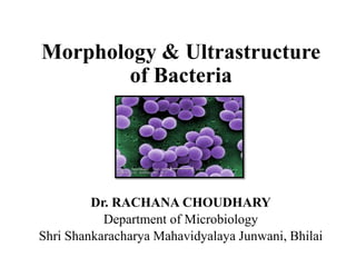
Morphology & Ultrastructure
of Bacteria
Dr. RACHANA CHOUDHARY
Department of Microbiology
Shri Shankaracharya Mahavidyalaya Junwani, Bhilai
INTRODUCTION
• Bacteria are the first organisms to
appear on earth
• That thrive in diverse environments.
• These organisms can live in soil, the
ocean and inside the human gut.
• Humans' relationship with bacteria is
complex.
• Some bacteria are harmful, but most
serve a useful purpose.
• They support many forms of life, both
plant and animal, and they are used in
industrial and medicinal processes.
GENERAL CHARACTERISTICS OF
BACTERIA
•Bacteria are Microscopic, Single-celled organisms.
•They lack organelles such as chloroplasts and
mitochondria, and they do not have the true nucleus.
•Bacteria also have a cell membrane and a cell wall .
•The cell membrane and cell wall are referred to as the cell
envelope.
•Asexual Reproduction by binary fission.
•However, some bacteria can also exchange genetic
material among one another in a process known as
horizontal gene transfer.
MORPHOLOGY OF BACTERIA
Morphology of Bacteria include:
•Size
•Shape
•Arrangement
Size of bacteria
•Bacterial cells are about one-tenth the size of
eukaryotic cells.
• typically 0.5–5.0 micrometers in length.
•Thiomargarita namibiensis is up to half a
millimeter long
•Epulopiscium fishelsoni reaches 0.7 mm
•Mycoplasma, which measure only 0.3 micrometers.
•E. coli , is 1.1 to 1.5 µm wide by 2.0 to 6.0 µm
long.
•Spirochaetes 500 µm in length.
•Cyanobacterium Oscillatoria is about 7 µm in
diameter.
Shapes of Bacteria
•Coccus
•Chain = Streptoccus
•Cluster = Staphylococcus
•Bacillus
•Chain = Streptobacillus
•Coccobacillus
•Vibrio = curved
•Spirillum
•Spirochete
•Square
•Star
Arrangement in bacteria
Chapter 4
Arrangement on the Basis of Presence of
flagella
1. Atrichpus :Absence of
flagella
2. Monotrichous; 1 flagella
3. Lophotrichous; tuft at one
end
4. Amphitrichous; both ends
5. Peritrichous; all around
bacteria
ULTRASTRUCTURE OF BACTERA
•Flagella
•Pili & Fimbriae
•Capsule & Slime Layers
•Cell Wall
•Plasma Membrane
•Mesosomes
•Cytoplasmic Inclusions
•Nucleoid
•Plasmids
Flagella
•Flagellum (singular) is hair like helical structure
emerges from cell wall and cell membrane
•It is responsible for motility of the bacteria
•Size: thin 15-20nm in diameter.
•Single flagella can be seen with light microscope
only after staining with special stain which increase
the diameter of flagella.
•Non-contractile
•Single protein subunit - flagellin
STRUCTURE OF FLAGELLA
Flagella has three parts. Basal body, Hook and filament
Basal body:
• it is composed of central rod inserted into series of rings which is
attached to cytoplasmic membrane and cell wall.
• L-ring: it is the outer ring present only in Gram -ve bacteria, it
anchored in lipopolysaccharide layer.
• P-ring: it is second ring anchored in peptidoglycan layer of cell wall.
• M-S ring: anchored in cytoplasmic membrane.
Hook:
• it is the wider region at the base of filament.it connects filament to the
motor protein in the base.length of hook is longer in gram +ve
bacteria than gram –ve bacteria
Filament:
• it is thin hair like structure arises from hook.
STRUCTURE OF FLAGELLA
FUNCTION OF FLAGELLA
1. That acts primarily as an organelle of
locomotion/movement in the cells.
2. They act as sensory organs to detect temperature
and pH changes.
3. Few eukaryotes use flagellum to increase
reproduction rates.
4. Recent researches have proved that flagella are
also used as a secretory organelle.
For eg., in Chlamydomonas
Pili & Fimbrae
• Both fimbriae and pili are hair like appendages on bacterial
cell wall. They originate from cytoplasm that protrudes
outside after penetrating the peptidoglycan layer of cell wall.
• Fimbriae are made up of 100% protein called fimbrilin or
pilin which consists of about 163 amino acids
• Smaller than flagella
• Adhere bacteria to surfaces
• E. coli has numerous types
• K88, K99, F41, etc.
• Antibodies to will block adherance
• F-pilus; used in conjugation
• Exchange of genetic information
• Flotation; increase
Chapter 4
Function of Pili & Fimbrae
•Fimbriae have the adhesive properties which attach
the organism to the natural substrate or to the other
organism.
•Fimbriae agglutinate the blood cells such as
erythrocytes, leucocytes, eplithelial cells, etc.
•Fimbriae are equipped with antigenic properties as
they act as thermolabile nonspecific agglutinogen.
•Fimbriae affect the metabolic activity.
• The sex pili make contact between two cells. Since
they posses hollow core, they act as conjugation
tube.
Chapter 4
Capsule & Slime Layers
• Some bacteria have an additional layer outside of the cell
wall called the glycocalyx. This coating of macromolecules
protects the cell and helps it adhere to surfaces.
• Slime Layer: glycoprotein molecules are loosely associated
with the cell wall. Bacteria that are covered with this loose
shield are protected from dehydration and loss of nutrients.
• Capsule: The glycocalyx is considered a capsule when the
polysaccharides are more firmly attached to the cell wall.
Capsules have a gummy, sticky consistency and provide
protection as well as adhesion to solid surfaces and to
nutrients in the environment.
Cell Wall
•Peptido-glycan Polymer (amino
acids + sugars)
•Unique to bacteria
•Sugars; NAG & NAM
•N-acetylglucosamine
•N-acetymuramic acid
•D form of Amino acids used not
L form
•Hard to break down D form
•Amino acids cross link NAG &
NAM
CHEMICAL STUCTURE OF BACTERIAL
CELL WALL
Chapter 4
Fig :-gram positive bacteria
(A) cell wall
(B) view after gram staining
A
B
FIG : gram negative bacteria
(A) cell wall
(B) view after gram staining
(A)
(B)
Difference - Gram positive and Gram
negative
Gram positive
•Composed of thick
layers peptidoglycan.
•Gram staining
procedure, gram-
positive cells retain the
purple coloured stain.
•Produce exotoxins
Gram negative
•Composed of thin layers
of peptidoglycan.
•Gram staining
procedure, gram-
negative cells do not
retain the purple
coloured stain.
•Produce endotoxins
Difference - Gram positive and Gram
negative
FUNCTION OF CELL WALL
• Determine shape of bacteria
• Strength prevents osmotic rupture
• 20-40% of bacteria
• Unique to bacteria
• Some antibiotics effect directly
• Penicillin
Plasma Membrane /Cell Membrane
•The bacterial cytoplasmic/Cell membrane is a
fluid phospholipid bilayer that encloses
the bacterial cytoplasm.
•The cytoplasmic membrane is semipermeable and
determines what molecules enter and leave
the bacterial cell.
•Water can penetrate
•Flexible
•Not strong, ruptures easily
•Osmotic Pressure created by cytoplasm
FLUID MOSAIC MODEL
• This model was first proposed by S.J. Singer and Garth L.
Nicolson in 1972.
• Describes the structure of the plasma membrane as a mosaic
of components-including phospholipids, cholesterol,
proteins, and carbohydrates.
• Range from 5 to 10 nm in thickness.
• The proportions of proteins, lipids, and carbohydrates in the
plasma membrane vary with cell type.
• For example,
• Myelin contains 18% protein and 76% lipid.
• The mitochondrial inner membrane contains 76% protein and
24% lipid.
FLUID MOSAIC MODEL
The structure of a phospholipid molecule:
Functions of the Plasma Membrane
•A Physical Barrier
•Selective Permeability
•Endocytosis and Exocytosis
•Cell Signaling
MESOSOMES
• Mesosome is formed by an extension
of the plasma membrane into the cell
wall.
• These extensions are usually in the
form of vesicles, tubules, and
lamellae.
The main use of mesosomes are
• Synthesis of a cell wall.
• DNA replication.
• Distribution of daughter cells,
respiration, secretions, etc.
CYTOPLASMIC INCLUSIONS
Cytoplasmic inclusions found in bacteria.
1. Ribosomes
2. Polyphosphates
3. Poly-β-hydroxybutyrate
4. Glycogen
5. Gas Vacuoles
6. Magnetosomes
7. Sulfur Globules
8. Carboxysomes.
1.RIBOSOMES
• Small granular bodies of 10-20 nm in diameter freely lying
in the cytoplasm and composed of ribosomal ribonucleic
acid (rRNA) and proteins.
• Bacterial ribosomes are thought to contain about 80-85% of
the bacterial RNA.
• Sometimes, they are found in small groups called
polyribosomes ox polysomes.
• Generally, the ribosomes are a few hundred in number in
each bacterial cell, but when the cell undertakes active
protein synthesis, they increase in number to as many as
15,000-20,000 per cell about 15% of the cell mass.
Chapter 4
Structure of Ribosome
• Sedimentation coefficient of 70S ( 50S + 30S),
• Ribosomes are functional only when the two subunits are
combined together.
• The association and dissociation of two subunits of ribosomes
depend on the concentration of Mg++ ions.
50S subunit
• 23S rRNA , 5S rRNA and 34 different proteins ( L1 to L34)
30S subunit
• 16 rRNA and 21 different proteins (S1 to S21).
Models of Ribosome
Functions of Ribosome
• Ribosomes are the sites of protein synthesis in bacteria
• There are three sites on the ribosome: the acceptor site, where
the charged tRNA first combines; the peptide site, where the
growing polypeptide chain is held; and exite site.
• During each step of amino acid addition, the ribosome
advances three nucleotides (one codon) along the mRNA and
the tRNA moves from the acceptor to the peptide site.
• Termination of protein synthesis takes place when a nonsense
codon, which does not encode an amino acid, is reached.
2. Polyphosphates (Volutin Granules or
Metachromatin Granules):
• Many bacteria and microalgae accumulate inorganic phosphates in the
form of granules of polyphosphates.
• It is a liner polymer of orthrophosphates joined by ester bonds.
• Because they were first described in Spirillum volutans and they bring a
about metachromatic effect also called ‘volutin granules’ and
‘metachromatin granules’.
• They composed of polymetaphosphate, common in diphtheria bacillus
,certain lactic acid bacteria.
• Polyphosphates are also used as source of phosphate for phospholipids.
• In some cells the polyphosphates act as an energy reserve and can serve as
energy source in reactions.
3. Poly-β-hydroxybutyrate (PHB):
• Poly- β -hydroxybutyrate (PHB), is one of the most common lipid formed.
• the monomers (units) joined by easter-linkages of adjacent molecules.
• It readily stained with Sudan black.
• PHB, which is a long-term energy storage,
• is used as an energy and carbon source .
• Some bacteria produce co-polymers of PHB often referred to as poly-β-
hydroxy-alkanoate (PHA).
• a copolymer containing approximately equal amounts of poly-β-
hydroxybutyrate (PHB) and poly-β- hydroxyvalerate (PHV) has had the
greatest market success thus far.
• they are more cost-effective, the conventional petroleum-based plastics
still make up virtually the entire plastics market today.
4. Glycogen:
• It is a polymer of glucose units composed of long
chains.
• It is dispersed more evenly throughout the cytoplasmic
matrix as small (about 20 – 100 nm in diameter) and is
a storage reservoir tor carbon and energy.
• Glycogen is also known as ‘animal starch’ and, besides
prokaryotes, is found in fungi.
5. Gas Vacuoles:
• Gas vacuoles, small, hollow, cylindrical structures.
• These structures confer buoyancy on cells by decreasing their
density and live a floating existence.
• Each about 75 nm in diameter with conical ends & 200-1,000
nm in length.
• floatation due to gas vacuoles are seen in cyanobacteria
(blooms).
• Two different proteins, GvpA and GvpC ,compose the gas
vesicle wall.
• GvpA composes 97%, GvpC, the protein in minor amount of
3%, of total gas vesicle protein.
Gas Vacuoles
6. Magnetosomes:
• Magnetosomes are the inorganic inclusion bodies of iron usually
in the form chains of magnetite (Fe3O4).
• Some species magnetosomes containing greigite (Fe3S4) and
pyrite (FeS2).
• Magnetosome bacteria are called magnetotactic bacteria (e.g.,
Aquaspirillum magnetotacticum).
• It is vary in shape from square to rectangular to spike-shaped .
• They are 40 to 100 nm in diameter , made up of phospholipids,
proteins, and glycoproteins.
• Its proteins probably play a role in precipitating F3+ as Fe3O4 in
the developing magnetosome.
7. Sulphur Globules:
• Sulphur globules present in the bacterial cells
growing In H2S rich environment ( purple
sulfur bacteria (Beggiatoa and Thiothrix).
• They oxidize H2S into elemental sulfur (H2S
→ S°) which accumulates sulfur globules.
• elemental sulfur remain until the H2S source is
reduced.
• sulfur globules occur in the periplasm rather
than the cytoplasm.
8. Carboxysomes
• Carboxysomes are polyhedrical bodies ,100 nm in diameter.
• They contain, apart from a little DNA, the enzyme ribulose-1,
5- bisphosphate carboxylase (RUBISCO).
• Rapid CO2 fixation .
• The carboxysome is insoluble.
• Photoautotrophic (cyanobacteria) and chemolithoautotrophic
(sulfur bacteria, nitrifying bacteria) that use Calvin cycle for
CO2 fixation produce carboxysomes.
• The carboxysomes appear to be an evolutionary adaptation to
bacteria under strict autotrophic environment.
Nucleoid
•The nucleoid (meaning nucleus-like) is an
irregularly-shaped region within the cell of a
prokaryote that contains all or most of the genetic
material.
•In contrast to the nucleus of a eukaryotic cell, it is
not surrounded by a nuclear membrane.
Plasmids
•A plasmid is a small, extrachromosomal DNA
molecule within a cell.
•It is physically separated from chromosomal DNA
and can replicate independently.
•They are most commonly found as small circular,
double-stranded DNA molecules in bacteria;
however, plasmids are sometimes present in archaea
and eukaryotic organisms.
Types of plasmid on the basis of function
•Fertility F-plasmids
•Resistance Plasmids
•Virulence Plasmids
•Degradative Plasmids
•Col Plasmids
CONCLUSI0N
• Most of the Bacteria in humans live a harmonious existence
with human cells, but disease and infection can be caused
when this balance is disrupted or when the body or immune
system is weakened.
• The beneficial uses of bacteria include the production of
traditional foods such as yogurt, cheese, and vinegar.
• Also important in agriculture for the compost and fertilizer
production.
• Bacteria are used in genetic engineering and genetic changes.
• have an important role to play in the breakdown of human
waste in sewage plants.
REFERENCES
•Textbook of Microbiology by R.P.Singh .
•Textbook of Microbiology by R.C. Dubey &
Maheshwari.
•Textbook of Botany by Dr. Y.D.Tyagi.
•www. Google.com
THANK YOU

More Related Content

What's hot

Reproduction in fungi
Reproduction in fungiReproduction in fungi
Reproduction in fungi
AnurAg Kerketta
 
Classification of fungi
Classification of fungiClassification of fungi
Classification of fungi
swethamohan17
 
Life cycle of yeast
Life cycle of yeastLife cycle of yeast
Life cycle of yeast
Rebika Rajbanshi
 
Bacteria, Bacteria Structure
Bacteria, Bacteria StructureBacteria, Bacteria Structure
Bacteria, Bacteria Structure
Umesh Maskare
 
Measurement of microbial growth
Measurement of microbial growthMeasurement of microbial growth
Measurement of microbial growth
NOOR ARSHIA
 
Structure of bacterial cell
Structure of bacterial cellStructure of bacterial cell
Structure of bacterial cell
gohil sanjay bhagvanji
 
sporulation
sporulationsporulation
General Characteristics of Fungi
General Characteristics of FungiGeneral Characteristics of Fungi
General Characteristics of Fungi
Saajida Sultaana
 
History & Scope of Microbiology SMG
History &  Scope of Microbiology   SMGHistory &  Scope of Microbiology   SMG
History & Scope of Microbiology SMG
sajigeorge64
 
bacteriophage.ppt
bacteriophage.pptbacteriophage.ppt
bacteriophage.ppt
NCRIMS, Meerut
 
Flagella
Flagella Flagella
Classification of algae
Classification of algaeClassification of algae
Classification of algae
saran96
 
Bacterial cell wall
Bacterial cell wallBacterial cell wall
Bacterial cell wall
Ashfaq Ahmad
 
tobacco mosaic virus
tobacco mosaic virustobacco mosaic virus
tobacco mosaic virus
University of Allahabad
 
Viruses structure and classification
Viruses structure and classificationViruses structure and classification
Viruses structure and classification
HARINATHA REDDY ASWARTHA
 
Ultrastructure of fungal cell and different type of
Ultrastructure of fungal cell and different type ofUltrastructure of fungal cell and different type of
Ultrastructure of fungal cell and different type of
jeeva raj
 
Bacteriophages
Bacteriophages  Bacteriophages
Bacteriophages
SKYFALL
 
Archaebacteria
ArchaebacteriaArchaebacteria
Archaebacteria
Praveen Garg
 
MEASURMENTS OF BACTERIAL GROWTH
MEASURMENTS OF BACTERIAL GROWTHMEASURMENTS OF BACTERIAL GROWTH
MEASURMENTS OF BACTERIAL GROWTH
AnkitaSoni57
 
Pure culture techniques
Pure culture techniquesPure culture techniques
Pure culture techniques
vinaya warad
 

What's hot (20)

Reproduction in fungi
Reproduction in fungiReproduction in fungi
Reproduction in fungi
 
Classification of fungi
Classification of fungiClassification of fungi
Classification of fungi
 
Life cycle of yeast
Life cycle of yeastLife cycle of yeast
Life cycle of yeast
 
Bacteria, Bacteria Structure
Bacteria, Bacteria StructureBacteria, Bacteria Structure
Bacteria, Bacteria Structure
 
Measurement of microbial growth
Measurement of microbial growthMeasurement of microbial growth
Measurement of microbial growth
 
Structure of bacterial cell
Structure of bacterial cellStructure of bacterial cell
Structure of bacterial cell
 
sporulation
sporulationsporulation
sporulation
 
General Characteristics of Fungi
General Characteristics of FungiGeneral Characteristics of Fungi
General Characteristics of Fungi
 
History & Scope of Microbiology SMG
History &  Scope of Microbiology   SMGHistory &  Scope of Microbiology   SMG
History & Scope of Microbiology SMG
 
bacteriophage.ppt
bacteriophage.pptbacteriophage.ppt
bacteriophage.ppt
 
Flagella
Flagella Flagella
Flagella
 
Classification of algae
Classification of algaeClassification of algae
Classification of algae
 
Bacterial cell wall
Bacterial cell wallBacterial cell wall
Bacterial cell wall
 
tobacco mosaic virus
tobacco mosaic virustobacco mosaic virus
tobacco mosaic virus
 
Viruses structure and classification
Viruses structure and classificationViruses structure and classification
Viruses structure and classification
 
Ultrastructure of fungal cell and different type of
Ultrastructure of fungal cell and different type ofUltrastructure of fungal cell and different type of
Ultrastructure of fungal cell and different type of
 
Bacteriophages
Bacteriophages  Bacteriophages
Bacteriophages
 
Archaebacteria
ArchaebacteriaArchaebacteria
Archaebacteria
 
MEASURMENTS OF BACTERIAL GROWTH
MEASURMENTS OF BACTERIAL GROWTHMEASURMENTS OF BACTERIAL GROWTH
MEASURMENTS OF BACTERIAL GROWTH
 
Pure culture techniques
Pure culture techniquesPure culture techniques
Pure culture techniques
 

Similar to Morphology & Ultrastructure of Bacteria

Bacterial morphology and classification
Bacterial morphology and classificationBacterial morphology and classification
Bacterial morphology and classification
Hariharan R
 
Architechture of microbial cell
Architechture of microbial cellArchitechture of microbial cell
Architechture of microbial cell
Vikas CJ Vikki
 
Morphology of Bacteria
Morphology of BacteriaMorphology of Bacteria
Morphology of Bacteria
Dr. Rakesh Prasad Sah
 
Bacterial cell structure and composition
Bacterial cell structure and composition Bacterial cell structure and composition
Bacterial cell structure and composition
NAGALAKSHMI R
 
Morphology of bacteria
Morphology of bacteriaMorphology of bacteria
Morphology of bacteria
Snehal Patel
 
Morphologyof Bacteria.pptx
Morphologyof Bacteria.pptxMorphologyof Bacteria.pptx
Morphologyof Bacteria.pptx
OsmanHassan35
 
Ultra structure of bateria
Ultra structure of bateriaUltra structure of bateria
Ultra structure of bateria
HemantKurmi3
 
cell biology, cell theory, bacterial cell
cell biology, cell theory, bacterial cellcell biology, cell theory, bacterial cell
cell biology, cell theory, bacterial cell
vidan biology
 
bacteriastructureandfunction-201109055324.pdf
bacteriastructureandfunction-201109055324.pdfbacteriastructureandfunction-201109055324.pdf
bacteriastructureandfunction-201109055324.pdf
MohamedSaiduMansaray
 
Bacteria structure and function
Bacteria structure and functionBacteria structure and function
Bacteria structure and function
Praveen Garg
 
Bacterial morphology
Bacterial morphologyBacterial morphology
Bacterial morphology
Ashfaq Ahmad
 
Bacterial amatomy.ppt
Bacterial amatomy.pptBacterial amatomy.ppt
Bacterial amatomy.ppt
BasraJaswinder
 
Bacterial morphology
Bacterial morphology Bacterial morphology
Bacterial morphology
MAULIK CHAUDHARI
 
structure of bacterial cell dr. ihsan alsaimary
structure of bacterial cell dr. ihsan alsaimarystructure of bacterial cell dr. ihsan alsaimary
structure of bacterial cell dr. ihsan alsaimary
dr.Ihsan alsaimary
 
Chapter 4 functional anatomy of prok and euk partial
Chapter 4 functional anatomy of prok and euk partialChapter 4 functional anatomy of prok and euk partial
Chapter 4 functional anatomy of prok and euk partial
BilalHoushaymi
 
1_prokaryote and eukaryote.pptx
1_prokaryote and eukaryote.pptx1_prokaryote and eukaryote.pptx
1_prokaryote and eukaryote.pptx
AKHILRDONGA
 
Bacterial Morphology.ppt
Bacterial Morphology.pptBacterial Morphology.ppt
Bacterial Morphology.ppt
Dr. Rakesh Prasad Sah
 
Morophology of bacteria
Morophology of bacteriaMorophology of bacteria

Similar to Morphology & Ultrastructure of Bacteria (20)

Bacterial morphology and classification
Bacterial morphology and classificationBacterial morphology and classification
Bacterial morphology and classification
 
Architechture of microbial cell
Architechture of microbial cellArchitechture of microbial cell
Architechture of microbial cell
 
Morphology of Bacteria
Morphology of BacteriaMorphology of Bacteria
Morphology of Bacteria
 
Bacterial cell structure and composition
Bacterial cell structure and composition Bacterial cell structure and composition
Bacterial cell structure and composition
 
Morphology of bacteria
Morphology of bacteriaMorphology of bacteria
Morphology of bacteria
 
Morphologyof Bacteria.pptx
Morphologyof Bacteria.pptxMorphologyof Bacteria.pptx
Morphologyof Bacteria.pptx
 
Ultra structure of bateria
Ultra structure of bateriaUltra structure of bateria
Ultra structure of bateria
 
cell biology, cell theory, bacterial cell
cell biology, cell theory, bacterial cellcell biology, cell theory, bacterial cell
cell biology, cell theory, bacterial cell
 
bacteriastructureandfunction-201109055324.pdf
bacteriastructureandfunction-201109055324.pdfbacteriastructureandfunction-201109055324.pdf
bacteriastructureandfunction-201109055324.pdf
 
Bacteria structure and function
Bacteria structure and functionBacteria structure and function
Bacteria structure and function
 
2
22
2
 
Bacterial morphology
Bacterial morphologyBacterial morphology
Bacterial morphology
 
Bacterial amatomy.ppt
Bacterial amatomy.pptBacterial amatomy.ppt
Bacterial amatomy.ppt
 
Bacterial morphology
Bacterial morphology Bacterial morphology
Bacterial morphology
 
Bacteria
BacteriaBacteria
Bacteria
 
structure of bacterial cell dr. ihsan alsaimary
structure of bacterial cell dr. ihsan alsaimarystructure of bacterial cell dr. ihsan alsaimary
structure of bacterial cell dr. ihsan alsaimary
 
Chapter 4 functional anatomy of prok and euk partial
Chapter 4 functional anatomy of prok and euk partialChapter 4 functional anatomy of prok and euk partial
Chapter 4 functional anatomy of prok and euk partial
 
1_prokaryote and eukaryote.pptx
1_prokaryote and eukaryote.pptx1_prokaryote and eukaryote.pptx
1_prokaryote and eukaryote.pptx
 
Bacterial Morphology.ppt
Bacterial Morphology.pptBacterial Morphology.ppt
Bacterial Morphology.ppt
 
Morophology of bacteria
Morophology of bacteriaMorophology of bacteria
Morophology of bacteria
 

More from Rachana Choudhary

Introduction & Description on Cancer.pptx
Introduction & Description on Cancer.pptxIntroduction & Description on Cancer.pptx
Introduction & Description on Cancer.pptx
Rachana Choudhary
 
A BRIEF ABOUT ATOMIC ABSORPTION SPECTROSCOPY.ppt
A BRIEF ABOUT ATOMIC ABSORPTION  SPECTROSCOPY.pptA BRIEF ABOUT ATOMIC ABSORPTION  SPECTROSCOPY.ppt
A BRIEF ABOUT ATOMIC ABSORPTION SPECTROSCOPY.ppt
Rachana Choudhary
 
Microbial Fermentation(Strain Improvement)
Microbial  Fermentation(Strain Improvement)Microbial  Fermentation(Strain Improvement)
Microbial Fermentation(Strain Improvement)
Rachana Choudhary
 
TRANSCRIPTION IN EUKARYOTES
TRANSCRIPTION IN EUKARYOTES TRANSCRIPTION IN EUKARYOTES
TRANSCRIPTION IN EUKARYOTES
Rachana Choudhary
 
MUTATION.pptx
MUTATION.pptxMUTATION.pptx
MUTATION.pptx
Rachana Choudhary
 
DNA Vaccine.pptx
DNA Vaccine.pptxDNA Vaccine.pptx
DNA Vaccine.pptx
Rachana Choudhary
 
bioremediation.pptx
bioremediation.pptxbioremediation.pptx
bioremediation.pptx
Rachana Choudhary
 
Radioactivity.pptx
Radioactivity.pptxRadioactivity.pptx
Radioactivity.pptx
Rachana Choudhary
 
hypersensitivity.pptx
hypersensitivity.pptxhypersensitivity.pptx
hypersensitivity.pptx
Rachana Choudhary
 
DIPTHERIA.ppt
DIPTHERIA.pptDIPTHERIA.ppt
DIPTHERIA.ppt
Rachana Choudhary
 
Clostridium.pptx
Clostridium.pptxClostridium.pptx
Clostridium.pptx
Rachana Choudhary
 
Transposons2.pptx
Transposons2.pptxTransposons2.pptx
Transposons2.pptx
Rachana Choudhary
 
Gene Regulation.pptx
Gene Regulation.pptxGene Regulation.pptx
Gene Regulation.pptx
Rachana Choudhary
 
GENETIC RECOMBINATION.pptx
GENETIC RECOMBINATION.pptxGENETIC RECOMBINATION.pptx
GENETIC RECOMBINATION.pptx
Rachana Choudhary
 
Molds and Mycotoxin.pptx
Molds and Mycotoxin.pptxMolds and Mycotoxin.pptx
Molds and Mycotoxin.pptx
Rachana Choudhary
 
History of Microbiology.pptx
History of Microbiology.pptxHistory of Microbiology.pptx
History of Microbiology.pptx
Rachana Choudhary
 
Mushroom Product.pptx
Mushroom Product.pptxMushroom Product.pptx
Mushroom Product.pptx
Rachana Choudhary
 
Role of Microorganism in Human Welfare.pptx
Role of Microorganism in Human Welfare.pptxRole of Microorganism in Human Welfare.pptx
Role of Microorganism in Human Welfare.pptx
Rachana Choudhary
 
Genetically Modified Organism.ppt
Genetically Modified Organism.pptGenetically Modified Organism.ppt
Genetically Modified Organism.ppt
Rachana Choudhary
 
Biofertilizer
BiofertilizerBiofertilizer
Biofertilizer
Rachana Choudhary
 

More from Rachana Choudhary (20)

Introduction & Description on Cancer.pptx
Introduction & Description on Cancer.pptxIntroduction & Description on Cancer.pptx
Introduction & Description on Cancer.pptx
 
A BRIEF ABOUT ATOMIC ABSORPTION SPECTROSCOPY.ppt
A BRIEF ABOUT ATOMIC ABSORPTION  SPECTROSCOPY.pptA BRIEF ABOUT ATOMIC ABSORPTION  SPECTROSCOPY.ppt
A BRIEF ABOUT ATOMIC ABSORPTION SPECTROSCOPY.ppt
 
Microbial Fermentation(Strain Improvement)
Microbial  Fermentation(Strain Improvement)Microbial  Fermentation(Strain Improvement)
Microbial Fermentation(Strain Improvement)
 
TRANSCRIPTION IN EUKARYOTES
TRANSCRIPTION IN EUKARYOTES TRANSCRIPTION IN EUKARYOTES
TRANSCRIPTION IN EUKARYOTES
 
MUTATION.pptx
MUTATION.pptxMUTATION.pptx
MUTATION.pptx
 
DNA Vaccine.pptx
DNA Vaccine.pptxDNA Vaccine.pptx
DNA Vaccine.pptx
 
bioremediation.pptx
bioremediation.pptxbioremediation.pptx
bioremediation.pptx
 
Radioactivity.pptx
Radioactivity.pptxRadioactivity.pptx
Radioactivity.pptx
 
hypersensitivity.pptx
hypersensitivity.pptxhypersensitivity.pptx
hypersensitivity.pptx
 
DIPTHERIA.ppt
DIPTHERIA.pptDIPTHERIA.ppt
DIPTHERIA.ppt
 
Clostridium.pptx
Clostridium.pptxClostridium.pptx
Clostridium.pptx
 
Transposons2.pptx
Transposons2.pptxTransposons2.pptx
Transposons2.pptx
 
Gene Regulation.pptx
Gene Regulation.pptxGene Regulation.pptx
Gene Regulation.pptx
 
GENETIC RECOMBINATION.pptx
GENETIC RECOMBINATION.pptxGENETIC RECOMBINATION.pptx
GENETIC RECOMBINATION.pptx
 
Molds and Mycotoxin.pptx
Molds and Mycotoxin.pptxMolds and Mycotoxin.pptx
Molds and Mycotoxin.pptx
 
History of Microbiology.pptx
History of Microbiology.pptxHistory of Microbiology.pptx
History of Microbiology.pptx
 
Mushroom Product.pptx
Mushroom Product.pptxMushroom Product.pptx
Mushroom Product.pptx
 
Role of Microorganism in Human Welfare.pptx
Role of Microorganism in Human Welfare.pptxRole of Microorganism in Human Welfare.pptx
Role of Microorganism in Human Welfare.pptx
 
Genetically Modified Organism.ppt
Genetically Modified Organism.pptGenetically Modified Organism.ppt
Genetically Modified Organism.ppt
 
Biofertilizer
BiofertilizerBiofertilizer
Biofertilizer
 

Recently uploaded

Seminar of U.V. Spectroscopy by SAMIR PANDA
 Seminar of U.V. Spectroscopy by SAMIR PANDA Seminar of U.V. Spectroscopy by SAMIR PANDA
Seminar of U.V. Spectroscopy by SAMIR PANDA
SAMIR PANDA
 
Astronomy Update- Curiosity’s exploration of Mars _ Local Briefs _ leadertele...
Astronomy Update- Curiosity’s exploration of Mars _ Local Briefs _ leadertele...Astronomy Update- Curiosity’s exploration of Mars _ Local Briefs _ leadertele...
Astronomy Update- Curiosity’s exploration of Mars _ Local Briefs _ leadertele...
NathanBaughman3
 
Nutraceutical market, scope and growth: Herbal drug technology
Nutraceutical market, scope and growth: Herbal drug technologyNutraceutical market, scope and growth: Herbal drug technology
Nutraceutical market, scope and growth: Herbal drug technology
Lokesh Patil
 
Large scale production of streptomycin.pptx
Large scale production of streptomycin.pptxLarge scale production of streptomycin.pptx
Large scale production of streptomycin.pptx
Cherry
 
Mammalian Pineal Body Structure and Also Functions
Mammalian Pineal Body Structure and Also FunctionsMammalian Pineal Body Structure and Also Functions
Mammalian Pineal Body Structure and Also Functions
YOGESH DOGRA
 
Anemia_ different types_causes_ conditions
Anemia_ different types_causes_ conditionsAnemia_ different types_causes_ conditions
Anemia_ different types_causes_ conditions
muralinath2
 
Orion Air Quality Monitoring Systems - CWS
Orion Air Quality Monitoring Systems - CWSOrion Air Quality Monitoring Systems - CWS
Orion Air Quality Monitoring Systems - CWS
Columbia Weather Systems
 
Earliest Galaxies in the JADES Origins Field: Luminosity Function and Cosmic ...
Earliest Galaxies in the JADES Origins Field: Luminosity Function and Cosmic ...Earliest Galaxies in the JADES Origins Field: Luminosity Function and Cosmic ...
Earliest Galaxies in the JADES Origins Field: Luminosity Function and Cosmic ...
Sérgio Sacani
 
Lab report on liquid viscosity of glycerin
Lab report on liquid viscosity of glycerinLab report on liquid viscosity of glycerin
Lab report on liquid viscosity of glycerin
ossaicprecious19
 
NuGOweek 2024 Ghent - programme - final version
NuGOweek 2024 Ghent - programme - final versionNuGOweek 2024 Ghent - programme - final version
NuGOweek 2024 Ghent - programme - final version
pablovgd
 
ESR_factors_affect-clinic significance-Pathysiology.pptx
ESR_factors_affect-clinic significance-Pathysiology.pptxESR_factors_affect-clinic significance-Pathysiology.pptx
ESR_factors_affect-clinic significance-Pathysiology.pptx
muralinath2
 
Circulatory system_ Laplace law. Ohms law.reynaults law,baro-chemo-receptors-...
Circulatory system_ Laplace law. Ohms law.reynaults law,baro-chemo-receptors-...Circulatory system_ Laplace law. Ohms law.reynaults law,baro-chemo-receptors-...
Circulatory system_ Laplace law. Ohms law.reynaults law,baro-chemo-receptors-...
muralinath2
 
Hemoglobin metabolism_pathophysiology.pptx
Hemoglobin metabolism_pathophysiology.pptxHemoglobin metabolism_pathophysiology.pptx
Hemoglobin metabolism_pathophysiology.pptx
muralinath2
 
The ASGCT Annual Meeting was packed with exciting progress in the field advan...
The ASGCT Annual Meeting was packed with exciting progress in the field advan...The ASGCT Annual Meeting was packed with exciting progress in the field advan...
The ASGCT Annual Meeting was packed with exciting progress in the field advan...
Health Advances
 
RNA INTERFERENCE: UNRAVELING GENETIC SILENCING
RNA INTERFERENCE: UNRAVELING GENETIC SILENCINGRNA INTERFERENCE: UNRAVELING GENETIC SILENCING
RNA INTERFERENCE: UNRAVELING GENETIC SILENCING
AADYARAJPANDEY1
 
GBSN- Microbiology (Lab 3) Gram Staining
GBSN- Microbiology (Lab 3) Gram StainingGBSN- Microbiology (Lab 3) Gram Staining
GBSN- Microbiology (Lab 3) Gram Staining
Areesha Ahmad
 
Citrus Greening Disease and its Management
Citrus Greening Disease and its ManagementCitrus Greening Disease and its Management
Citrus Greening Disease and its Management
subedisuryaofficial
 
insect morphology and physiology of insect
insect morphology and physiology of insectinsect morphology and physiology of insect
insect morphology and physiology of insect
anitaento25
 
SCHIZOPHRENIA Disorder/ Brain Disorder.pdf
SCHIZOPHRENIA Disorder/ Brain Disorder.pdfSCHIZOPHRENIA Disorder/ Brain Disorder.pdf
SCHIZOPHRENIA Disorder/ Brain Disorder.pdf
SELF-EXPLANATORY
 
platelets- lifespan -Clot retraction-disorders.pptx
platelets- lifespan -Clot retraction-disorders.pptxplatelets- lifespan -Clot retraction-disorders.pptx
platelets- lifespan -Clot retraction-disorders.pptx
muralinath2
 

Recently uploaded (20)

Seminar of U.V. Spectroscopy by SAMIR PANDA
 Seminar of U.V. Spectroscopy by SAMIR PANDA Seminar of U.V. Spectroscopy by SAMIR PANDA
Seminar of U.V. Spectroscopy by SAMIR PANDA
 
Astronomy Update- Curiosity’s exploration of Mars _ Local Briefs _ leadertele...
Astronomy Update- Curiosity’s exploration of Mars _ Local Briefs _ leadertele...Astronomy Update- Curiosity’s exploration of Mars _ Local Briefs _ leadertele...
Astronomy Update- Curiosity’s exploration of Mars _ Local Briefs _ leadertele...
 
Nutraceutical market, scope and growth: Herbal drug technology
Nutraceutical market, scope and growth: Herbal drug technologyNutraceutical market, scope and growth: Herbal drug technology
Nutraceutical market, scope and growth: Herbal drug technology
 
Large scale production of streptomycin.pptx
Large scale production of streptomycin.pptxLarge scale production of streptomycin.pptx
Large scale production of streptomycin.pptx
 
Mammalian Pineal Body Structure and Also Functions
Mammalian Pineal Body Structure and Also FunctionsMammalian Pineal Body Structure and Also Functions
Mammalian Pineal Body Structure and Also Functions
 
Anemia_ different types_causes_ conditions
Anemia_ different types_causes_ conditionsAnemia_ different types_causes_ conditions
Anemia_ different types_causes_ conditions
 
Orion Air Quality Monitoring Systems - CWS
Orion Air Quality Monitoring Systems - CWSOrion Air Quality Monitoring Systems - CWS
Orion Air Quality Monitoring Systems - CWS
 
Earliest Galaxies in the JADES Origins Field: Luminosity Function and Cosmic ...
Earliest Galaxies in the JADES Origins Field: Luminosity Function and Cosmic ...Earliest Galaxies in the JADES Origins Field: Luminosity Function and Cosmic ...
Earliest Galaxies in the JADES Origins Field: Luminosity Function and Cosmic ...
 
Lab report on liquid viscosity of glycerin
Lab report on liquid viscosity of glycerinLab report on liquid viscosity of glycerin
Lab report on liquid viscosity of glycerin
 
NuGOweek 2024 Ghent - programme - final version
NuGOweek 2024 Ghent - programme - final versionNuGOweek 2024 Ghent - programme - final version
NuGOweek 2024 Ghent - programme - final version
 
ESR_factors_affect-clinic significance-Pathysiology.pptx
ESR_factors_affect-clinic significance-Pathysiology.pptxESR_factors_affect-clinic significance-Pathysiology.pptx
ESR_factors_affect-clinic significance-Pathysiology.pptx
 
Circulatory system_ Laplace law. Ohms law.reynaults law,baro-chemo-receptors-...
Circulatory system_ Laplace law. Ohms law.reynaults law,baro-chemo-receptors-...Circulatory system_ Laplace law. Ohms law.reynaults law,baro-chemo-receptors-...
Circulatory system_ Laplace law. Ohms law.reynaults law,baro-chemo-receptors-...
 
Hemoglobin metabolism_pathophysiology.pptx
Hemoglobin metabolism_pathophysiology.pptxHemoglobin metabolism_pathophysiology.pptx
Hemoglobin metabolism_pathophysiology.pptx
 
The ASGCT Annual Meeting was packed with exciting progress in the field advan...
The ASGCT Annual Meeting was packed with exciting progress in the field advan...The ASGCT Annual Meeting was packed with exciting progress in the field advan...
The ASGCT Annual Meeting was packed with exciting progress in the field advan...
 
RNA INTERFERENCE: UNRAVELING GENETIC SILENCING
RNA INTERFERENCE: UNRAVELING GENETIC SILENCINGRNA INTERFERENCE: UNRAVELING GENETIC SILENCING
RNA INTERFERENCE: UNRAVELING GENETIC SILENCING
 
GBSN- Microbiology (Lab 3) Gram Staining
GBSN- Microbiology (Lab 3) Gram StainingGBSN- Microbiology (Lab 3) Gram Staining
GBSN- Microbiology (Lab 3) Gram Staining
 
Citrus Greening Disease and its Management
Citrus Greening Disease and its ManagementCitrus Greening Disease and its Management
Citrus Greening Disease and its Management
 
insect morphology and physiology of insect
insect morphology and physiology of insectinsect morphology and physiology of insect
insect morphology and physiology of insect
 
SCHIZOPHRENIA Disorder/ Brain Disorder.pdf
SCHIZOPHRENIA Disorder/ Brain Disorder.pdfSCHIZOPHRENIA Disorder/ Brain Disorder.pdf
SCHIZOPHRENIA Disorder/ Brain Disorder.pdf
 
platelets- lifespan -Clot retraction-disorders.pptx
platelets- lifespan -Clot retraction-disorders.pptxplatelets- lifespan -Clot retraction-disorders.pptx
platelets- lifespan -Clot retraction-disorders.pptx
 

Morphology & Ultrastructure of Bacteria

  • 1. Morphology & Ultrastructure of Bacteria Dr. RACHANA CHOUDHARY Department of Microbiology Shri Shankaracharya Mahavidyalaya Junwani, Bhilai
  • 2. INTRODUCTION • Bacteria are the first organisms to appear on earth • That thrive in diverse environments. • These organisms can live in soil, the ocean and inside the human gut. • Humans' relationship with bacteria is complex. • Some bacteria are harmful, but most serve a useful purpose. • They support many forms of life, both plant and animal, and they are used in industrial and medicinal processes.
  • 3. GENERAL CHARACTERISTICS OF BACTERIA •Bacteria are Microscopic, Single-celled organisms. •They lack organelles such as chloroplasts and mitochondria, and they do not have the true nucleus. •Bacteria also have a cell membrane and a cell wall . •The cell membrane and cell wall are referred to as the cell envelope. •Asexual Reproduction by binary fission. •However, some bacteria can also exchange genetic material among one another in a process known as horizontal gene transfer.
  • 4. MORPHOLOGY OF BACTERIA Morphology of Bacteria include: •Size •Shape •Arrangement
  • 5. Size of bacteria •Bacterial cells are about one-tenth the size of eukaryotic cells. • typically 0.5–5.0 micrometers in length. •Thiomargarita namibiensis is up to half a millimeter long •Epulopiscium fishelsoni reaches 0.7 mm •Mycoplasma, which measure only 0.3 micrometers. •E. coli , is 1.1 to 1.5 µm wide by 2.0 to 6.0 µm long. •Spirochaetes 500 µm in length. •Cyanobacterium Oscillatoria is about 7 µm in diameter.
  • 6. Shapes of Bacteria •Coccus •Chain = Streptoccus •Cluster = Staphylococcus •Bacillus •Chain = Streptobacillus •Coccobacillus •Vibrio = curved •Spirillum •Spirochete •Square •Star
  • 8. Arrangement on the Basis of Presence of flagella 1. Atrichpus :Absence of flagella 2. Monotrichous; 1 flagella 3. Lophotrichous; tuft at one end 4. Amphitrichous; both ends 5. Peritrichous; all around bacteria
  • 9. ULTRASTRUCTURE OF BACTERA •Flagella •Pili & Fimbriae •Capsule & Slime Layers •Cell Wall •Plasma Membrane •Mesosomes •Cytoplasmic Inclusions •Nucleoid •Plasmids
  • 10. Flagella •Flagellum (singular) is hair like helical structure emerges from cell wall and cell membrane •It is responsible for motility of the bacteria •Size: thin 15-20nm in diameter. •Single flagella can be seen with light microscope only after staining with special stain which increase the diameter of flagella. •Non-contractile •Single protein subunit - flagellin
  • 11. STRUCTURE OF FLAGELLA Flagella has three parts. Basal body, Hook and filament Basal body: • it is composed of central rod inserted into series of rings which is attached to cytoplasmic membrane and cell wall. • L-ring: it is the outer ring present only in Gram -ve bacteria, it anchored in lipopolysaccharide layer. • P-ring: it is second ring anchored in peptidoglycan layer of cell wall. • M-S ring: anchored in cytoplasmic membrane. Hook: • it is the wider region at the base of filament.it connects filament to the motor protein in the base.length of hook is longer in gram +ve bacteria than gram –ve bacteria Filament: • it is thin hair like structure arises from hook.
  • 13. FUNCTION OF FLAGELLA 1. That acts primarily as an organelle of locomotion/movement in the cells. 2. They act as sensory organs to detect temperature and pH changes. 3. Few eukaryotes use flagellum to increase reproduction rates. 4. Recent researches have proved that flagella are also used as a secretory organelle. For eg., in Chlamydomonas
  • 14. Pili & Fimbrae • Both fimbriae and pili are hair like appendages on bacterial cell wall. They originate from cytoplasm that protrudes outside after penetrating the peptidoglycan layer of cell wall. • Fimbriae are made up of 100% protein called fimbrilin or pilin which consists of about 163 amino acids • Smaller than flagella • Adhere bacteria to surfaces • E. coli has numerous types • K88, K99, F41, etc. • Antibodies to will block adherance • F-pilus; used in conjugation • Exchange of genetic information • Flotation; increase Chapter 4
  • 15. Function of Pili & Fimbrae •Fimbriae have the adhesive properties which attach the organism to the natural substrate or to the other organism. •Fimbriae agglutinate the blood cells such as erythrocytes, leucocytes, eplithelial cells, etc. •Fimbriae are equipped with antigenic properties as they act as thermolabile nonspecific agglutinogen. •Fimbriae affect the metabolic activity. • The sex pili make contact between two cells. Since they posses hollow core, they act as conjugation tube. Chapter 4
  • 16. Capsule & Slime Layers • Some bacteria have an additional layer outside of the cell wall called the glycocalyx. This coating of macromolecules protects the cell and helps it adhere to surfaces. • Slime Layer: glycoprotein molecules are loosely associated with the cell wall. Bacteria that are covered with this loose shield are protected from dehydration and loss of nutrients. • Capsule: The glycocalyx is considered a capsule when the polysaccharides are more firmly attached to the cell wall. Capsules have a gummy, sticky consistency and provide protection as well as adhesion to solid surfaces and to nutrients in the environment.
  • 17. Cell Wall •Peptido-glycan Polymer (amino acids + sugars) •Unique to bacteria •Sugars; NAG & NAM •N-acetylglucosamine •N-acetymuramic acid •D form of Amino acids used not L form •Hard to break down D form •Amino acids cross link NAG & NAM
  • 18. CHEMICAL STUCTURE OF BACTERIAL CELL WALL
  • 19.
  • 21. Fig :-gram positive bacteria (A) cell wall (B) view after gram staining A B
  • 22. FIG : gram negative bacteria (A) cell wall (B) view after gram staining (A) (B)
  • 23. Difference - Gram positive and Gram negative Gram positive •Composed of thick layers peptidoglycan. •Gram staining procedure, gram- positive cells retain the purple coloured stain. •Produce exotoxins Gram negative •Composed of thin layers of peptidoglycan. •Gram staining procedure, gram- negative cells do not retain the purple coloured stain. •Produce endotoxins
  • 24. Difference - Gram positive and Gram negative
  • 25. FUNCTION OF CELL WALL • Determine shape of bacteria • Strength prevents osmotic rupture • 20-40% of bacteria • Unique to bacteria • Some antibiotics effect directly • Penicillin
  • 26. Plasma Membrane /Cell Membrane •The bacterial cytoplasmic/Cell membrane is a fluid phospholipid bilayer that encloses the bacterial cytoplasm. •The cytoplasmic membrane is semipermeable and determines what molecules enter and leave the bacterial cell. •Water can penetrate •Flexible •Not strong, ruptures easily •Osmotic Pressure created by cytoplasm
  • 27.
  • 28. FLUID MOSAIC MODEL • This model was first proposed by S.J. Singer and Garth L. Nicolson in 1972. • Describes the structure of the plasma membrane as a mosaic of components-including phospholipids, cholesterol, proteins, and carbohydrates. • Range from 5 to 10 nm in thickness. • The proportions of proteins, lipids, and carbohydrates in the plasma membrane vary with cell type. • For example, • Myelin contains 18% protein and 76% lipid. • The mitochondrial inner membrane contains 76% protein and 24% lipid.
  • 30.
  • 31. The structure of a phospholipid molecule:
  • 32. Functions of the Plasma Membrane •A Physical Barrier •Selective Permeability •Endocytosis and Exocytosis •Cell Signaling
  • 33. MESOSOMES • Mesosome is formed by an extension of the plasma membrane into the cell wall. • These extensions are usually in the form of vesicles, tubules, and lamellae. The main use of mesosomes are • Synthesis of a cell wall. • DNA replication. • Distribution of daughter cells, respiration, secretions, etc.
  • 34. CYTOPLASMIC INCLUSIONS Cytoplasmic inclusions found in bacteria. 1. Ribosomes 2. Polyphosphates 3. Poly-β-hydroxybutyrate 4. Glycogen 5. Gas Vacuoles 6. Magnetosomes 7. Sulfur Globules 8. Carboxysomes.
  • 35. 1.RIBOSOMES • Small granular bodies of 10-20 nm in diameter freely lying in the cytoplasm and composed of ribosomal ribonucleic acid (rRNA) and proteins. • Bacterial ribosomes are thought to contain about 80-85% of the bacterial RNA. • Sometimes, they are found in small groups called polyribosomes ox polysomes. • Generally, the ribosomes are a few hundred in number in each bacterial cell, but when the cell undertakes active protein synthesis, they increase in number to as many as 15,000-20,000 per cell about 15% of the cell mass. Chapter 4
  • 36. Structure of Ribosome • Sedimentation coefficient of 70S ( 50S + 30S), • Ribosomes are functional only when the two subunits are combined together. • The association and dissociation of two subunits of ribosomes depend on the concentration of Mg++ ions. 50S subunit • 23S rRNA , 5S rRNA and 34 different proteins ( L1 to L34) 30S subunit • 16 rRNA and 21 different proteins (S1 to S21).
  • 37.
  • 39. Functions of Ribosome • Ribosomes are the sites of protein synthesis in bacteria • There are three sites on the ribosome: the acceptor site, where the charged tRNA first combines; the peptide site, where the growing polypeptide chain is held; and exite site. • During each step of amino acid addition, the ribosome advances three nucleotides (one codon) along the mRNA and the tRNA moves from the acceptor to the peptide site. • Termination of protein synthesis takes place when a nonsense codon, which does not encode an amino acid, is reached.
  • 40. 2. Polyphosphates (Volutin Granules or Metachromatin Granules): • Many bacteria and microalgae accumulate inorganic phosphates in the form of granules of polyphosphates. • It is a liner polymer of orthrophosphates joined by ester bonds. • Because they were first described in Spirillum volutans and they bring a about metachromatic effect also called ‘volutin granules’ and ‘metachromatin granules’. • They composed of polymetaphosphate, common in diphtheria bacillus ,certain lactic acid bacteria. • Polyphosphates are also used as source of phosphate for phospholipids. • In some cells the polyphosphates act as an energy reserve and can serve as energy source in reactions.
  • 41. 3. Poly-β-hydroxybutyrate (PHB): • Poly- β -hydroxybutyrate (PHB), is one of the most common lipid formed. • the monomers (units) joined by easter-linkages of adjacent molecules. • It readily stained with Sudan black. • PHB, which is a long-term energy storage, • is used as an energy and carbon source . • Some bacteria produce co-polymers of PHB often referred to as poly-β- hydroxy-alkanoate (PHA). • a copolymer containing approximately equal amounts of poly-β- hydroxybutyrate (PHB) and poly-β- hydroxyvalerate (PHV) has had the greatest market success thus far. • they are more cost-effective, the conventional petroleum-based plastics still make up virtually the entire plastics market today.
  • 42. 4. Glycogen: • It is a polymer of glucose units composed of long chains. • It is dispersed more evenly throughout the cytoplasmic matrix as small (about 20 – 100 nm in diameter) and is a storage reservoir tor carbon and energy. • Glycogen is also known as ‘animal starch’ and, besides prokaryotes, is found in fungi.
  • 43. 5. Gas Vacuoles: • Gas vacuoles, small, hollow, cylindrical structures. • These structures confer buoyancy on cells by decreasing their density and live a floating existence. • Each about 75 nm in diameter with conical ends & 200-1,000 nm in length. • floatation due to gas vacuoles are seen in cyanobacteria (blooms). • Two different proteins, GvpA and GvpC ,compose the gas vesicle wall. • GvpA composes 97%, GvpC, the protein in minor amount of 3%, of total gas vesicle protein.
  • 45. 6. Magnetosomes: • Magnetosomes are the inorganic inclusion bodies of iron usually in the form chains of magnetite (Fe3O4). • Some species magnetosomes containing greigite (Fe3S4) and pyrite (FeS2). • Magnetosome bacteria are called magnetotactic bacteria (e.g., Aquaspirillum magnetotacticum). • It is vary in shape from square to rectangular to spike-shaped . • They are 40 to 100 nm in diameter , made up of phospholipids, proteins, and glycoproteins. • Its proteins probably play a role in precipitating F3+ as Fe3O4 in the developing magnetosome.
  • 46.
  • 47. 7. Sulphur Globules: • Sulphur globules present in the bacterial cells growing In H2S rich environment ( purple sulfur bacteria (Beggiatoa and Thiothrix). • They oxidize H2S into elemental sulfur (H2S → S°) which accumulates sulfur globules. • elemental sulfur remain until the H2S source is reduced. • sulfur globules occur in the periplasm rather than the cytoplasm.
  • 48. 8. Carboxysomes • Carboxysomes are polyhedrical bodies ,100 nm in diameter. • They contain, apart from a little DNA, the enzyme ribulose-1, 5- bisphosphate carboxylase (RUBISCO). • Rapid CO2 fixation . • The carboxysome is insoluble. • Photoautotrophic (cyanobacteria) and chemolithoautotrophic (sulfur bacteria, nitrifying bacteria) that use Calvin cycle for CO2 fixation produce carboxysomes. • The carboxysomes appear to be an evolutionary adaptation to bacteria under strict autotrophic environment.
  • 49. Nucleoid •The nucleoid (meaning nucleus-like) is an irregularly-shaped region within the cell of a prokaryote that contains all or most of the genetic material. •In contrast to the nucleus of a eukaryotic cell, it is not surrounded by a nuclear membrane.
  • 50. Plasmids •A plasmid is a small, extrachromosomal DNA molecule within a cell. •It is physically separated from chromosomal DNA and can replicate independently. •They are most commonly found as small circular, double-stranded DNA molecules in bacteria; however, plasmids are sometimes present in archaea and eukaryotic organisms.
  • 51.
  • 52. Types of plasmid on the basis of function •Fertility F-plasmids •Resistance Plasmids •Virulence Plasmids •Degradative Plasmids •Col Plasmids
  • 53. CONCLUSI0N • Most of the Bacteria in humans live a harmonious existence with human cells, but disease and infection can be caused when this balance is disrupted or when the body or immune system is weakened. • The beneficial uses of bacteria include the production of traditional foods such as yogurt, cheese, and vinegar. • Also important in agriculture for the compost and fertilizer production. • Bacteria are used in genetic engineering and genetic changes. • have an important role to play in the breakdown of human waste in sewage plants.
  • 54. REFERENCES •Textbook of Microbiology by R.P.Singh . •Textbook of Microbiology by R.C. Dubey & Maheshwari. •Textbook of Botany by Dr. Y.D.Tyagi. •www. Google.com