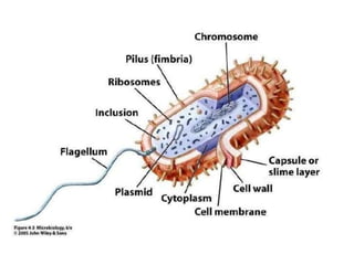
Bacteria have a simple cellular structure compared to eukaryotes. They lack internal membrane-bound organelles. Their cellular components include a cytoplasm containing a nucleoid, plasmids, ribosomes and other structures. The cell envelope includes a plasma membrane, cell wall, and in some cases an outer membrane. Some bacteria have external structures like flagella, fimbriae or pili that allow movement or attachment. Gram staining distinguishes bacteria based on differences in their cell wall composition and thickness.
Bacteria have a simple structure compared to eukaryotic cells, lacking organelles. Their small size allows rapid growth and inhabitation of diverse environments. Bacterial cells contain a cytoplasm surrounded by a cell membrane and cell wall. The cytoplasm holds the circular chromosome, ribosomes for protein production, and storage structures. Some bacteria have flagella for mobility or pili for attachment. Gram-positive bacteria have a thick peptidoglycan cell wall, while Gram-negatives have a thin wall and an outer membrane. This membrane structure contributes to differences in antibiotic susceptibility between Gram-positive and Gram-negative bacteria.
The document summarizes the structure of bacteria. It discusses that bacteria have no organelles and carry out all activities within the cell. It then describes the three main categories of bacterial structure:
1) Internal structures like the cytoplasm, nucleoid, plasmids, and ribosomes.
2) The cell envelope including the cell membrane, cell wall, and outer membrane in gram-negative bacteria.
3) External structures such as flagella, fimbriae, and capsule layers.
Cell is the fundamental structural and functional unit of all living organisms. Unicellular organisms can live independently while multicellular organisms are made of many cells that work together. Cells come in various shapes and sizes depending on their function. Prokaryotic cells like bacteria are generally smaller and simpler than eukaryotic cells found in plants, animals and fungi. Both have a cell membrane but eukaryotic cells also have organelles like the nucleus, mitochondria and Golgi apparatus that are not present in prokaryotes.
A Power point presentation on General Features of Prokaryotes.
This ppt covers brief information of "General Features of Prokaryotes" useful for introduction lecture as well as for seminar purpose.
Bacteria have a variety of shapes and arrangements. Their cells are surrounded by a cell wall and cytoplasmic membrane. The cell wall provides shape and protection, and its structure differs between gram-positive and gram-negative bacteria. Bacteria may also have extra structures like a capsule outside the cell wall or fimbriae. These extra structures help bacteria attach to surfaces and sometimes contribute to virulence.
This document provides information on cells at both the prokaryotic and eukaryotic levels. It discusses cell theory and key aspects of prokaryotic cells including their shape, structures like plasmids and mesosomes, and cell envelopes. For eukaryotic cells, it describes the membrane-bound organelles like the nucleus, mitochondria, plastids, Golgi apparatus, endoplasmic reticulum, lysosomes and vacuoles. It also discusses cytoskeletal elements and membrane properties.
Bacteria come in a variety of shapes and sizes. Their cell structure also varies between types. Gram positive bacteria have a thicker peptidoglycan cell wall without an outer membrane, while gram negative bacteria have a thinner cell wall but also an outer membrane containing lipopolysaccharides. Some bacteria like mycobacteria lack a cell wall entirely. Bacterial cells may also contain additional structures like flagella, pili, capsules, spores, and inclusions that serve important functions. Characterizing bacterial morphology provides insight into their phylogeny and potential pathogenicity.
This document summarizes the structure and morphology of bacterial cells. It discusses that bacteria are unicellular and microscopic, between 0.4-1.5 micrometers in size. Bacteria have characteristic shapes including cocci (spherical), bacilli (rod-shaped), and spirals. They contain DNA, RNA, ribosomes, and in some cases plasmids and mesosomes. Bacteria have a cell wall, plasma membrane, flagella or pili for motility, and may contain a capsule or endospores. The cell wall structure differs between gram-positive and gram-negative bacteria.
This document summarizes the ultrastructure of bacterial cells. It describes that bacteria are unicellular and microscopic. They have characteristic shapes including cocci, bacilli, vibrio and spirals. Their structures include a cell wall, plasma membrane, flagella for motility, pili for adhesion, capsules for protection, ribosomes for protein synthesis, and some can form endospores. The cell wall structure differs between gram-positive and gram-negative bacteria. The cytoplasm contains ribosomes and DNA but not membrane-bound organelles.
The bacterial cell has a small size between 0.5-2um and lacks membrane-bound organelles. It possesses a cell wall, plasma membrane, circular DNA located in the nucleoid region, ribosomes, and can contain inclusion bodies. The cell wall structure differs between gram-positive and gram-negative bacteria, with gram-positives having a thick peptidoglycan layer and gram-negatives having an additional outer membrane. Some bacteria possess appendages like flagella, pili, or fimbriae and structures like endospores, capsules, or S-layers may also be present outside the cell wall.
Cell Anatomy and physiology ( structure and function for NEET asparients, Biology, MBBS, BPT, Allied, nursing , medical and paramedical students. This is the easiest form of slide share to understand the context better.
Eukaryotic cells contain organelles that carry out specialized functions. The organelles include the cell membrane, cytoplasm, nucleus, mitochondria, endoplasmic reticulum, Golgi apparatus, lysosomes, ribosomes, and in plant cells, a cell wall, chloroplasts, and central vacuole. The cell membrane forms the boundary of the cell and is selectively permeable. The nucleus houses DNA and controls the cell's activities. Mitochondria generate energy for the cell. The endoplasmic reticulum and Golgi apparatus help synthesize and transport proteins and lipids. Lysosomes break down materials and recycle cell components. Ribosomes produce proteins using instructions from DNA. Plant cells have a cell wall for structure and
Biology Class 11 Chapter 8
FOR FURTHER DETAILS YOU CAN WATCH THE RELATED VIDEO AT THE GIVEN LINK
https://www.youtube.com/channel/UCxo06Nj-QWo_7SNvMyDnJCQ?view_as=subscriber
CELL BIOLOGY LECTURE. Cells described in the purest of forms.ndugbu34
The document provides information on basic cell biology, summarizing the key differences between prokaryotic and eukaryotic cells. Prokaryotic cells are simpler and smaller than eukaryotic cells, lacking membrane-bound organelles and having DNA in the nucleoid region of the cytoplasm. Eukaryotic cells are larger and more complex, having a nucleus that houses DNA and various membrane-bound organelles that compartmentalize functions. Both cell types have a plasma membrane and cytoplasm, but prokaryotes lack internal structures while eukaryotes have organelles like mitochondria and chloroplasts.
Bacteria are unicellular, procaryotic microorganisms which have diverse shape size and structures. Bacteria are found almost everywhere on Earth. Even the human body is full of bacteria, and in fact is estimated to contain more bacterial cells than human cells. Most bacteria in the body are harmless, and some are even helpful. A relatively small number of species cause disease.
This document summarizes the structure of bacterial cells. It describes the key components including the cell wall, plasma membrane, and intracellular and extracellular structures. The cell wall differs between gram-positive and gram-negative bacteria. Gram-positive walls are thicker and contain higher amounts of peptidoglycan while gram-negative walls are thinner and contain an outer lipopolysaccharide membrane. Intracellular structures discussed include plasmids, ribosomes, and the nucleoid region containing DNA. Extracellular structures include fimbriae, flagella, and capsules.
This document summarizes the structure of bacterial cells. It describes the key components including the cell wall, plasma membrane, and intracellular and extracellular structures. The cell wall differs between gram-positive and gram-negative bacteria. Gram-positive walls are thicker and contain higher amounts of peptidoglycan while gram-negative walls are thinner and contain an outer lipopolysaccharide membrane. Intracellular structures discussed include the nucleoid, ribosomes, plasmids, and mesosomes. Extracellular structures include flagella, pili, and capsules. Bacteria range in size from 0.4 to 1.5 micrometers and have characteristic shapes including cocci, bacilli, spirilla and spirochetes.
This document summarizes the structure of bacterial cells. It describes the key components of the bacterial cell including the cell wall, plasma membrane, and intracellular and extracellular structures. The cell wall differs between gram-positive and gram-negative bacteria in its thickness and composition. The plasma membrane controls what enters and exits the cell. Extracellular structures include fimbriae, flagella, and capsules. Intracellular structures discussed are plasmids, ribosomes, mesosomes, and the nucleoid, which contains the bacterial DNA.
Bacteria are classified based on taxonomy, nomenclature, and observational techniques. Morphology, staining properties, motility, growth characteristics, biochemical activities, and genetics are used to classify and identify bacteria. Bacterial cells have a cell envelope consisting of a capsule, cell wall, and cell membrane. The cell envelope encloses cellular elements like ribosomes, nucleoid, and mesosomes. Some bacteria also have extracellular appendages like flagella and pili.
CELL FEATURES presentation.pptx andrew.pptxkitati1
Cells have several key features that allow them to carry out essential functions. These include a cell membrane, cytoplasm, nucleus, and various organelles. Plant and animal cells share many structures but plant cells also contain chloroplasts. Cells reproduce, metabolize, maintain homeostasis, pass on hereditary information, respond to stimuli, grow and develop, and evolve adaptations. Prokaryotic and eukaryotic cells differ in their structures, with eukaryotes generally being larger and containing membrane-bound organelles.
Cell biology is the study of the structure and function of cells, the fundamental units of life. There are two main types of cells - prokaryotic and eukaryotic. Eukaryotic cells are larger, more complex, and contain organelles like the nucleus, endoplasmic reticulum, Golgi apparatus, mitochondria, chloroplasts, lysosomes, and peroxisomes, each with specialized functions. The animal cell is surrounded by a plasma membrane and contains a large nucleus housing the DNA, as well as various membrane-bound organelles that carry out specific functions necessary for cell survival and homeostasis.
The document summarizes key information about cells, including their structure, components, and main differences between prokaryotic and eukaryotic cells. It describes that cells are the basic unit of life, and can be unicellular like bacteria or multicellular like humans. The main organelles of animal cells are then outlined, including the nucleus, mitochondria, endoplasmic reticulum, Golgi apparatus, ribosomes, lysosomes, peroxisomes, and centrosomes. The last section highlights differences between simpler prokaryotic cells versus more complex eukaryotic cells.
This document summarizes the morphology and structures of bacterial cells. It describes key differences between prokaryotic and eukaryotic cells, as well as differences between gram-positive and gram-negative bacterial cell walls. The structures discussed include the cell envelope, cell wall, cell membrane, cytoplasm, ribosomes, nucleoid, capsules, flagella, fimbriae, endospores, and inclusions. Functions and methods of demonstrating various structures are also presented.
Bacteria are microscopic, unicellular organisms that lack nuclei and organelles. They display a diversity of shapes and sizes, and structures like flagella and pili enable motility and genetic exchange. The bacterial cell wall provides structural integrity and protection, and differs between gram-positive and gram-negative bacteria in its chemical composition and layers. Flagella are helical filaments that rotate to propel bacteria and allow for swimming movement in different directions.
Bacteria are single-celled organisms that exist in communities of millions. They come in various shapes such as cocci, bacilli, and spirals. Bacteria have a cell wall, cell membrane, and sometimes a capsule, flagella, and endospore. The cell wall gives the cell its shape and protects it from bursting due to osmotic pressure. Bacteria are classified as gram-positive or gram-negative based on differences in their cell wall composition and thickness. The cell membrane regulates what passes in and out of the cell.
Anatomy of prokaryotic cells and eukaryotic cells with differencesHassanLatif15
A complete comprehensive details of functions and functional anatomy of prokaryotic cells and eukaryotic cells according to microbiology, biotechnology and pharmacy medicine
Prokaryotes have relatively simple structures compared to eukaryotes. They lack membrane-bound organelles and have a plasma membrane, cell wall, and genetic material not enclosed within a nucleus. Bacteria come in various shapes including cocci, bacilli, and spirilla. Their cell walls differ between gram-positive and gram-negative bacteria. Prokaryotes also possess external structures like flagella, pili, and capsules. They reproduce through binary fission and some form resistant endospores.
This document provides information about cell structure and organization. It discusses the key components and features of prokaryotic and eukaryotic cells. Some of the main points covered include:
- Prokaryotic cells like bacteria have no nucleus or membrane-bound organelles. They have a cell envelope, cytoplasm, ribosomes, and a nucleoid containing DNA.
- Eukaryotic cells contain membrane-bound organelles like the nucleus, mitochondria, endoplasmic reticulum, Golgi bodies, lysosomes, and vacuoles. Their genetic material is contained within the nucleus.
- Cell membranes are semipermeable and made of a phospholipid bilayer. They regulate the passage of materials
This document discusses the tree of life and the three domains of life: Bacteria, Archaea, and Eukarya. It provides details on the domains of Bacteria and Archaea, which are both prokaryotes, and notes some of their differences. The domain Eukarya is then discussed, including the kingdoms of plants, fungi, and animals. Key aspects of fungi and plants are summarized, such as fungi obtaining nutrients outside their bodies, and plants producing food through photosynthesis using chloroplasts in leaves.
FFA Judging Plant pathology study guide.pptDawitGetahun6
This document is a study guide for a plant pathology judging contest that was assembled by Tom McCutcheon and Mike Shamblin from various internet sources for educational purposes. It may be copied and distributed without financial compensation as long as the efforts of the authors are recognized. The study guide contains over 50 slides that provide pictures of plant diseases along with their common name and 3 sentences or less about recommended control methods.
This document summarizes the ultrastructure of bacterial cells. It describes that bacteria are unicellular and microscopic. They have characteristic shapes including cocci, bacilli, vibrio and spirals. Their structures include a cell wall, plasma membrane, flagella for motility, pili for adhesion, capsules for protection, ribosomes for protein synthesis, and some can form endospores. The cell wall structure differs between gram-positive and gram-negative bacteria. The cytoplasm contains ribosomes and DNA but not membrane-bound organelles.
The bacterial cell has a small size between 0.5-2um and lacks membrane-bound organelles. It possesses a cell wall, plasma membrane, circular DNA located in the nucleoid region, ribosomes, and can contain inclusion bodies. The cell wall structure differs between gram-positive and gram-negative bacteria, with gram-positives having a thick peptidoglycan layer and gram-negatives having an additional outer membrane. Some bacteria possess appendages like flagella, pili, or fimbriae and structures like endospores, capsules, or S-layers may also be present outside the cell wall.
Cell Anatomy and physiology ( structure and function for NEET asparients, Biology, MBBS, BPT, Allied, nursing , medical and paramedical students. This is the easiest form of slide share to understand the context better.
Eukaryotic cells contain organelles that carry out specialized functions. The organelles include the cell membrane, cytoplasm, nucleus, mitochondria, endoplasmic reticulum, Golgi apparatus, lysosomes, ribosomes, and in plant cells, a cell wall, chloroplasts, and central vacuole. The cell membrane forms the boundary of the cell and is selectively permeable. The nucleus houses DNA and controls the cell's activities. Mitochondria generate energy for the cell. The endoplasmic reticulum and Golgi apparatus help synthesize and transport proteins and lipids. Lysosomes break down materials and recycle cell components. Ribosomes produce proteins using instructions from DNA. Plant cells have a cell wall for structure and
Biology Class 11 Chapter 8
FOR FURTHER DETAILS YOU CAN WATCH THE RELATED VIDEO AT THE GIVEN LINK
https://www.youtube.com/channel/UCxo06Nj-QWo_7SNvMyDnJCQ?view_as=subscriber
CELL BIOLOGY LECTURE. Cells described in the purest of forms.ndugbu34
The document provides information on basic cell biology, summarizing the key differences between prokaryotic and eukaryotic cells. Prokaryotic cells are simpler and smaller than eukaryotic cells, lacking membrane-bound organelles and having DNA in the nucleoid region of the cytoplasm. Eukaryotic cells are larger and more complex, having a nucleus that houses DNA and various membrane-bound organelles that compartmentalize functions. Both cell types have a plasma membrane and cytoplasm, but prokaryotes lack internal structures while eukaryotes have organelles like mitochondria and chloroplasts.
Bacteria are unicellular, procaryotic microorganisms which have diverse shape size and structures. Bacteria are found almost everywhere on Earth. Even the human body is full of bacteria, and in fact is estimated to contain more bacterial cells than human cells. Most bacteria in the body are harmless, and some are even helpful. A relatively small number of species cause disease.
This document summarizes the structure of bacterial cells. It describes the key components including the cell wall, plasma membrane, and intracellular and extracellular structures. The cell wall differs between gram-positive and gram-negative bacteria. Gram-positive walls are thicker and contain higher amounts of peptidoglycan while gram-negative walls are thinner and contain an outer lipopolysaccharide membrane. Intracellular structures discussed include plasmids, ribosomes, and the nucleoid region containing DNA. Extracellular structures include fimbriae, flagella, and capsules.
This document summarizes the structure of bacterial cells. It describes the key components including the cell wall, plasma membrane, and intracellular and extracellular structures. The cell wall differs between gram-positive and gram-negative bacteria. Gram-positive walls are thicker and contain higher amounts of peptidoglycan while gram-negative walls are thinner and contain an outer lipopolysaccharide membrane. Intracellular structures discussed include the nucleoid, ribosomes, plasmids, and mesosomes. Extracellular structures include flagella, pili, and capsules. Bacteria range in size from 0.4 to 1.5 micrometers and have characteristic shapes including cocci, bacilli, spirilla and spirochetes.
This document summarizes the structure of bacterial cells. It describes the key components of the bacterial cell including the cell wall, plasma membrane, and intracellular and extracellular structures. The cell wall differs between gram-positive and gram-negative bacteria in its thickness and composition. The plasma membrane controls what enters and exits the cell. Extracellular structures include fimbriae, flagella, and capsules. Intracellular structures discussed are plasmids, ribosomes, mesosomes, and the nucleoid, which contains the bacterial DNA.
Bacteria are classified based on taxonomy, nomenclature, and observational techniques. Morphology, staining properties, motility, growth characteristics, biochemical activities, and genetics are used to classify and identify bacteria. Bacterial cells have a cell envelope consisting of a capsule, cell wall, and cell membrane. The cell envelope encloses cellular elements like ribosomes, nucleoid, and mesosomes. Some bacteria also have extracellular appendages like flagella and pili.
CELL FEATURES presentation.pptx andrew.pptxkitati1
Cells have several key features that allow them to carry out essential functions. These include a cell membrane, cytoplasm, nucleus, and various organelles. Plant and animal cells share many structures but plant cells also contain chloroplasts. Cells reproduce, metabolize, maintain homeostasis, pass on hereditary information, respond to stimuli, grow and develop, and evolve adaptations. Prokaryotic and eukaryotic cells differ in their structures, with eukaryotes generally being larger and containing membrane-bound organelles.
Cell biology is the study of the structure and function of cells, the fundamental units of life. There are two main types of cells - prokaryotic and eukaryotic. Eukaryotic cells are larger, more complex, and contain organelles like the nucleus, endoplasmic reticulum, Golgi apparatus, mitochondria, chloroplasts, lysosomes, and peroxisomes, each with specialized functions. The animal cell is surrounded by a plasma membrane and contains a large nucleus housing the DNA, as well as various membrane-bound organelles that carry out specific functions necessary for cell survival and homeostasis.
The document summarizes key information about cells, including their structure, components, and main differences between prokaryotic and eukaryotic cells. It describes that cells are the basic unit of life, and can be unicellular like bacteria or multicellular like humans. The main organelles of animal cells are then outlined, including the nucleus, mitochondria, endoplasmic reticulum, Golgi apparatus, ribosomes, lysosomes, peroxisomes, and centrosomes. The last section highlights differences between simpler prokaryotic cells versus more complex eukaryotic cells.
This document summarizes the morphology and structures of bacterial cells. It describes key differences between prokaryotic and eukaryotic cells, as well as differences between gram-positive and gram-negative bacterial cell walls. The structures discussed include the cell envelope, cell wall, cell membrane, cytoplasm, ribosomes, nucleoid, capsules, flagella, fimbriae, endospores, and inclusions. Functions and methods of demonstrating various structures are also presented.
Bacteria are microscopic, unicellular organisms that lack nuclei and organelles. They display a diversity of shapes and sizes, and structures like flagella and pili enable motility and genetic exchange. The bacterial cell wall provides structural integrity and protection, and differs between gram-positive and gram-negative bacteria in its chemical composition and layers. Flagella are helical filaments that rotate to propel bacteria and allow for swimming movement in different directions.
Bacteria are single-celled organisms that exist in communities of millions. They come in various shapes such as cocci, bacilli, and spirals. Bacteria have a cell wall, cell membrane, and sometimes a capsule, flagella, and endospore. The cell wall gives the cell its shape and protects it from bursting due to osmotic pressure. Bacteria are classified as gram-positive or gram-negative based on differences in their cell wall composition and thickness. The cell membrane regulates what passes in and out of the cell.
Anatomy of prokaryotic cells and eukaryotic cells with differencesHassanLatif15
A complete comprehensive details of functions and functional anatomy of prokaryotic cells and eukaryotic cells according to microbiology, biotechnology and pharmacy medicine
Prokaryotes have relatively simple structures compared to eukaryotes. They lack membrane-bound organelles and have a plasma membrane, cell wall, and genetic material not enclosed within a nucleus. Bacteria come in various shapes including cocci, bacilli, and spirilla. Their cell walls differ between gram-positive and gram-negative bacteria. Prokaryotes also possess external structures like flagella, pili, and capsules. They reproduce through binary fission and some form resistant endospores.
This document provides information about cell structure and organization. It discusses the key components and features of prokaryotic and eukaryotic cells. Some of the main points covered include:
- Prokaryotic cells like bacteria have no nucleus or membrane-bound organelles. They have a cell envelope, cytoplasm, ribosomes, and a nucleoid containing DNA.
- Eukaryotic cells contain membrane-bound organelles like the nucleus, mitochondria, endoplasmic reticulum, Golgi bodies, lysosomes, and vacuoles. Their genetic material is contained within the nucleus.
- Cell membranes are semipermeable and made of a phospholipid bilayer. They regulate the passage of materials
Similar to bacteriacellstructure-200914164803.pdf (20)
This document discusses the tree of life and the three domains of life: Bacteria, Archaea, and Eukarya. It provides details on the domains of Bacteria and Archaea, which are both prokaryotes, and notes some of their differences. The domain Eukarya is then discussed, including the kingdoms of plants, fungi, and animals. Key aspects of fungi and plants are summarized, such as fungi obtaining nutrients outside their bodies, and plants producing food through photosynthesis using chloroplasts in leaves.
FFA Judging Plant pathology study guide.pptDawitGetahun6
This document is a study guide for a plant pathology judging contest that was assembled by Tom McCutcheon and Mike Shamblin from various internet sources for educational purposes. It may be copied and distributed without financial compensation as long as the efforts of the authors are recognized. The study guide contains over 50 slides that provide pictures of plant diseases along with their common name and 3 sentences or less about recommended control methods.
Fungi are a diverse group of organisms that are ubiquitous in terrestrial and freshwater environments. They play important ecological roles in recycling organic matter as saprotrophs, symbionts, and parasites. Fungi can be unicellular yeasts or multicellular molds composed of threadlike filaments called hyphae. Hyphae may be septate or coenocytic, and form together into a mat-like structure called a mycelium. Fungi are classified based on their sexual reproduction structures and spores as well as hyphal morphology. The four main classes of medically important fungi are Ascomycetes, Basidiomycetes, Deuteromycetes, and Phy
plant pathology and plant disease 1516297.pptDawitGetahun6
This document provides an overview of plant pathology principles organized into jeopardy game format. It covers topics including Oomycota, Ascomycota, symptoms and signs of disease, disease management, terminology used in plant pathology, and general mycology. For each topic, there are 5 questions ranging from 100 to 500 points about key concepts within that category.
Lecture 3- Bacterial Nutrition and Growth-.pptDawitGetahun6
This document discusses various environmental factors that affect microbial growth, including temperature, pH, osmotic pressure, and oxygen levels. It describes how microorganisms are classified based on their optimal and maximum temperature ranges, as well as their ability to grow under acidic, alkaline, high salt, or anaerobic conditions. The document also covers microbial nutrition requirements, discussing the main macronutrients of carbon, nitrogen, phosphorus, and others needed for growth, and how organisms are categorized based on their carbon and energy sources.
Nutrient cycles occur at both the local ecosystem level through biological processes, and the global level through geological processes and atmospheric circulation. Key nutrients like carbon, oxygen, hydrogen, nitrogen, and phosphorus cycle through biotic and abiotic compartments of ecosystems, including organisms, soils, atmosphere, and oceans. At the global scale, the hydrosphere and atmosphere drive the water cycle, while carbon and nitrogen cycle through oceans, soils, atmosphere, and biosphere. Human activities are altering global nutrient cycles, such as through the addition of nitrogen to ecosystems from fossil fuel combustion.
Rhizobacteria are root-colonizing bacteria that form symbiotic relationships with plants. They are often referred to as plant growth-promoting rhizobacteria (PGPR) as they enhance plant growth through mechanisms such as nitrogen fixation, phosphate solubilization, and inducing systemic resistance against pathogens. Rhizobacteria have various applications as bioinoculants, biofertilizers, and biocontrol agents to promote plant growth and reduce the need for chemical fertilizers and pesticides. Future prospects include genetically engineering PGPR to overexpress beneficial traits and improve their biocontrol efficacy through synergistic effects.
The document discusses three key biogeochemical cycles - carbon, nitrogen, and phosphorus. It provides details on each cycle, including:
1) The carbon cycle involves the movement of carbon between the atmosphere, organisms, and fossils fuels. Plants and animals exchange carbon via photosynthesis and respiration. Human emissions impact the cycle.
2) Nitrogen circulates between the air, soil, plants and animals through nitrogen fixation, plant/animal uptake, and denitrification. It is essential for proteins but scarce without bacterial conversion.
3) Phosphorus cycles slowly between rocks, soil and organisms and is important for energy transfer and genetic material. Excess fertilizer runoff impacts nitrogen and phosphorus cycles in waterways
The document summarizes microbiology techniques for culturing and observing microbes. It describes the five steps for culturing as inoculation, isolation, incubation, inspection, and identification. Inoculation introduces microbes to growth media. Isolation produces pure cultures on agar plates. Incubation allows growth under optimal conditions. Inspection involves observing colony morphology and staining for microscopy. Identification correlates observed characteristics to identify the microbe. It also explains light and electron microscopy, describing their components, resolutions, and sample preparation techniques like staining.
metabolsim of microorganism 1664107090.pptDawitGetahun6
Microbial metabolism involves the breakdown and synthesis of complex molecules. There are two main types of metabolism: catabolism which breaks down molecules and releases energy, and anabolism which uses energy to build complex molecules. Microbes metabolize carbohydrates, lipids, and proteins through various pathways. During aerobic respiration, glucose is completely oxidized using oxygen as the final electron acceptor to generate ATP. Fermentation pathways produce ATP without oxygen by using organic molecules as electron acceptors. The study of microbial metabolism is important for food production and preservation.
agri microbiology for Micro Hort PPT.pptxDawitGetahun6
1. The document discusses various microscopy techniques used to study microorganisms, including bright-field, dark-field, phase-contrast, and fluorescent microscopy. It also covers electron microscopy.
2. Microorganisms can be classified as prokaryotes or eukaryotes. Prokaryotes include bacteria and archaea and lack a nucleus, while eukaryotes such as protists, fungi, plants and animals have membrane-bound organelles including a nucleus.
3. The key differences between prokaryotic and eukaryotic cells are that eukaryotic cells have a nucleus enclosed in a membrane, linear DNA, histones, chromosomes, and membrane-bound organelles, while
method of studing microbial 13114527.pptDawitGetahun6
The document discusses various types of culture media used for growing microorganisms in the laboratory, including chemically defined media which has an exact known composition, complex media made from extracts of yeasts and meats which provide nutrients like carbon, nitrogen and vitamins, and solid media which uses agar as a solidifying agent to allow bacterial growth on surfaces like test tubes and Petri dishes. The different media types are selected based on the specific microorganism being cultured and whether a solid or liquid growth surface is needed.
growth of microorganism chp 7-8 ppt.pptDawitGetahun6
1. Bacterial growth occurs through binary fission and can happen rapidly depending on environmental conditions like temperature and nutrient levels. The number of bacterial cells increases exponentially.
2. Microbial growth is affected by environmental factors like temperature and pH as well as chemical factors including proper concentrations of carbon, hydrogen, oxygen, nitrogen, phosphorus, and trace elements.
3. A bacterial growth curve consists of four phases - lag, log, stationary, and death - and is used to understand microbial growth under different conditions and the effect of various chemicals and temperatures. Growth is typically measured through turbidity using a spectrophotometer.
This document outlines the historical development of microbiology from the 17th century to present day. It discusses the key early discoveries including van Leeuwenhoek's discovery of microorganisms under the microscope in the 1670s. Later sections cover the development of vaccines and antimicrobial therapies like antibiotics. It also addresses the role of microbes in industry and emerging diseases in the post-antibiotic era.
in the soil micriobial bioremediation2.pptDawitGetahun6
This document defines and describes various methods of bioremediation. It discusses in situ bioremediation techniques like bioventing, biodegradation and bioaugmentation that treat contamination on-site. Ex situ techniques like land farming and composting remove contamination for off-site treatment. Phytoremediation uses plants to extract, degrade, stabilize or filter contaminants. The effectiveness of bioremediation depends on microorganisms, environmental factors and the contaminant type.
Soil is a complex medium that provides nutrients and support for plant growth. It is composed of minerals, air, water, and organic matter. Soil forms over long periods through weathering and the activity of organisms. Key factors that influence soil formation are parent material, climate, organisms, topography, and time. Soil provides nutrients, water, and anchorage for plants through complex nutrient cycles and soil profiles that vary in texture, structure, pH, and organic content. Proper soil management through practices like tillage, no-till, and conservation methods helps maintain healthy soil that supports plant growth.
Lecture 2- on Cell Structures.ppt biologyDawitGetahun6
Fungi are eukaryotic, spore-bearing organisms that produce extracellular enzymes and absorb nutrients. Their basic cellular unit is the hypha, which can be septate or coenocytic. Fungi have typical eukaryotic organelles like mitochondria and nuclei, as well as fungal-specific structures involved in cell wall growth. They reproduce both sexually through spores formed via meiosis and asexually via mitotic spores. Fungal life cycles can be predominantly haploid, dikaryotic, or diploid depending on when karyogamy and meiosis occur. Important model fungi like Neurospora crassa were influential in developing concepts in genetics.
The document provides an overview of plant pathology and the history of several important plant diseases. It discusses causal agents of disease including fungi, bacteria, nematodes and viruses. Several significant diseases that have impacted agriculture throughout history are summarized, including late blight of potato in Ireland which caused the Irish Potato Famine, chestnut blight which wiped out the American chestnut, Dutch elm disease which devastated street trees, and sudden oak death first discovered in California in the 1990s.
This document summarizes key aspects of the kingdom Fungi. It describes the characteristics of fungi, including their heterotrophic nutrition and modes of growth. It discusses fungal life cycles and roles as decomposers, symbionts through mycorrhizae and lichens, and parasites. The evolution and classification of fungi is covered, focusing on the four main phyla: Chytridiomycota, Zygomycota, Ascomycota, and Basidiomycota. Representative fungi from each phylum are described along with their reproductive structures and life cycles.
what is plant pathology and plant disease .pptDawitGetahun6
Plant pathology is the study of plant diseases caused by pathogens such as fungi, bacteria, viruses and nematodes. Throughout history, devastating plant diseases like the Irish Potato Famine caused by late blight have resulted in millions of deaths. Today, plant diseases cause $8 billion in annual losses to U.S. crops. Plant pathologists work to understand and manage diseases through research and developing control methods to protect food and plant health.
This presentation includes basic of PCOS their pathology and treatment and also Ayurveda correlation of PCOS and Ayurvedic line of treatment mentioned in classics.
How to Manage Your Lost Opportunities in Odoo 17 CRMCeline George
Odoo 17 CRM allows us to track why we lose sales opportunities with "Lost Reasons." This helps analyze our sales process and identify areas for improvement. Here's how to configure lost reasons in Odoo 17 CRM
ISO/IEC 27001, ISO/IEC 42001, and GDPR: Best Practices for Implementation and...PECB
Denis is a dynamic and results-driven Chief Information Officer (CIO) with a distinguished career spanning information systems analysis and technical project management. With a proven track record of spearheading the design and delivery of cutting-edge Information Management solutions, he has consistently elevated business operations, streamlined reporting functions, and maximized process efficiency.
Certified as an ISO/IEC 27001: Information Security Management Systems (ISMS) Lead Implementer, Data Protection Officer, and Cyber Risks Analyst, Denis brings a heightened focus on data security, privacy, and cyber resilience to every endeavor.
His expertise extends across a diverse spectrum of reporting, database, and web development applications, underpinned by an exceptional grasp of data storage and virtualization technologies. His proficiency in application testing, database administration, and data cleansing ensures seamless execution of complex projects.
What sets Denis apart is his comprehensive understanding of Business and Systems Analysis technologies, honed through involvement in all phases of the Software Development Lifecycle (SDLC). From meticulous requirements gathering to precise analysis, innovative design, rigorous development, thorough testing, and successful implementation, he has consistently delivered exceptional results.
Throughout his career, he has taken on multifaceted roles, from leading technical project management teams to owning solutions that drive operational excellence. His conscientious and proactive approach is unwavering, whether he is working independently or collaboratively within a team. His ability to connect with colleagues on a personal level underscores his commitment to fostering a harmonious and productive workplace environment.
Date: May 29, 2024
Tags: Information Security, ISO/IEC 27001, ISO/IEC 42001, Artificial Intelligence, GDPR
-------------------------------------------------------------------------------
Find out more about ISO training and certification services
Training: ISO/IEC 27001 Information Security Management System - EN | PECB
ISO/IEC 42001 Artificial Intelligence Management System - EN | PECB
General Data Protection Regulation (GDPR) - Training Courses - EN | PECB
Webinars: https://pecb.com/webinars
Article: https://pecb.com/article
-------------------------------------------------------------------------------
For more information about PECB:
Website: https://pecb.com/
LinkedIn: https://www.linkedin.com/company/pecb/
Facebook: https://www.facebook.com/PECBInternational/
Slideshare: http://www.slideshare.net/PECBCERTIFICATION
Exploiting Artificial Intelligence for Empowering Researchers and Faculty, In...Dr. Vinod Kumar Kanvaria
Exploiting Artificial Intelligence for Empowering Researchers and Faculty,
International FDP on Fundamentals of Research in Social Sciences
at Integral University, Lucknow, 06.06.2024
By Dr. Vinod Kumar Kanvaria
Executive Directors Chat Leveraging AI for Diversity, Equity, and InclusionTechSoup
Let’s explore the intersection of technology and equity in the final session of our DEI series. Discover how AI tools, like ChatGPT, can be used to support and enhance your nonprofit's DEI initiatives. Participants will gain insights into practical AI applications and get tips for leveraging technology to advance their DEI goals.
Main Java[All of the Base Concepts}.docxadhitya5119
This is part 1 of my Java Learning Journey. This Contains Custom methods, classes, constructors, packages, multithreading , try- catch block, finally block and more.
How to Build a Module in Odoo 17 Using the Scaffold MethodCeline George
Odoo provides an option for creating a module by using a single line command. By using this command the user can make a whole structure of a module. It is very easy for a beginner to make a module. There is no need to make each file manually. This slide will show how to create a module using the scaffold method.
Walmart Business+ and Spark Good for Nonprofits.pdfTechSoup
"Learn about all the ways Walmart supports nonprofit organizations.
You will hear from Liz Willett, the Head of Nonprofits, and hear about what Walmart is doing to help nonprofits, including Walmart Business and Spark Good. Walmart Business+ is a new offer for nonprofits that offers discounts and also streamlines nonprofits order and expense tracking, saving time and money.
The webinar may also give some examples on how nonprofits can best leverage Walmart Business+.
The event will cover the following::
Walmart Business + (https://business.walmart.com/plus) is a new shopping experience for nonprofits, schools, and local business customers that connects an exclusive online shopping experience to stores. Benefits include free delivery and shipping, a 'Spend Analytics” feature, special discounts, deals and tax-exempt shopping.
Special TechSoup offer for a free 180 days membership, and up to $150 in discounts on eligible orders.
Spark Good (walmart.com/sparkgood) is a charitable platform that enables nonprofits to receive donations directly from customers and associates.
Answers about how you can do more with Walmart!"
How to Fix the Import Error in the Odoo 17Celine George
An import error occurs when a program fails to import a module or library, disrupting its execution. In languages like Python, this issue arises when the specified module cannot be found or accessed, hindering the program's functionality. Resolving import errors is crucial for maintaining smooth software operation and uninterrupted development processes.
2. STRUCTURE OF BACTERIA
• Smaller and simpler in structure than
eukaryotic cells, with no recognizable
organelles.
• All of the activities performed by organelles
also take place in bacteria, but they are not
carried out by specialized structures.
• The small size, simple design, and broad
metabolic capabilities of bacteria allow them
to grow and divide very rapidly and to inhabit
and flourish in almost any environment.
3. STRUCTURE OF BACTERIA
• They were first seen under a microscope by Anton
van Leeuwenhoek in 1676.
• As microscopes have improved, scientists have
come to understand bacterial cell structure better.
4.
5.
6. Bacterial cell structure
• organized into 3 categories :
• Internal Structures: Cytoplasm, nucleoid,
bacterial chromosome, plasmid, ribosomes, and
storage granules
• Cell envelope: cell membrane, peptidoglycan cell
wall or an outer lipid membrane (only found in
Gram-negative cells)
• External structures (appendages & coverings):
flagella, fimbriae, sex pilus and glycocalyx
8. Cytoplasm
• Portion of the cell that lies within the PM
• substances within the plasma membrane, excluding
the genetic material.
• Gel-like matrix composed of mostly water(4/5 th ),
enzymes, nutrients, wastes, and gases
• Contains cell structures - ribosomes, chromosome,
and plasmids , as well as the components necessary
for bacterial metabolism.
• It is relatively featureless by electron microscope -
although small granules can be seen.
• carries out very important functions for the cell -
growth, metabolism, and replication .
9. Constituents
– Proteins including enzymes
– Vitamins
– Ions
– Nucleic acids and their precursors
– Amino acids and their precursors
– Sugars, carbohydrates and their derivatives
– Fatty acids and their derivatives
10. Nucleoid
• Unlike the eukaryotic (true) cells, bacteria do not
have a membrane enclosed nucleus.
• The nucleoid is a region of cytoplasm where the
chromosomal DNA is located.
• It is not a membrane bound nucleus, but simply an
area of the cytoplasm where the strands of DNA
are found.
11. Plasmids
• small extra-chromosomal DNA
• contain genes for antibiotic resistance or virulence.
• Structure Similar to most bacterial chromosomes, but
considerably smaller.
• plasmids are covalently closed circular DNA
• In a few species linear plasmids have been found.
• Size : Chromosomal DNA is typically about 4000 kb,
• plasmid DNA ranges from 1-200 kb.
• Number of plasmids: 1-700 copies of plasmid in a
cell.
12. Plasmid Function
• The function of plasmids is not always known, but they
are not normally essential for survival of host, although
their presence generally gives the host some
advantage.
• Antibiotic resistance - Some plasmids code for proteins
that degrade antibiotics-a big advantage for pathogens.
• Some encode for proteins which confer virulence
factors on the host. For example- E. coli plasmid Ent
P307 codes for an enterotoxin which makes E. coli
pathogenic.
• Conjugative plasmids - These allow exchange of DNA
between bacterial cells.
13. Plasmids
• Plasmids and the associated traits can be transferred
between bacteria, even from one bacterial species to
another.
• Plasmids are not involved in reproduction.
• Plasmids replicate independently of the chromosome.
• Plasmids are passed to other bacteria by two means.
• For most plasmid types, copies in the cytoplasm are
passed on to daughter cells during binary fission.
14. Plasmids
• Other types of plasmids ,form tube like structure at the
surface called a pilus that passes copies of the plasmid
to other bacteria during conjugation, a process by
which bacteria exchange genetic information.
• Plasmids have been shown to be instrumental in the
transmission of special properties, such as antibiotic
drug resistance, resistance to heavy metals, and
virulence factors necessary for infection of animal or
plant hosts.
• The ability to insert specific genes into plasmids have
made them extremely useful tools in the area of
genetic engineering/RDNA Technology .
15. Ribosomes- protein synthesis machinery
• Consists of RNA and protein
• Abundant in cytoplasm
• Often grouped in long chains called polyribosomes.
• give the cytoplasm of bacteria a granular appearance
in EM.
• smaller than the ribosomes in eukaryotic cells-but
have a similar function
• Bacterial ribosomes have sedimentation rate of 70S;
their subunits have rates of 30S and 50S.
• The unit used to measure sedimentation velocity is
Svedberg
16.
17. Ribosomes
• They translate the genetic code from the molecular language of nucleic
acid to that of amino acids—the building blocks of proteins.
• Bacterial ribosomes are similar to those of eukaryotes, but are smaller and
have a slightly different composition and molecular structure.
• Bacterial ribosomes are never bound to other organelles as they
sometimes are bound to the endoplasmic reticulum in eukaryotes, but are
free-standing structures distributed throughout the cytoplasm.
• There are sufficient differences between bacterial ribosomes and
eukaryotic ribosomes that some antibiotics will inhibit the functioning of
bacterial ribosomes, but not a eukaryote's, thus killing bacteria but not the
eukaryotic organisms they are infecting.
• Streptomycin binds 70S ribosome and stops protein synthesis but it can
not bind 80S ribosome of eukaryotes and thereby eukaryotic cell remains
unaffected.
18. Bacterial Chromosome - Genophore
• The bacterial chromosome consists of a single,
circle of deoxyribonucleic acid.
• DNA is double stranded- two strands line up
antiparrallel to each other and the bases are
linked together with hydrogen bonds.
• It includes most of the genetic material of the
organism .
19.
20. Bacterial Chromosome
• Unlike the DNA in eukaryotic cells, which resides
in the nucleus, DNA in bacterial cells is not
sequestered in a membrane-bound organelle but
appears as a long coil distributed through the
cytoplasm.
• In many bacteria the DNA is present as a single,
circular chromosome and in some cases the DNA
is linear rather than circular.
• some bacteria may contain two chromosomes
21. Bacterial Chromosome
• As in all organisms, bacterial DNA contains the
four nitrogenous bases adenine (A), cytosine
(C), guanine (G), and t
• The amount of DNA in bacterial chromosomes
ranges from 580,000 base pairs in
Mycoplasma gallinarum to 4,700,000 base
pairs in E. coli to 9,140,000 base pairs in
Myxococcus xanthus.
22. Inclusion bodies
• Inclusion bodies: Bacteria can have within their
cytoplasm a variety of small bodies collectively referred
to as inclusion bodies.
• Some are called granules and other are called vesicles.
• Inclusions are considered to be nonliving components
of the cell that do not possess metabolic activity and
are not bounded by membranes.
• The most common inclusions are glycogen, lipid
droplets, crystals, and pigments.
23. Inclusion bodies - Granules
• Granules: Densely compacted substances without a
membrane covering.
• Nutrients and reserves may be stored in the cytoplasm
in the form of glycogen, lipids, polyphosphate, or in
some cases, sulfur or nitrogen for later use.
• Each granule contains specific substances, such as
glycogen (glucose polymer) and polyphosphate
(phosphate polymer, supplies energy to metabolic
processes).
• Sulfur bacteria contains reserve granules of sulfur.
• These granules are depleted in starvation.
24. Inclusion bodies-vesicles
• Some aquatic photosynthetic bacteria and cyano
bacteria have rigid gas-filled vacuoles and it helps in
floating at a certain level - allowing them to move
up or down into water layers with different light
intensities and nutrient levels.
• Some magnetotactic bacterium, eg. Aquaspirillium
magnetotacticum , stores Magnetitite (Ferric oxide).
The presence of such magnetic inclusions enables
these bacteria to responds to magnetic fields.
25. Microcompartments
• Microcompartments are widespread, membrane-
bound organelles that are made of a protein shell
that surrounds and encloses various enzymes.
• Carboxysomes are protein-enclosed bacterial
microcompartments that contain enzymes involved
in carbon fixation.
• Magnetosomes are bacterial microcompartments,
present in magnetotactic bacteria, that contain
magnetic crystals.
27. Plasma Membrane
• Phospholipid bilayer surrounding the cytoplasm and
regulates the flow of substances in and out of the cell.
• Consists of both lipids and proteins.
• Protects the cell from its surroundings.
• Selectively permeable to ions and organic molecules
and controls the movement of substances in and out.
• numerous proteins moving within or upon this layer are
primarily responsible for transport of ions, nutrients
and waste across the membrane.
28.
29.
30. Periplasmic space
• Gram-negative bacteria : space between the
cytoplasmic membrane and the cell wall and space
found between cell wall and the outer membrane
• Periplasm may constitute up to 40% of the total
cell volume in G-ve species.
• Gram-positive bacteria : space between the
cytoplasmic membrane and the cell wall.
• The periplasm is filled with water and proteins
and is reminiscent of the cytoplasm.
31. Periplasmic Space
• However periplasm contains proteins and other
molecules distinct from those in the cytoplasm
because the membrane prevents the free exchange
between these two compartments.
• Periplasmic proteins have various functions in
cellular processes including: transport, degradation,
and motility.
• Periplasm controls molecular traffic entering and
leaving the cell.
32.
33. Cell wall
• Outer covering of most cells that protects the
bacterial cell and gives it shape (spherical, rod and
spiral).
• Composed of peptidoglycan (polysaccharides +
protein)
• Mycoplasma are bacteria that have no cell wall
and therefore have no definite shape.
34. Cell wall
• Peptidoglycan - molecule found only in bacterial cell
walls.
• The rigid structure of peptidoglycan gives the bacterial
cell shape, surrounds the plasma membrane and
provides prokaryotes with protection from the
environment
• From the peptidoglycan inwards all bacterial cells are
similar.
• Going further out, the bacterial world divides into two
major classes: Gram-positive and Gram-negative .
• Amount and location of peptidoglycan in the cell wall
determines whether a bacterium is G+ve or G-ve
35. Peptidoglycan = (polysaccharides + protein),
• Peptidoglycan (murein) - huge polymer of interlocking
chains composed of similar monomers.
• peptidoglycan is made from polysaccharide chains
cross-linked by peptides containing D-amino acids
• The backbone of the peptidoglycan molecule is
composed of two derivatives of glucose:
• N-acetylglucosamine (NAG)
• N-acetlymuramic acid (NAM).
• The NAG and NAM strands are connected by inter
peptide bridges.
36.
37.
38. Gram-positive Cells
• G+ve bacteria possess thick cell wall containing
many layers of peptidoglycan and teichoic acids.
• In G+ ve cells, peptidoglycan is the outermost
structure and makes up as much as 90% of the
thick compact cell wall.
39. Gram-negative
• G-ve bacteria have relatively thin cell wall
consisting of few layers of peptidoglycan
surrounded by a second lipid membrane containing
lipopolysaccharides and lipoproteins
• Peptidoglycan makes up only 5 – 20% of the cell
wall and is not the outermost layer, but lies
between the plasma membrane and an outer
membrane.
40. Gram Staining
• Developed in 1884 by Danish scientist Christian
Gram.
• It is a differential stain.
• In this, bacteria are first stained with crystal violet,
then treated with a mordant - a solution that fixes
the stain inside the cell.
• Bacteria are then washed with a decolorizing agent,
such as alcohol, and counterstained with safranin, a
light red dye.
41. Gram Staining
• Gram-positive bacteria are those that are stained
dark blue or violet by Gram staining.
• Gram-negative bacteria cannot retain the crystal
violet stain, instead take up the counterstain and
appearred or pink.
• The walls of gram-positive bacteria have more
peptidoglycans than do gram-negative bacteria.
Thus, gram-positive bacteria retain the original
violet dye and cannot be counterstained.
42. Cell wall
• If the bacterial cell wall is entirely removed, it is
called a protoplast while if it's partially removed, it
is called a spheroplast.
• Antibiotics such as penicillin inhibit the formation of
peptidoglycan cross-links in the bacterial cell wall.
• The enzyme lysozyme, found in human tears, also
digests the cell wall of bacteria and is the body's
main defense against eye infections.
43. outer membrane
• Similar to the plasma membrane, but is less permeable .
• This membrane has tiny holes or openings called porins.
• Porins block the entrance of harmful chemicals and antibiotics,
making G-ve bacteria much more difficult to treat than G+ve cells.
• Composed of lipopolysaccharides (LPS).
• LPS is a harmful substance classified as an endotoxin.
• Lipopolysaccharides, which acts as an endotoxin, are composed of
polysaccharides and lipid A (responsible for much of the toxicity of
G-ve bacteria).
• These differences in structure can produce differences in antibiotic
susceptibility
• Ex: vancomycin can kill only Gram +ve bacteria and is ineffective
against Gram -ve pathogens, such as Haemophilus influenzae or
Pseudomonas aeruginosa.
45. Flagella
• Singular: flagellum
• Long, whip-like semi-rigid cylindrical structures that
aids in cellular locomotion
• Function much like the propeller on a ship.
• about 20 nm in diameter and up to 20 micromts in
length.
• Diameter of a prokaryotic flagellum is about 1/10 th
of that of eukaryotic.
• Flagella are driven by the energy released by the
transfer of ions down an electrochemical gradient
across the cell membrane.
46. Flagella
• Made up of protein subunits called flagellin.
• Each flagellum is attached to cell membrane with
the help of proteins other than flagellin.
• The basal region has a hook like structure and a
complex basal body. The basal body consists of a
central rod or shaft surrounded by a set of rings.
47. Flagella
• Bacterial spp differ in the number and arrangement
of flagella on their surface.
• Bacteria may have one, a few, or many flagella in
different positions on the cell.
• Monotrichous - single flagellum
• amphitrichous a flagellum at each end
lophotrichous - clusters of flagella at the poles of
the cell
• peritrichous - flagella distributed over the entire
surface of the cell .
49. Flagella
• Motile bacteria are attracted or repelled by certain
stimuli in behaviors called taxis: these include
chemotaxis, phototaxis, and magnetotaxis.
• The flagella beat in a propeller-like motion to help
the bacterium move toward nutrients; away from
toxic chemicals; towards the light (photosynthetic
cyanobacteria).
• Prokaryotes exhibit a variety of movements:
move , swim ,tumble ,glide, swarm in response to
environmental stimuli.
50. FIMBRIAE AND PILI
• Hollow, hair like structures made of protein
• Involved in attachment to solid surfaces or to other
cells and are essential for the virulence of some
bacterial pathogens.
• Fimbriae fine filaments of protein just 2–10 nm in
diameter and up to several micrometers in length.
• They are distributed over the surface of the cell,
and resemble fine hairs when seen under the
electron microscope.
51. FIMBRIAE AND PILI
• Pili: (sing. pilus) are cellular appendages, slightly
larger than fimbriae
• Involved in attachment to surfaces.
• Specialized pili, the sex pili, allows the transfer of
genetic material from one bacteria to another in a
process called conjugation where they are called
conjugation pili or "sex pili".
• type IV pili - generate movement.
• Helps in colonization and pathogenicity.
52. Glycocalyx
• Glycocalyx : sticky coating produced by many
bacteria covering the surface of cell.
• The glycocalyx is composed of polysaccharides
(sugars) and proteins.
• The bacterial glycocalyx has 2 forms
• a highly structured rigid capsule
• a disorganised loose slime layer -
• Capsules are found on many pathogenic bacteria
53. Glycocalyx
• The glycocalyx has several functions including :
protection, attachment to surfaces and formation
of biofilms.
• The glycocalyx helps protect the bacteria cell by
preventing immune cells from attaching to it and
destroying it through phagocytosis.
54. Bacterial reproduction
• Cell growth and reproduction by cell division are
tightly linked in unicellular organisms.
• Bacteria grow to a fixed size and then reproduce
through binary fission, a form of asexual reproduction
• Under optimal conditions, bacteria can grow and
divide extremely rapidly, and bacterial populations can
double as quickly as every 9.8 minutes.
• In cell division, two identical clone daughter cells are
produced.
• Budding involves a cell forming a protrusion that
breaks away and produces a daughter cell
55. Binary fission
• Most prokaryotes reproduce by a process of binary fission, in
which the cell grows in volume until it divides in half to yield two
identical daughter cells.
• Each daughter cell can continue to grow at the same rate as its
parent.
• For this process to occur, the cell must grow over its entire
surface until the time of cell division, when a new hemispherical
pole forms at the division septum in the middle of the cell.
• The septum grows inward from the plasma membrane along the
midpoint and forms as the side wall which pinches inward,
dividing the cell in two.
• In order for the cell to divide in half, the peptidoglycan structure
must be different in the hemispherical cap than in the straight
portion of the cell wall, and different wall-cross-linking enzymes
must be active at the septum than elsewhere.
56. Binary fission
• Binary fission begins with the single DNA molecule
replicating and both copies attaching to the cell
membrane.
• Next, the cell membrane begins to grow between
the two DNA molecules. Once the bacterium just
about doubles its original size, the cell membrane
begins to pinch inward.
• A cell wall then forms between the two DNA
molecules dividing the original cell into two identical
daughter cells
57.
58. Budding
• A group of environmental bacteria reproduces by budding.
• In this process a small bud forms at one end of the mother
cell
• As growth proceeds, the size of the mother cell remains
about constant, but the bud enlarges.
• When the bud is about the same size as the mother cell, it
separates. This type of reproduction is analogous to that in
budding fungi, such as brewer’s yeast (Saccharomyces
cerevisiae).
• One difference between fission and budding is that, in the
latter, the mother cell often has different properties from the
offspring.
• Ex: In some strains, mother cells have a flagellum and are
motile, whereas the daughter buds lack flagella.
61. Three mechanisms of genetic recombination
• Conjugation
• Transformation
• Transduction
62. CONJUGATION
• Two bacterial cells come together and mate such that a gene transfer occurs
between them.
• Can only occur between cells of opposite mating types.
– The donor (or "male") carries a fertility factor (F+).
– The recipient ("female") does not (F−).
• One cell, the donor cell (F+), gives up DNA; and another cell, the recipient
cell (F−), receives the DNA.
• The transfer is nonreciprocal, and a special pilus called the sex pilus joins
the donor and recipient during the transfer.
• The channel for transfer is usually a special conjugation tube formed during
contact between the two cells.
• The DNA most often transferred is a copy of the F factor plasmid.
• The factor moves to the recipient, and when it enters the recipient, it is
copied to produce a double-stranded DNA for integration.
63.
64. BACTERIAL TRANSFORMATION
• Discovered by Frederick Griffith in 1928.
• Many bacteria can acquire new genes by taking up DNA
molecules (ex: plasmid) from their surroundings.
• When bacteria undergo lysis, they release considerable
amounts of DNA into the environment.
• This DNA may be picked up by a competent cell- one
capable of taking up the DNA and undergoing a
transformation.
• To be competent, bacteria must be in the logarithmic
stage of growth, and a competence factor needed for the
transformation must be present.
65. BACTERIAL TRANSDUCTION
• Bacterial viruses ( bacteriophages) transfer
DNA fragments from one bacterium (the
donor) to another bacterium (the recipient).
• The viruses involved contain a strand of DNA
enclosed in an outer coat of protein.
66.
67. After a bacteriophage enters a bacterium, it may encourage the
bacterium to make copies of the phage.
At the conclusion of the process, the host bacterium undergoes
lysis and releases new phages. This cycle is called the lytic cycle.
Under other circumstances, the virus may attach to the bacterial
chromosome and integrate its DNA into the bacterial DNA. It may
remain here for a period of time before detaching and continuing its
replicative process. This cycle is known as the lysogenic cycle.
Under these conditions, the virus does not destroy the host
bacterium, but remains in a lysogenic condition with it. The virus is
called a temperate phage, also known as a prophage.
At a later time, the virus can detach, and the lytic cycle will
ensue.
It will express not only its genes, but also the genes acquired
from the donor bacterium.
69. Bacterial Growth
• Growth of Bacteria is the orderly increase of
all the chemical constituents of the bacteria.
• Multiplication is the consequence of growth.
• Death of bacteria is the irreversible loss of
ability to reproduce.
70. Generation /doubling time
• Generation time (g) : The time it takes the cells to
double.
• The average generative time is about 20-30 minutes
in majority of medically important bacteria.
• They are some exceptions among pathogenic
bacteria.
• Mycobacterium tuberculosis - 18 hrs.
• Mycobacterium leprae -10-20 days
• Length of generative time is in direct dependence
on the length of incubation period of infections.
71. Growth Kinetics
• Bacterial growth follows four phases.
• lag phase
• log phase
• stationary phase
• death phase
72.
73. Lag phase
• Immediately following the seeding of a culture medium.
• A period of adaptation for the cells to their new environment
• cells are adapting to the high-nutrient environment and
preparing for fast growth.
• The lag phase has high biosynthesis rates, as proteins
and metabolic intermediates are built up in adequate
quantities for rapid growth & multiplication to proceed.
• New enzymes are synthesized.
• A slight increase in cell mass and volume, but no increase
in cell number.
74. Duration of the lag phase varies with
- the species
- size of inoculum - Prolonged by low inoculum volume,
poor inoculum condition (high % of dead cells)
- age of inoculum
- Nature of the culture medium (Prolonged by nutrient-
poor medium)
- And environmental factors like temperature, pH etc
75. Log/Exponential growth phase
• In this phase, the cells have adjusted to their new
environment and multiply rapidly (exponentially)
• The bacteria will grow and divide at a doubling time
characteristic of the strains and determined by the
conditions during the exponential phase.
• During this phase, the number of bacteria will increase to
2n, in which n is the no.of generations.
• Balanced growth –all components of a cell grow at the
same rate.
76. Deceleration growth phase
Very short phase, during which growth decelerates due
to either:
• Depletion of one or more essential nutrients
• The accumulation of toxic by-products of growth (e.g.
Ethanol in yeast fermentations)
• Period of unbalanced growth: Cells undergo internal
restructuring to increase their chances of survival
77. Stationary Phase
With the exhaustion of nutrients or build-up of toxic waste
substances and secondary metabolic products in the
medium , the bacteria stop growing and enter the
stationary phase.
- The growth rate equals the death rate – The number of progeny cells
formed is just enough to replace the number of cells that die.
- There is no net growth in the organism population – The viable count
remains stationary as an equilibrium exists between the dying cells
and newly formed cells.
78. Death Phase
- Phase of decline
- The living organism population decreases with time,
due to a lack of nutrients and accumulation of toxic
metabolic by-products.
- Cell death may also be caused by autolytic enzymes.
84. (B) Sources of Metabolic Energy
• Mainly three mechanisms generate metabolic
energy. These are
• Fermentation
• Respiration and
• Photosynthesis.
An organism to grow, at least one of these
mechanisms must be used.
86. • Balanced (exponential) growth occurs when
all cellular components are synthesized at
constant rates relative to one another
• Unbalanced growth occurs when the rate of
synthesis of some components change
relative to the rate of synthesis of other
components.
– This usually occurs when the environmental
conditions change
88. MORPHOLOGY
• Bacteria display a wide diversity of shapes and sizes called
morphologies
• Cannot be seen with human eyes (microscopic)
• Their presence was only first recognized in 1677, when the Dutch
naturalist Antonie van Leeuwenhoek saw microscopic organisms
in a variety of substances with the aid of primitive microscopes.
• Now bacteria are usually examined under light microscopes
capable of more than 1,000-fold magnification
• Details of their internal structure can be observed only with the
aid of much more powerful transmission electron microscopes.
• Unless special phase-contrast microscopes are used, bacteria
have to be stained with a coloured dye so that they will stand
out from their background.
89. Size
• Bacteria are the smallest living creatures
• Most bacteria are 0.2 um in diameter and 2-8 um in length.
• Bacterial cells are about one tenth the size of eukaryotic cells
• are typically 0.5 – 5.0 micrometres in length.
• Giant bacteria for example, Thiomargarita namibiensis,
Titanospirillum namibiensis and Epulopiscium fishelsoni — are
up to half a mm long and are visible to the unaided eye
• E. fishelsoni reaches 0.7 mm.
• Among the smallest bacteria are members of the genus
Mycoplasma, which measure about 0.1 to 0.25 μm in
diameter, as small as the largest viruses.
• Some bacteria may be even smaller, but these ultramicro
bacteria are not well-studied.
90. Size
• E. coli, a normal inhabitant of the intestinal tract of humans
and animals, is about 2 μm long and 0.5 μm in diameter
• spherical cells of Staphylococcus aureus - up to 1 μm in
diameter.
• the rod-shaped Bordetella pertussis, causative agent of
whooping cough - 0.2 to 0.5 μm in diameter and 0.5 to 1 μm
in length
• corkscrew-shaped Treponema pallidum, causative agent of
syphilis averaging only 0.15 μm in diameter but 10 to 13 μm
in length.
• Some bacteria are relatively large, such as Azotobacter, which
has diameters of 2 to 5 μm or more
• cyanobacterium Synechococcus averages 6 μm by 12 μm
• Achromatium, which has a minimum width of 5 μm and a
maximum length of 100 μm, depending on the species.
91. Cell Shape
• Bacteria come in a wide variety of shapes.
• Coccus – are spherical or oval cells.
• Bacillus - are round-ended cylinder shaped cells.
• Vibrios comma shaped ,curved rods and derive the
name from their characteristic vibratory motility
• Spirilla – are rigid spiral forms(coil).
• Spirochetes - are long, slender, and flexible spiral
forms(from speira meaning coil and chaite meaning
hair)
• Filamentous – resembles radiating rays of sun
92. Cell Shape
• coccobacilli - Some bacilli are so short and fat that
they look like cocci and are referred to as
coccobacilli.
• A small number of species even have tetrahedral
or cuboidal shapes.
• More recently, bacteria were discovered deep
under the Earth's crust that grow as long rods with
a star-shaped cross-section.
• The large surface area to volume ratio of this
morphology may give these bacteria an advantage
in nutrient-poor environments.
93. Cell Shape
• is generally characteristic of a given bacterial species
• but can vary depending on growth conditions.
• Some bacteria have complex life cycles involving the
production of stalks and appendages (e.g. Caulobacter)
and some produce elaborate structures bearing
reproductive spores (e.g. Myxococcus, Streptomyces).
• Bacteria generally form distinctive cell morphologies
when examined by light microscopy and distinct colony
morphologies when grown on Petri plates.
• These are often the first characteristics observed by a
microbiologist to determine the identity of an unknown
bacterial culture
94. Cell Shape
• This wide variety of shapes is determined by
the bacterial cell wall and cytoskeleton
• Shape of the cell is important because it can
influence the ability of bacteria to acquire
nutrients, attach to surfaces, swim through
liquids and escape predators
95. Arrangement of cells
• Cellular arrangements occur singularly, in pairs,
in chains and in clusters.
103. • Culture techniques are designed to promote the
growth and identify particular bacteria,while
restricting the growth of the other bacteria in the
sample.
• In the laboratory, bacteria are usually grown using
solid growth media such as agar plates or liquid
media such as broth.
• Solid, agar-based media can be used to identify
colonial characteristics (shape, size, elevation, margin
type) and to isolate pure cultures of a bacterial strain
• liquid growth media are used when measurement of
growth or large volumes of cells are required.
104. • Growth in stirred liquid media occurs as an even cell
suspension, making the cultures easy to divide and
transfer
• isolating single bacteria from liquid media is difficult.
• The use of selective media (media with specific
nutrients added or deficient, or with antibiotics added)
can help identify specific org’s.
• Most laboratory techniques for growing bacteria use
high levels of nutrients to produce large amounts of
cells cheaply and quickly.
• However, in natural environments nutrients are limited,
meaning that bacteria cannot continue to reproduce
indefinitely
105. Cultural characteristics
Basic conditions for cultivation
• Optimum environmental moisture. It is possible to
cultivate bacteria in liquid media or solid media with a
gelling agent (agar) binding about 90% of water.
• Optimum temperature for cultivation of bacteria of
medical importance is about 370C. Saprophytic bacteria
are able to grow at lower temperatures.
• Optimum pH of culture media is usually 7.2-7.4
Lactobacillus spp need acid pH and vibrio cholera needs
alkaline pH for the growth.
• Optimum constituents of bacteriological culture media.
• All culture media share a number of common constituents
necessary to enable bacteria to grow in vitro.
106. Optimum Quantity of oxygen in
cultivation environment.
• Bacteria obtain energy either by oxidation or by fermentation i.e.,
oxidation – reduction procedure without oxygen.
• Bacteria are classified into four basic groups according to their
relation to atmospheric oxygen:
• Obligate aerobes: Reproduce only in the presence of oxygen
• Facultative anaerobes : reproduce in both aerobic and anaerobic
environments. Their complete enzymatic equipment allows them
to live and grow in the presence or absence of oxygen.
• Obligate anaerobes: grow only in the absence of free oxygen (i,e
unable to grow and reproduce in the presence of oxygen). Some
species are so sensitive that they die if exposed to oxygen.
• Anaerobic Aerotolerant: microbes do not need oxygen for their
growth and it is not fatal for them
107. Colony morphology
• Form - the basic shape of the colony
ex: circular, filamentous etc.
• Size – The diameter of the colony.
• Elevation - This describes the side view of a colony.
Turn the Petri dish on end.
• Margin/border - magnified shape of the edge of the
colony
• Surface - colony appearance
ex: smooth, glistening, rough, wrinkled or dull.
• Opacity - ex transparent (clear), opaque, translucent
(like looking through frosted glass), etc.
• Colour (pigmentation) ex: white, buff, red, purple, etc.
108.
109. Colony morphology
• Colony morphology is a method that scientists use
to describe the characteristics of an individual
colony of bacteria growing on agar in a Petri dish. It
can be used to help to identify them.
• Each distinct colony represents an individual
bacterial cell or group that has divided repeatedly.
Being kept in one place, the resulting cells have
accumulated to form a visible patch.
• Most bacterial colonies appear white or a creamy
yellow in colour, and are fairly circular in shape.
110. Effect of media
• different types of media, which contain different
nutrients can affect the cultural characteristics of
bacteria.
• Some types of media are much more nutritive and will
encourage hearty growth. Some types of media may
restrict growth.
• Colonial morphology may also be affected by the
temperature at which the bacteria is incubated. Some
bacteria grow better at body temperature and grow
weakly at room temperature, or vice versa.
• Some bacteria express certain characteristics, such as
the formation of pigment, more strongly at some
temperatures than at others.
112. Bacterial Classification Based on Shapes
• Bacilli: Rod shaped bacteria.
• Diplobacilli, tetrad , palisade (two cells arranged parallel) or
sterptobacilli (chain arrangement). e.g. E.Coli and Salmonella
• Coccus: Spherical or oval cells shaped bacteria which is further
classified as monococcus, diplococci, streptococci ,Staphylcocci
e.g. Staphylococcus and Streptococcus
• Spiral: Spiral shaped bacteria are called spirilla
e.g. Treponema and Borellia
sub divided into spirilla (rigid spiral forms) and
spirochetes(flexible spiral forms).
• Comma shaped: Vibrio
• Branching filamentous forms : Actinomycetes
113. Bacterial Classification Based on
Staining Methods
• Gram positive bacteria - take up crystal violet dye
and retain their blue or violet color.
Gram negative bacteria - do not take up crystal
violet dye, and thus appear red or pink.
114. Classification Based on Respiration
• Aerobic Respiration : sugars are broken down in
the presence of oxygen to produce carbon dioxide,
water, and energy.
• Anaerobic Respiration : anaerobic respiration
breaks down sugars and releases energy in the
absence of oxygen.
• anaerobic respiration is typically slower and less
efficient than aerobic respiration.
• anaerobic respiration involves chemicals other
than oxygen and carbondioxide.
115. Classification Based on Respiration
• Facultative Anaerobic Respiration : Facultative
Anaerobes are able to perform either aerobic /
anaerobic respiration depending on the oxygen
content of their environment.
• Ex: Coliform bacteria
• Microaerophiles : sugars are broken down in the
presence of minute amounts of oxygen to
produce energy.
116. Classification Based on Environment
• Mesophiles - which require moderate temp to survive.
Neutrophiles - require moderate conditions to survive.
Extremophiles - can survive in extreme conditions.
Acidophiles - which can tolerate low pH conditions.
Alkaliphiles - which can tolerate high pH conditions.
Thermophiles - which can resist high temperature.
Psychrophiles - can survive extremely cold conditions.
Halophiles - can survive in highly saline conditions.
Osmophiles - can survive in high sugar osmotic
conditions.
117. Classification Based on Flagella
• Atrichous (no flagella),
• monotrichous (uni flagella)
• amphitrichous (bi flagella)
• polytrichous (more flagella)
120. Nutritional Source
• bacteria are also classified based on the type of
energy source utilized by them for survival.
• Autotrophs: obtain the carbon it requires from
carbon dioxide
• Photoautotrophs: directly use sunlight in order to
produce sugar from carbon dioxide.
• Chemoautotrophs : depend on various chemical
reactions.
• use inorganic energy sources, such as hydrogen
sulfide, elemental sulfur, ferrous iron, molecular
hydrogen, and ammonia.
121. Nutritional Source
• Heterotrophs : Heterotrophic bacteria obtain
sugar from the environment they are in (ex: the
living cells or organisms they are in).
• symbiotic
• saprophytes
• parasite
123. Classification of bacteria
• With over millions of bacteria present in the planet, it
is not an easy job to identify, isolate and study a
particular species or particular bacteria as such.
• Microbiologists categorized bacteria based on basic
and important factors making all the bacteria fall under
any one of the categories and thus making the process
of isolation and identification much easier.
• Bacteria are classified based on various factors like
shape (morphology), Cell wall structure, Respiration
(metabolism), type of nutritional source, characteristic
and environmental factor.
124. Bacteria are classified based on
various factors
• shape (morphology)
• Cell wall structure
• Respiration (metabolism)
• type of nutritional source
• characteristic
• environmental factor etc.
125. • Chemostat
– A continuous culture device that maintains a
constant growth rate by:
• supplying a medium containing a limited amount of an
essential nutrient at a fixed rate
• removing medium that contains microorganisms at the
same rate
– As fresh media is added to the chamber, bacteria
are removed
– Limiting nutrients control growth rates
– Cell density depends on nutrient concentration
126. • Turbidostat
A continuous culture device that regulates the flow rate of
media through the vessel in order to maintain a
predetermined turbidity or cell density
• There is no limiting nutrient
• Absorbance is measured by a photocell (optical sensing device)