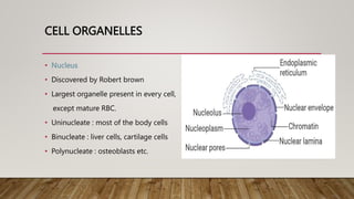
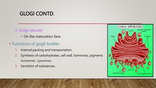
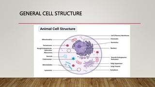
CELL STRUCTURE AND
FUNCTION
FOR MBBS/ NURSING / PHYSIOTHERAPY & OTHER PARAMEDICS.
DR. ZAHOOR MIR
CELL MEMBRANE
• Also called plasma membrane or plasmalemma or biological membrane.
• It is a thin, delicate, elastic and selectively/ semipermeable living boundary
that separates interior of the cell from its outside environment.
• Composition:
• Lipids (40%)_ two type:
1. Phospholipids – lollipop like structures in plasma membrane with
hydrophyllic head(water loving) made up of phosphate and glycerol and
hydrophobic tail (water repellent) made up of two long chain fatty acids.
CELL MEMBRANE
2. Cholestrol – provides regidity and stability to phospholipid bilayer.
• Proteins (50%) _ two types:
1. Integral proteins or intrensic proteins_ they are embedded in phospholipid
bilayer.
1. Totally embedded intrensic proteins or transmembrane proteins or
channels or tunnels. They help in the transportation of large molcules
across the membrane.
2. Partially embedded intrensic proteins.
2. Extrensic proteins_ present on the on the peripheries of phospholipid bilayer. It
mantains cell shape and catalyse the cellular reactions.
CELL MEMBRANE CONTD.
• Proteins act as transporter, enzyme, communication_ (cell signalling
by harmones, neurotransmitters etc,.), cell adhesion, recognition of
foreign cells.
• Carbohydrates (10%) two types:
1. Glycolipids_ when attached with lipid molecule.
2. Glycoproteins_ when attached with protein molecule.
• Carbohydrates helps in cell identification
CELL MEMBRANE CONTD.
• Functions of cell membrane:
1. Selectively Permeable I, e allow movement of some molecules only
across the membrane.
2. Cell Signalling / cellular communication
3. Cell recognition
4. Shape of cell
5. Anchoring of cells to form tissue
CELL MEMBRANE CONTD.
CELL ORGANELLES
• cell organelle is a tiny cellular structure that performs specific functions within a cell. OR
• An organelle is a subcellular structure that has one or more specific jobs to perform in the cell,
much like an organ does in the body
• Cell organelles are often enclosed by their own membranes, which divide the cell into many small
compartments for different biochemical reactions.
CYTOPLASM
• Cytoplasm
• Aqueous material (protoplasm) present between nuclear membrane
and cell membrane
• 90% water, appears transparent and structureless.
• Contains salts, sugers, aminoacids, fatty acids and cell organelles.
• Cytoplasmic streaming or cyclosis_ active mass movement of
cytoplasm in living cells. It helps in the formation of pseudopoda in
amobea.
• Note; cell – cell membrane = protoplasm
CYTOPLASM CONTD.
• Functions of cytoplasm
• Helps in intracellular distribution of nutrients, metabolites and
enzymes.
• Helps in the exchange of materials between cell organelles.
• Site of biochemical reactions like glycolysis, synthesis of fatty acids
etc.
• All the functions for cell expansion, growth and replication are carried out in the
cytoplasm of a cell.
CELL ORGANELLES
• Nucleus
• Discovered by Robert brown
• Largest organelle present in every cell,
except mature RBC.
• Uninucleate : most of the body cells
• Binucleate : liver cells, cartilage cells
• Polynucleate : osteoblasts etc.
NUCLEUS CONTD.
• Two nuclear membranes: outer rough due to presence of ribosomes and inner
soft.
• Space between outer and inner nuclear membranes is called perinuclear space.
• Nuclear pores allow the exchange of substances (mRNA, ribosomal units,
nucleotides, ribosomal proteins etc)
• Nucleoplasm_ gel like nuclear sap.
• Nucleus contains chromatin network. During cell division it condenses into thread
or rod like structure called chromosome.
NUCLEUS CONTD.
• Nucleolus _ site of formation of ribosomes. Every cell may possess one or few
nucleolus containing large amount of DNA and RNA. It controls all cellular
activities.
• Functions
• Control center of the cell.
• Stores Heridetry info (DNA)
• Makes RNA and proteins
• Makes ribosomes (nucleolus)
MITOCHONDRIA
• Singular –Mitochondrion, 🌭sausages in shape , counts one to many thousands.
• Power house of the cell as ATPs are produced here.
• Semi autonomous organelle (semi_ half, autonomus_dependent)
as it contain it’s own DNA, ribosomes, synthesize some structural
proteins but depend for food (glucose)
• Present in every cell except RBCs and prokeryotic cells
MITOCHONDRIA CONTD.
• Two membranes
• Outer membrane
• Inner highly folded membrane. Foldings called cristae.
• Intermembrane space_ outer compartment
• Matrix_ Fluid inside containing ribosomes and DNA.
• Oxysomes or F0-F1 complex or elementary particles:
Lollipop like structures.
• F1 or head helps in the synthesis of ATPs and F0 helps in electron transport chain (ETC)
MITOCHONDRIA CONTD.
• Functions of mitochondria
• Site of cellular respiration
• Site of Kreb‘s cycle
• Synthesize many amino acids
• Help in yolk formation during development of ovum
• Form middle peice of sperm during sperm maturation.
ENDOPLASMIC RETICULUM
• Series of interconnecting membrane canals in the cytoplasm.
• continues with outer membrane of the nuclear envelope.
• Present in all cells except RBC of mammals, eggs, embryotic cells.
• Exist in three morphological forms:
1. Cisternae
2. Vesicles ( Always smooth ER)
3. Tubules
ENDOPLASMIC RETICULUM CONTD.
• On the basis of appearance, endoplasmic reticulum has
two types:
1. Smooth Endoplasmic reticulum or SER.
2. Rough endoplasmic reticulum or RER.
ENDOPLASMIC RETICULUM CONTD.
• Rough endoplasmic reticulum
• Rough endoplasmic reticulum has rough and granular
appearance due to presence of ribosomes on its outer
surface.
• Mainly cisternae and very few tubular forms are rough.
• Smooth endoplasmic reticulum
• Smooth appearance because it lacks ribosomes on its
surface. It is continues with RER.
ENDOPLASMIC RETICULUM CONTD.
• Note ; In muscle cells, ER is called sarcoplasmic reticulum.
• Functions of Endoplasmic reticulum
1. RER helps in the synthesis of proteins.
2. SER helps in the synthesis of lipids and steroids.
3. Transport of proteins through cisternae.
4. In muscles, SER stores calcium ions and release them during muscle contraction.
5. In liver, SER detoxifies medication and alcohol.
RIBOSOMES
• Ribosome is a complex molecule made of ribosomal RNA
molecule and proteins so called ribosomal protein particle.
• Also called Protein factory as they synthesize proteins.
• Ribosomes have two sub-units, upper and lower. Both the
units exist freely in the cytoplasm.
RIBOSOMES CONTD.
• In Prokeryotic cells, ribosomes are 70 S and are scattered
in cytoplasm.
• Two sub units;
• Upper 30S
• Lower 50S
• Wher S stands for Svedberg‘s constant or sedimentation co-efficient. S is the unit
of time (it is not second). S = 10-13 seconds
RIBOSOMES CONTD.
• In Eukeryotic cells, ribosomes are scattered in cytoplasm and are also attached to
endoplasmic reticulum.
• It is 80S
• Two sub units;
• Upper 40S
• Lower 50S
• Where S is Svedberg’s constant.
RIBOSOMES CONTD.
• Both the sub units are freely scattering in the cytoplasm,
but they unite during protein synthesis.
• Functions of ribosomes
• Ribosomes are need to make large no. Of proteins for cells and
are the site for protein synthesis where RNA is translated into
proteins.
• Decode tRNA
GOLGI
• Observed by Camilo Golgi in nerve cells of barn owl
• Also called golgi apparatus or gogli bodies.
• Golgi exists in many forms, three major forms are:
1. Cisternae
• Elongated , flatned tubular sacs.
• Cisternae is formed constantly by budding off from SER and by the fusion of
vesicles.
• Cisternae breaks down into vesicles and vacuoles at maturing face.
2. Vesicles
• Present at convex side (I, e forming side) by budding off from SER.
GLOGI CONTD.
3. Golgi vacuole
• On the maturation face.
• Functions of gogli bodies
1. Internal packing and transportation.
2. Synthesis of carbohydrates, cell wall, harmones, pigments
Acrosomes, Lysosomes.
3. Secretion of substances.
LYSOSOMES
• Also called suicidal bags.
• Present in all animal cells
• It contains many digestive enzymes called acid hydrolases. I, e,
• Nuclease _ Digestion of DNA and RNA
• Protease_ Digestion of proteins
• Glycosidases_ Acts on polysaccharides
• Lipase_ Digest lipids.
• Phosphatases_ Break phosphate linked compounds
• Sulphatases_ Break sulphate linked compounds.
LYSOSOMES CONTD.
• They are formed directly from ER Or Golgi bodies.
• Lysosomes are of four types;
1. Primary lysosomes
2. Secondary lysosomes
3. Residual lysosomes
4. Autophagic vacuoles.
LYSOSOMES CONTD.
• Functions of lysosomes
1. Digestion of extracellular particles like food particles.
2. Digestion of intracellular organelles during starvation. This process is called
autolysis. By this self destruction takes place by releasing its contents hence the
name suicidal bags.
3. Digestion of harmful substances like bacteria, virus etc by WBCs. Hence help in
immune system
4. They move towards periphery and release their contents to dissolve cell
membrane during fertilization.
MICRO FILAMENTS
• They are long, thin, very fine protein filaments.
• Abundent in eukeryotic cells and contain protein actin.
• They occur as sheets or bundles just below the plasma membrane.
• Functions
• They are involved in endocytosis and exocytosis.
• Help in muscle contraction.
• Provide structural support and maintain the characteristic shape of the
cells.
MICROTUBULES
• Hollow, cylindrical, fine tubes.
• Made of helically arranged polymer chain of protein tubulin.
• Functions
• Form cytoskeleton and are responsible for the shape of cells.
• They are the contractile protein structures in the cytoplasm involved
in the movement of the cell and of the organelles within the cell.
• They form the skeleton of cilia and flegella and involves their
beating movements.
CILIA AND FLAGELLA
• They are the projections from the surface of some cells.
• They both are similar in some aspects but differ in other aspects.
• Similarties between cilia and flagella are:
1. Both are fine, hair like outgrowings, covered by cell membrane.
2. Both can move front and backwards for locomotion.
• Differances between cilia and flagella are:
1. Cilia is comperatively smaller then flagella.
2. Cilia are larger in no. While flegella are fewer in no.
CILIA AND FLAGELLA
• Functions
• Locomotion
• Movement of the substances.
VACUOLES
• Fluid filled sacs covered by a membrane (called tonoplast) are
called vacuoles.
• The fluid present in the sacs is called Cell-sap.
• Animal cells contain small but numerous vacuoles.
• Vacuoles have many types;
• Food vacuole
• Contractile vacuole
• Phagocytic vacuole. etc
VACUOLES CONTD.
• Functions of vacuoles
1. Contractile vacuoles are osmoregulatory in function (regulates
water in the body)
2. They store waste products.
3. They act as food reserve by storing sucrose and mineral salts.
CENTRIOLES
• Present in cytoplasm near nucleus.
• Each centriole is made up of 9 group of microtubules arranged in a circle. They exist
as triplets.
• Functions of centrioles
• Centrioles are used for the synthesis
of spindle fibres after nuclear division
• They act as a basal bodies at the cilia
and flegella.
GENERAL CELL STRUCTURE
THANK YOU
.
Thank you

Cell Structure and Function by Dr. Zahoor Mir.pptx

  • 1.
    CELL STRUCTURE AND FUNCTION FORMBBS/ NURSING / PHYSIOTHERAPY & OTHER PARAMEDICS. DR. ZAHOOR MIR
  • 10.
    CELL MEMBRANE • Alsocalled plasma membrane or plasmalemma or biological membrane. • It is a thin, delicate, elastic and selectively/ semipermeable living boundary that separates interior of the cell from its outside environment. • Composition: • Lipids (40%)_ two type: 1. Phospholipids – lollipop like structures in plasma membrane with hydrophyllic head(water loving) made up of phosphate and glycerol and hydrophobic tail (water repellent) made up of two long chain fatty acids.
  • 11.
    CELL MEMBRANE 2. Cholestrol– provides regidity and stability to phospholipid bilayer. • Proteins (50%) _ two types: 1. Integral proteins or intrensic proteins_ they are embedded in phospholipid bilayer. 1. Totally embedded intrensic proteins or transmembrane proteins or channels or tunnels. They help in the transportation of large molcules across the membrane. 2. Partially embedded intrensic proteins. 2. Extrensic proteins_ present on the on the peripheries of phospholipid bilayer. It mantains cell shape and catalyse the cellular reactions.
  • 12.
    CELL MEMBRANE CONTD. •Proteins act as transporter, enzyme, communication_ (cell signalling by harmones, neurotransmitters etc,.), cell adhesion, recognition of foreign cells. • Carbohydrates (10%) two types: 1. Glycolipids_ when attached with lipid molecule. 2. Glycoproteins_ when attached with protein molecule. • Carbohydrates helps in cell identification
  • 13.
    CELL MEMBRANE CONTD. •Functions of cell membrane: 1. Selectively Permeable I, e allow movement of some molecules only across the membrane. 2. Cell Signalling / cellular communication 3. Cell recognition 4. Shape of cell 5. Anchoring of cells to form tissue
  • 14.
  • 15.
    CELL ORGANELLES • cellorganelle is a tiny cellular structure that performs specific functions within a cell. OR • An organelle is a subcellular structure that has one or more specific jobs to perform in the cell, much like an organ does in the body • Cell organelles are often enclosed by their own membranes, which divide the cell into many small compartments for different biochemical reactions.
  • 16.
    CYTOPLASM • Cytoplasm • Aqueousmaterial (protoplasm) present between nuclear membrane and cell membrane • 90% water, appears transparent and structureless. • Contains salts, sugers, aminoacids, fatty acids and cell organelles. • Cytoplasmic streaming or cyclosis_ active mass movement of cytoplasm in living cells. It helps in the formation of pseudopoda in amobea. • Note; cell – cell membrane = protoplasm
  • 17.
    CYTOPLASM CONTD. • Functionsof cytoplasm • Helps in intracellular distribution of nutrients, metabolites and enzymes. • Helps in the exchange of materials between cell organelles. • Site of biochemical reactions like glycolysis, synthesis of fatty acids etc. • All the functions for cell expansion, growth and replication are carried out in the cytoplasm of a cell.
  • 18.
    CELL ORGANELLES • Nucleus •Discovered by Robert brown • Largest organelle present in every cell, except mature RBC. • Uninucleate : most of the body cells • Binucleate : liver cells, cartilage cells • Polynucleate : osteoblasts etc.
  • 19.
    NUCLEUS CONTD. • Twonuclear membranes: outer rough due to presence of ribosomes and inner soft. • Space between outer and inner nuclear membranes is called perinuclear space. • Nuclear pores allow the exchange of substances (mRNA, ribosomal units, nucleotides, ribosomal proteins etc) • Nucleoplasm_ gel like nuclear sap. • Nucleus contains chromatin network. During cell division it condenses into thread or rod like structure called chromosome.
  • 20.
    NUCLEUS CONTD. • Nucleolus_ site of formation of ribosomes. Every cell may possess one or few nucleolus containing large amount of DNA and RNA. It controls all cellular activities. • Functions • Control center of the cell. • Stores Heridetry info (DNA) • Makes RNA and proteins • Makes ribosomes (nucleolus)
  • 21.
    MITOCHONDRIA • Singular –Mitochondrion,🌭sausages in shape , counts one to many thousands. • Power house of the cell as ATPs are produced here. • Semi autonomous organelle (semi_ half, autonomus_dependent) as it contain it’s own DNA, ribosomes, synthesize some structural proteins but depend for food (glucose) • Present in every cell except RBCs and prokeryotic cells
  • 22.
    MITOCHONDRIA CONTD. • Twomembranes • Outer membrane • Inner highly folded membrane. Foldings called cristae. • Intermembrane space_ outer compartment • Matrix_ Fluid inside containing ribosomes and DNA. • Oxysomes or F0-F1 complex or elementary particles: Lollipop like structures. • F1 or head helps in the synthesis of ATPs and F0 helps in electron transport chain (ETC)
  • 23.
    MITOCHONDRIA CONTD. • Functionsof mitochondria • Site of cellular respiration • Site of Kreb‘s cycle • Synthesize many amino acids • Help in yolk formation during development of ovum • Form middle peice of sperm during sperm maturation.
  • 24.
    ENDOPLASMIC RETICULUM • Seriesof interconnecting membrane canals in the cytoplasm. • continues with outer membrane of the nuclear envelope. • Present in all cells except RBC of mammals, eggs, embryotic cells. • Exist in three morphological forms: 1. Cisternae 2. Vesicles ( Always smooth ER) 3. Tubules
  • 25.
  • 26.
    ENDOPLASMIC RETICULUM CONTD. •On the basis of appearance, endoplasmic reticulum has two types: 1. Smooth Endoplasmic reticulum or SER. 2. Rough endoplasmic reticulum or RER.
  • 27.
    ENDOPLASMIC RETICULUM CONTD. •Rough endoplasmic reticulum • Rough endoplasmic reticulum has rough and granular appearance due to presence of ribosomes on its outer surface. • Mainly cisternae and very few tubular forms are rough. • Smooth endoplasmic reticulum • Smooth appearance because it lacks ribosomes on its surface. It is continues with RER.
  • 28.
    ENDOPLASMIC RETICULUM CONTD. •Note ; In muscle cells, ER is called sarcoplasmic reticulum. • Functions of Endoplasmic reticulum 1. RER helps in the synthesis of proteins. 2. SER helps in the synthesis of lipids and steroids. 3. Transport of proteins through cisternae. 4. In muscles, SER stores calcium ions and release them during muscle contraction. 5. In liver, SER detoxifies medication and alcohol.
  • 29.
    RIBOSOMES • Ribosome isa complex molecule made of ribosomal RNA molecule and proteins so called ribosomal protein particle. • Also called Protein factory as they synthesize proteins. • Ribosomes have two sub-units, upper and lower. Both the units exist freely in the cytoplasm.
  • 30.
    RIBOSOMES CONTD. • InProkeryotic cells, ribosomes are 70 S and are scattered in cytoplasm. • Two sub units; • Upper 30S • Lower 50S • Wher S stands for Svedberg‘s constant or sedimentation co-efficient. S is the unit of time (it is not second). S = 10-13 seconds
  • 31.
    RIBOSOMES CONTD. • InEukeryotic cells, ribosomes are scattered in cytoplasm and are also attached to endoplasmic reticulum. • It is 80S • Two sub units; • Upper 40S • Lower 50S • Where S is Svedberg’s constant.
  • 32.
    RIBOSOMES CONTD. • Boththe sub units are freely scattering in the cytoplasm, but they unite during protein synthesis. • Functions of ribosomes • Ribosomes are need to make large no. Of proteins for cells and are the site for protein synthesis where RNA is translated into proteins. • Decode tRNA
  • 33.
    GOLGI • Observed byCamilo Golgi in nerve cells of barn owl • Also called golgi apparatus or gogli bodies. • Golgi exists in many forms, three major forms are: 1. Cisternae • Elongated , flatned tubular sacs. • Cisternae is formed constantly by budding off from SER and by the fusion of vesicles. • Cisternae breaks down into vesicles and vacuoles at maturing face. 2. Vesicles • Present at convex side (I, e forming side) by budding off from SER.
  • 34.
    GLOGI CONTD. 3. Golgivacuole • On the maturation face. • Functions of gogli bodies 1. Internal packing and transportation. 2. Synthesis of carbohydrates, cell wall, harmones, pigments Acrosomes, Lysosomes. 3. Secretion of substances.
  • 35.
    LYSOSOMES • Also calledsuicidal bags. • Present in all animal cells • It contains many digestive enzymes called acid hydrolases. I, e, • Nuclease _ Digestion of DNA and RNA • Protease_ Digestion of proteins • Glycosidases_ Acts on polysaccharides • Lipase_ Digest lipids. • Phosphatases_ Break phosphate linked compounds • Sulphatases_ Break sulphate linked compounds.
  • 36.
    LYSOSOMES CONTD. • Theyare formed directly from ER Or Golgi bodies. • Lysosomes are of four types; 1. Primary lysosomes 2. Secondary lysosomes 3. Residual lysosomes 4. Autophagic vacuoles.
  • 37.
    LYSOSOMES CONTD. • Functionsof lysosomes 1. Digestion of extracellular particles like food particles. 2. Digestion of intracellular organelles during starvation. This process is called autolysis. By this self destruction takes place by releasing its contents hence the name suicidal bags. 3. Digestion of harmful substances like bacteria, virus etc by WBCs. Hence help in immune system 4. They move towards periphery and release their contents to dissolve cell membrane during fertilization.
  • 38.
    MICRO FILAMENTS • Theyare long, thin, very fine protein filaments. • Abundent in eukeryotic cells and contain protein actin. • They occur as sheets or bundles just below the plasma membrane. • Functions • They are involved in endocytosis and exocytosis. • Help in muscle contraction. • Provide structural support and maintain the characteristic shape of the cells.
  • 39.
    MICROTUBULES • Hollow, cylindrical,fine tubes. • Made of helically arranged polymer chain of protein tubulin. • Functions • Form cytoskeleton and are responsible for the shape of cells. • They are the contractile protein structures in the cytoplasm involved in the movement of the cell and of the organelles within the cell. • They form the skeleton of cilia and flegella and involves their beating movements.
  • 40.
    CILIA AND FLAGELLA •They are the projections from the surface of some cells. • They both are similar in some aspects but differ in other aspects. • Similarties between cilia and flagella are: 1. Both are fine, hair like outgrowings, covered by cell membrane. 2. Both can move front and backwards for locomotion. • Differances between cilia and flagella are: 1. Cilia is comperatively smaller then flagella. 2. Cilia are larger in no. While flegella are fewer in no.
  • 41.
    CILIA AND FLAGELLA •Functions • Locomotion • Movement of the substances.
  • 42.
    VACUOLES • Fluid filledsacs covered by a membrane (called tonoplast) are called vacuoles. • The fluid present in the sacs is called Cell-sap. • Animal cells contain small but numerous vacuoles. • Vacuoles have many types; • Food vacuole • Contractile vacuole • Phagocytic vacuole. etc
  • 43.
    VACUOLES CONTD. • Functionsof vacuoles 1. Contractile vacuoles are osmoregulatory in function (regulates water in the body) 2. They store waste products. 3. They act as food reserve by storing sucrose and mineral salts.
  • 44.
    CENTRIOLES • Present incytoplasm near nucleus. • Each centriole is made up of 9 group of microtubules arranged in a circle. They exist as triplets. • Functions of centrioles • Centrioles are used for the synthesis of spindle fibres after nuclear division • They act as a basal bodies at the cilia and flegella.
  • 45.
  • 46.