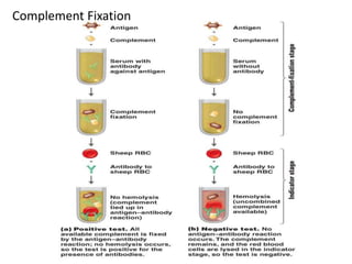
COMPLEMENT FIXATION TESTS
In general, complement fixation tests (CFT) are best
performed in reference laboratories where facilities exist for
the careful standardization and control of reagents, which
these tests require.
Principle of (CFT )
The complement fixation tests is a technique that has been
used over many years to detect and quantify antibody that
Serology does not agglutinate or precipitate when reacted
with its antigen, but can be demonstrated by its use, or
fixation, of complement.
Antigen-antibody reactions lead to immune complex
formation that produces complement fixation via the
classical pathway. That is when complement takes part
in antigen antibody reactions; it is bound or fixed to the
antigen antibody complexes. When these complexes are
on bacteria, red cells or other cells, the complement
brings about the lysis of the cells involved.
This may be exploited to determine the amount of
antigen or antibody present in the patient sample.
Complement fixation test can detect antibody at a level
of less than one microgram per milliliter.
CFT consists of two steps:
Step 1: a known antigen and inactivated patientโ€™s serum are
incubated with a standardized, limited amount of complement. If
the serum contains specific, complement activating antibody the
complement will be activated or fixed by the antigen-antibody
complex. However, if there is no antibody in the patientโ€™s serum,
there will be no formation of antigen-antibody complex, and
therefore complement will not be fixed. But will remain free.
Please note:
Complement (group of serum proteins) binds to antigen-antibody
complex and is used up usually guinea pig serum.
Complement Fixation
Step 2:
The second step detects whether complement has been
utilized in the first step or not. This is done by adding the
indicator system. If the complement is fixed in the first
step owing to the presence of antibody there will be no
complement left to fix to the indicator system. However,
if there is antibody in the patientโ€™s serum, there will be
no antigen-antibody complex, and therefore,
complement will be present free or unfixed in the
mixture.
This unfixed complement will now react with the antibody-
coated sheep red blood cells to bring about their lysis.
Thus, no lysis of sheep red blood cells (positive CFT)
indicates the presence of antibody in the presence of
antibody in the test serum, while lysis of sheep red blood
cells (Negative CFT) indicates the absence of antibody in
the serum.
Step 2 continued :
Interpretation
Positive test : The available complement is fixed by Ag-Ab complex
and no hemolysis of sheep RBCs occurs. So the test is positive for
presence of antibodies.
Negative test : No Ag-Ab reaction occurs and the complement is free.
Thisfree complement binds to the complex of sheep RBC and itโ€™s
antibody to cause hemolysis, causing the development of pink color.
Advantages of CFT
1. Ability to screen against a large number of viral and bacterial
infections at the same time.
2. Economical.
Disadvantages of CFT
1. Not sensitive - cannot be used for immunity screening
2. 2. Time consuming and labor intensive
3. Often non-specific.
Tertiary binding tests
Tertiary binding tests measure the consequences of
immune responses in vivo. These tests are much
more complex than primary and secondary tests, but
their results reflect the practical significance of the
immune response. E.g. measurement of the
protective effects of antibody.
Many factors affect the interaction between antigen
and antibody; these include specificity, cross reactivity,
temperature, pH, ionic strength, concentration, and
intermolecular specificity.
Factors affecting antigen antibody reaction
Specificity: The ability of a particular antibody to
combine with one antigen instead of another is referred
to as specificity. This property depends on the antigen โ€“
binding fragment of an immunoglobulin molecule.
Antigen โ€“ antibody reactions can show a high level of
specificity.
Cross reactivity: Unrelated molecules can have antigens
with similar antigenic determinants. This means a
proportion of the antibodies directed against one kind of
antigen will also react with the other kind of antigen.
This is called cross reactivity.
Temperature: The optimum temperature needed to
reach equilibrium in an antibody โ€“ antigen reaction
differs for different antibodies. IgM antibodies are cold
reacting, with a thermal range of 4-220C, and IgG
antibodies are warm reacting with an optimum
temperature of reaction of 370C.
pH: Although the optimum pH for all reactions has not
been determined, a pH of 7.0 is used for routine
laboratory testing.
Ionic strength: The concentration of salt in the reaction
medium has an effect on antibody uptake by the
membrane bound erythrocyte antigens. Sodium and
chloride ions in solution have an inhibitory effect.
These ions cluster around the opposite charges on
antigen and antibody molecules which partially
neutralizes them. This hinders the association of
antibody with antigen. Reducing or lowering the ionic
strength of a reaction medium, such as low-ionic
strength salt, can enhance antibody uptake.
Concentration: Under normal conditions, the concentration of
antigen and antibody should be optimal, but sometime this is not
the case. Excess antibody or antigen concentration will result in a
false reaction, sometimes known as zonal reaction when the
concentration of antigen is excess it is known as a Serology post
zone reaction;
excess antibody is referred to as a prozone reaction. This
phenomenon can be overcome by serial dilutions unil the
optimum amount of antigen and antibody is present.
Bonding of an antigen to an antibody takes place because of the
formation of multiple, reversible, intermolecular attraction between
an antigen and amino acids of the binding site. The bonding of
antigen to antibody is exclusively noncovalent.
The attractive force of noncovalent bonds is weak compared to
covalent bonds, but the formation of multiple noncovalent bonds
produces considerable total โ€“ binding energy. The strength of a
single antigen โ€“ antibody bond is termed as antibody affinity.
The strongest bonding develops when antigens and antibodies are
close to each other and when the shapes of both the antigenic
determinate and the antigen โ€“ binding site conform to each other.
This complementary matching is referred to as โ€œgoodness of fitโ€.
Bond strength and intermolecular attractive force

Complement fixation tests

  • 1.
  • 2.
    In general, complementfixation tests (CFT) are best performed in reference laboratories where facilities exist for the careful standardization and control of reagents, which these tests require. Principle of (CFT ) The complement fixation tests is a technique that has been used over many years to detect and quantify antibody that Serology does not agglutinate or precipitate when reacted with its antigen, but can be demonstrated by its use, or fixation, of complement.
  • 3.
    Antigen-antibody reactions leadto immune complex formation that produces complement fixation via the classical pathway. That is when complement takes part in antigen antibody reactions; it is bound or fixed to the antigen antibody complexes. When these complexes are on bacteria, red cells or other cells, the complement brings about the lysis of the cells involved. This may be exploited to determine the amount of antigen or antibody present in the patient sample. Complement fixation test can detect antibody at a level of less than one microgram per milliliter.
  • 4.
    CFT consists oftwo steps: Step 1: a known antigen and inactivated patientโ€™s serum are incubated with a standardized, limited amount of complement. If the serum contains specific, complement activating antibody the complement will be activated or fixed by the antigen-antibody complex. However, if there is no antibody in the patientโ€™s serum, there will be no formation of antigen-antibody complex, and therefore complement will not be fixed. But will remain free. Please note: Complement (group of serum proteins) binds to antigen-antibody complex and is used up usually guinea pig serum.
  • 5.
  • 6.
    Step 2: The secondstep detects whether complement has been utilized in the first step or not. This is done by adding the indicator system. If the complement is fixed in the first step owing to the presence of antibody there will be no complement left to fix to the indicator system. However, if there is antibody in the patientโ€™s serum, there will be no antigen-antibody complex, and therefore, complement will be present free or unfixed in the mixture.
  • 7.
    This unfixed complementwill now react with the antibody- coated sheep red blood cells to bring about their lysis. Thus, no lysis of sheep red blood cells (positive CFT) indicates the presence of antibody in the presence of antibody in the test serum, while lysis of sheep red blood cells (Negative CFT) indicates the absence of antibody in the serum. Step 2 continued : Interpretation Positive test : The available complement is fixed by Ag-Ab complex and no hemolysis of sheep RBCs occurs. So the test is positive for presence of antibodies. Negative test : No Ag-Ab reaction occurs and the complement is free. Thisfree complement binds to the complex of sheep RBC and itโ€™s antibody to cause hemolysis, causing the development of pink color.
  • 8.
    Advantages of CFT 1.Ability to screen against a large number of viral and bacterial infections at the same time. 2. Economical. Disadvantages of CFT 1. Not sensitive - cannot be used for immunity screening 2. 2. Time consuming and labor intensive 3. Often non-specific.
  • 9.
    Tertiary binding tests Tertiarybinding tests measure the consequences of immune responses in vivo. These tests are much more complex than primary and secondary tests, but their results reflect the practical significance of the immune response. E.g. measurement of the protective effects of antibody.
  • 10.
    Many factors affectthe interaction between antigen and antibody; these include specificity, cross reactivity, temperature, pH, ionic strength, concentration, and intermolecular specificity. Factors affecting antigen antibody reaction Specificity: The ability of a particular antibody to combine with one antigen instead of another is referred to as specificity. This property depends on the antigen โ€“ binding fragment of an immunoglobulin molecule. Antigen โ€“ antibody reactions can show a high level of specificity.
  • 11.
    Cross reactivity: Unrelatedmolecules can have antigens with similar antigenic determinants. This means a proportion of the antibodies directed against one kind of antigen will also react with the other kind of antigen. This is called cross reactivity. Temperature: The optimum temperature needed to reach equilibrium in an antibody โ€“ antigen reaction differs for different antibodies. IgM antibodies are cold reacting, with a thermal range of 4-220C, and IgG antibodies are warm reacting with an optimum temperature of reaction of 370C.
  • 12.
    pH: Although theoptimum pH for all reactions has not been determined, a pH of 7.0 is used for routine laboratory testing. Ionic strength: The concentration of salt in the reaction medium has an effect on antibody uptake by the membrane bound erythrocyte antigens. Sodium and chloride ions in solution have an inhibitory effect. These ions cluster around the opposite charges on antigen and antibody molecules which partially neutralizes them. This hinders the association of antibody with antigen. Reducing or lowering the ionic strength of a reaction medium, such as low-ionic strength salt, can enhance antibody uptake.
  • 13.
    Concentration: Under normalconditions, the concentration of antigen and antibody should be optimal, but sometime this is not the case. Excess antibody or antigen concentration will result in a false reaction, sometimes known as zonal reaction when the concentration of antigen is excess it is known as a Serology post zone reaction; excess antibody is referred to as a prozone reaction. This phenomenon can be overcome by serial dilutions unil the optimum amount of antigen and antibody is present.
  • 14.
    Bonding of anantigen to an antibody takes place because of the formation of multiple, reversible, intermolecular attraction between an antigen and amino acids of the binding site. The bonding of antigen to antibody is exclusively noncovalent. The attractive force of noncovalent bonds is weak compared to covalent bonds, but the formation of multiple noncovalent bonds produces considerable total โ€“ binding energy. The strength of a single antigen โ€“ antibody bond is termed as antibody affinity. The strongest bonding develops when antigens and antibodies are close to each other and when the shapes of both the antigenic determinate and the antigen โ€“ binding site conform to each other. This complementary matching is referred to as โ€œgoodness of fitโ€. Bond strength and intermolecular attractive force