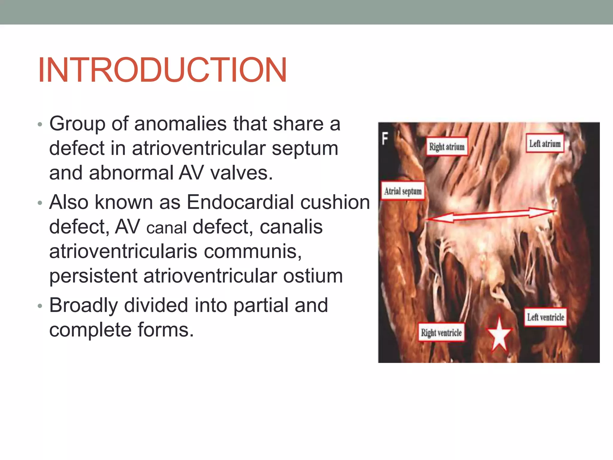
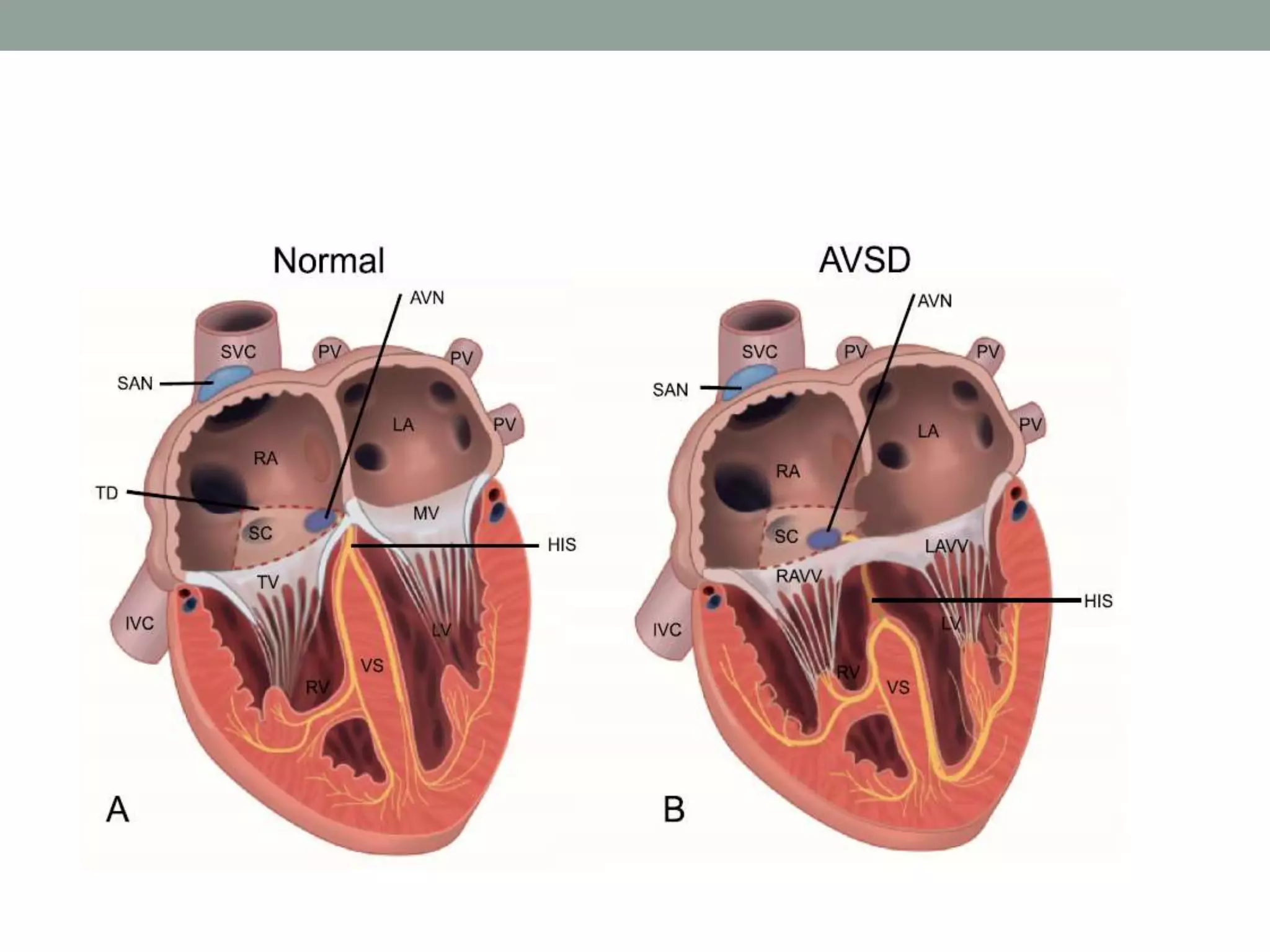
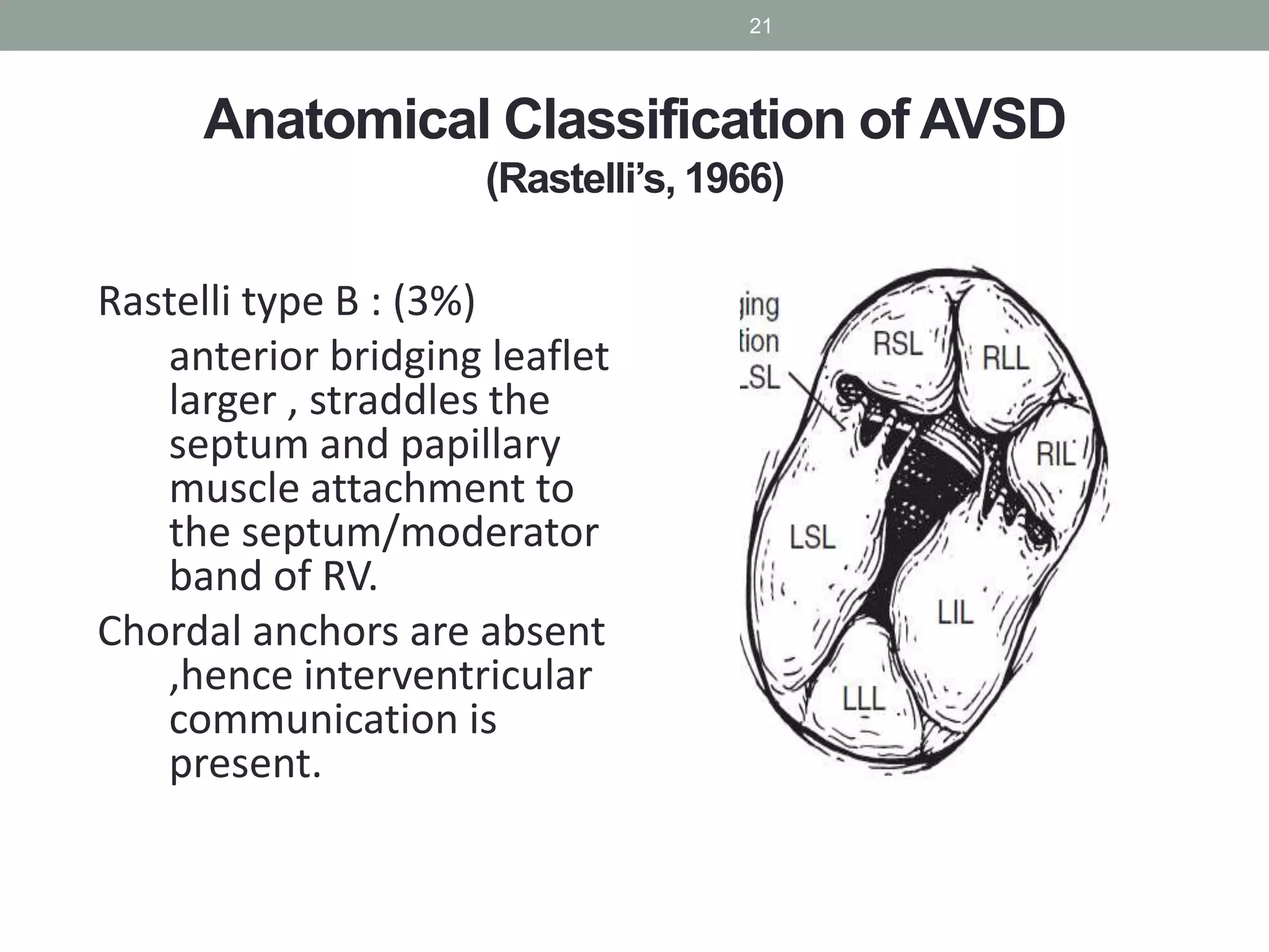
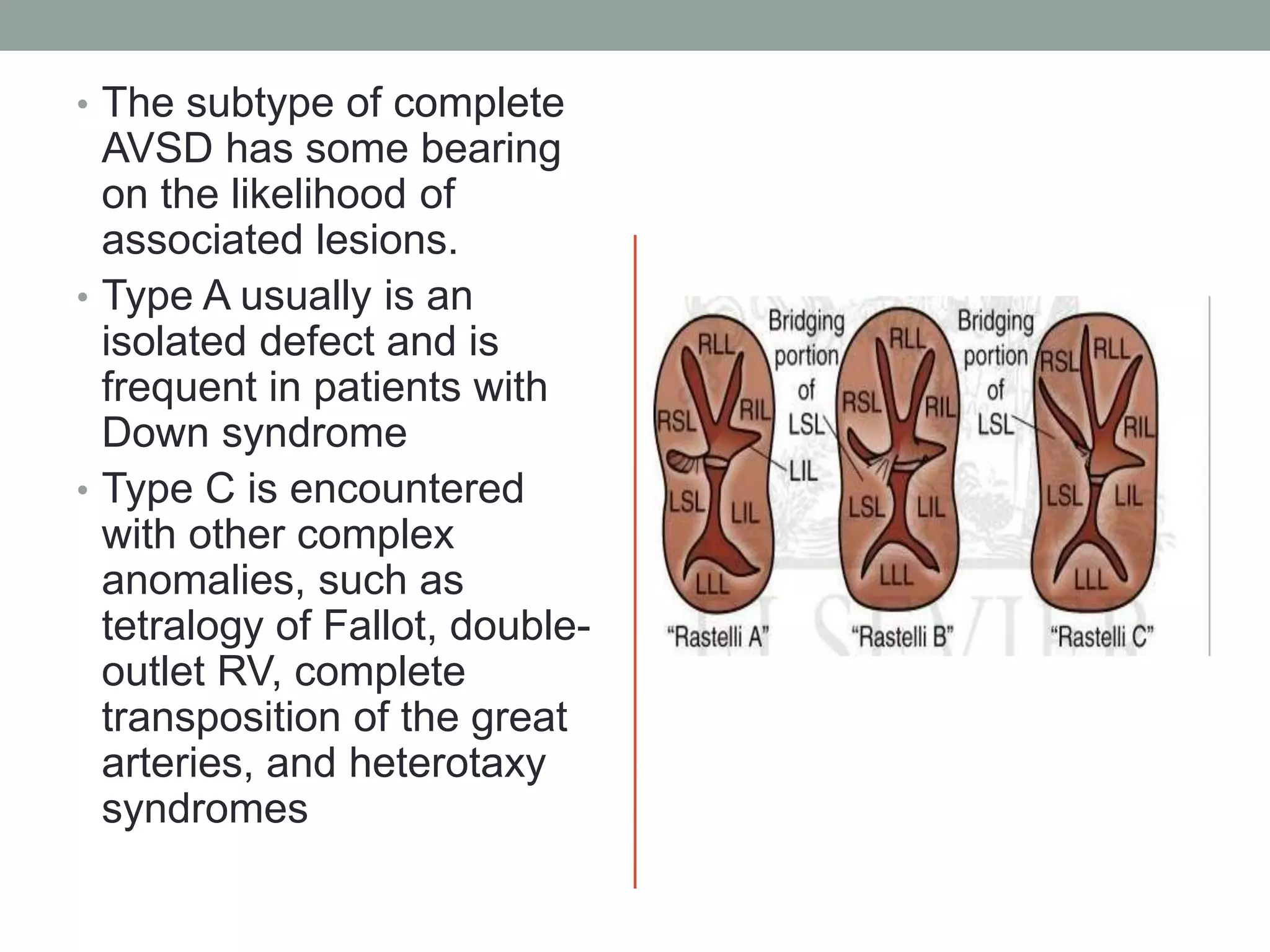
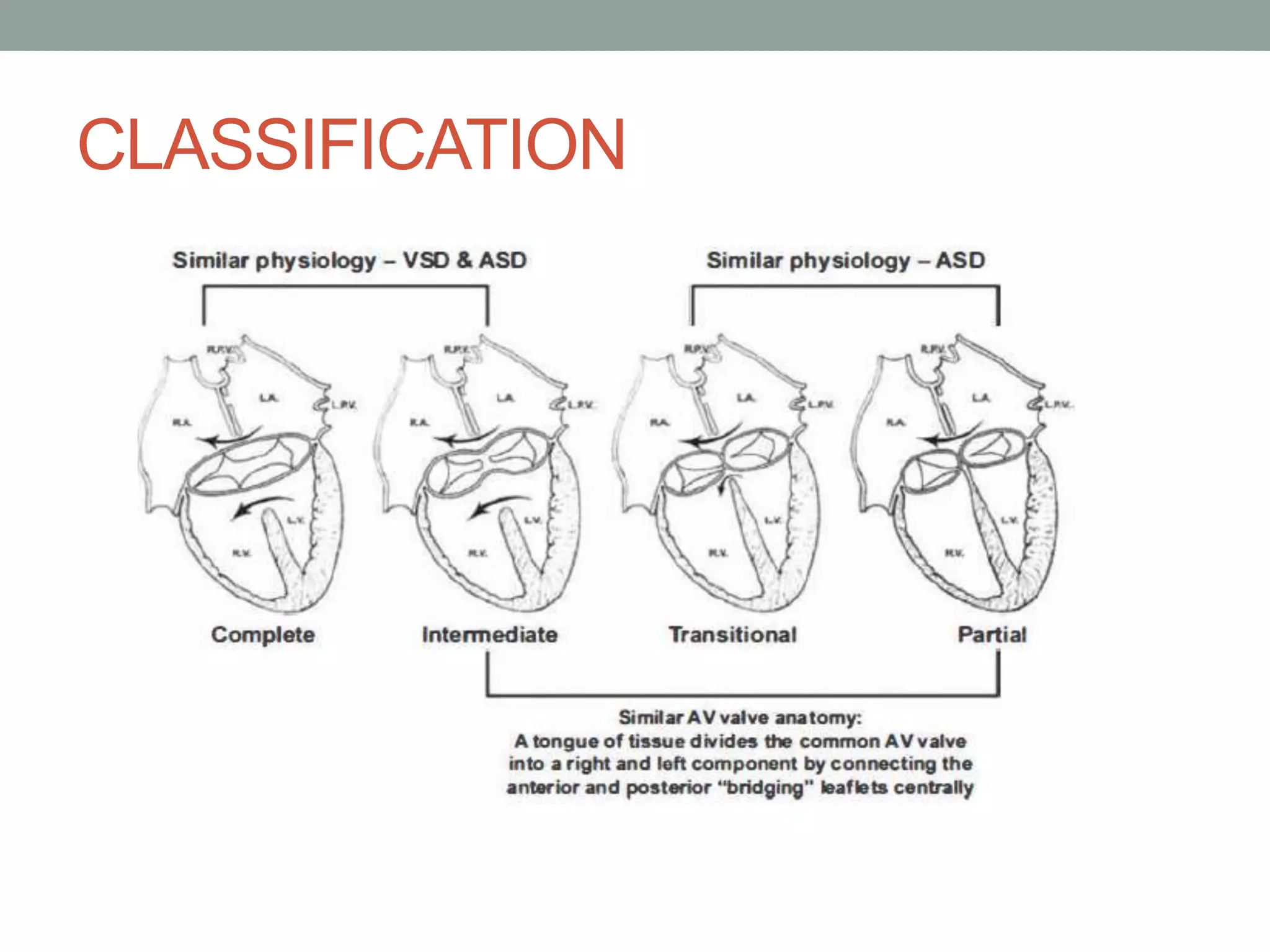
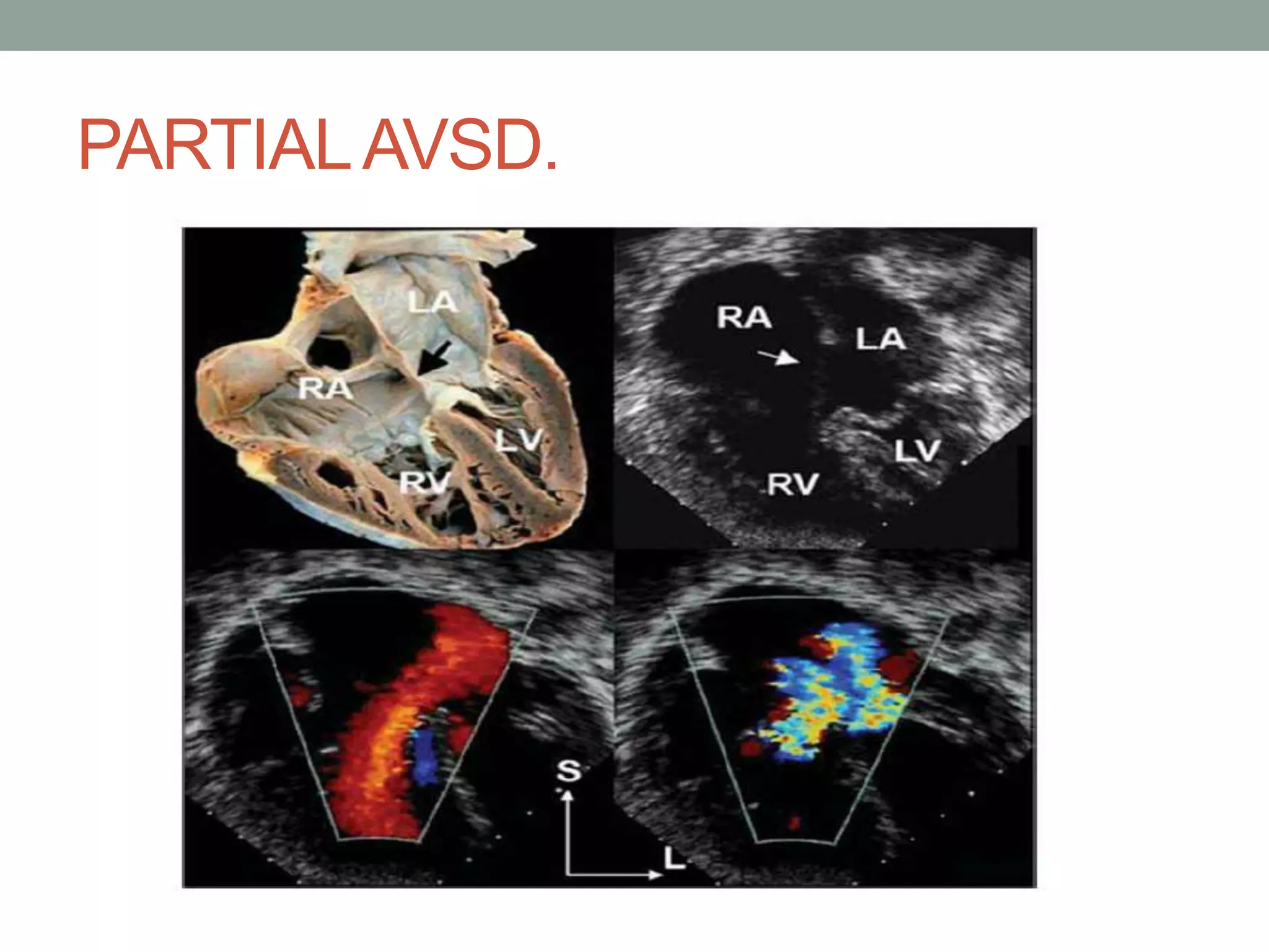
SlideShare a Scribd company logo
ATRIOVENTRICULAR
SEPTAL DEFECTS.
INTRODUCTION
• Group of anomalies that share a
defect in atrioventricular septum
and abnormal AV valves.
• Also known as Endocardial cushion
defect, AV canal defect, canalis
atrioventricularis communis,
persistent atrioventricular ostium
• Broadly divided into partial and
complete forms.
DEMOGRAPICS
• 4 TO 5 % Of congenital heart defects.
• Estimated occurrence of 0.19 in 1,000 live births
• Male = female or slight female preponderance.
• Downs syndrome: 40 to 45% have heart disease.
• Of which 45% have avsd.
• > 75 % of these are the complete form.
• Conversely approximately 50% of avsd patients have
downs syndrome.
HISTORY
 Rogers, Edwards : Recognised morphological similarity
of ostium primum ASD and complete defect in 1948
 Wakai, Edwards : Term of partial and complete AV
canal defect in 1956
 Bharati & Lev : Term of Intermediate & Transitional in
1980
 Rastelli: Described the of common anterior leaflet in
1966
 Lillehei : 1st repair of AVSD in 1954
 Kirklin, Watkin, Gross: Open repair using oxygenator
EMBRYOGENESIS.
• Defect in endocardial cushion development and fusion.
• Additional pathways have now been illustrated, which
shows that “dorsal mesenchymal protrusion”(vestibular
spine) is responsible for formation of avsd.
• In partial AVSDs, incomplete fusion of the superior and
inferior endocardial cushions results in a cleft in the mid-
portion of the left AV valve anterior leaflet often associated
with regurgitation.
• In contrast, complete AVSD is associated with lack of
fusion between the superior and inferior cushions and,
consequently, with the formation of separate anterior and
posterior bridging leaflets along the subjacent ventricular
septum
• Since the dextrodorsal conus cushion contributes to the
development of the right AV valve and the outflow tracts lie
adjacent to their respective inflow tracts, AVSDs may be
associated with conotruncal anomalies, such as tetralogy of
Fallot and double-outlet right ventricle (RV).
In addition, shift of the AV valve orifice may result in
connection of the valve primarily to only one ventricle,
creating disproportionate or unbalanced ventricles.
MORPHOLOGY
Pathology
 In the normal
heart, the aortic
valve is wedged.
 In AVSD the
aortic valve is
displaced
anteriorly and
creates an
elongated, k/a
gooseneck
deformity of the
LVOT
14
VALVE MORPHOLOGY
• Normal MV –
Posterior leaflet : 2/3
circumference
• AVSD MV – Left
lateral leaflet : 1/5
circumference
CONDUCTION SYSTEM
ANATOMICAL CLASSIFICATION
(RASTELLI)
• Prior to 1964, hospital mortality
for patients with AVSD was 60 %.
• Rastelli et al. from the Mayo
Clinic published their work in
1968 and operative mortality
between 1964 and 1967
decreased to 20 % .
Anatomical Classification of AVSD
(Rastelli’s, 1966)
Based bridging of LSL across
IVS
Rastelli type A : (55%) Anterior
bridging leaflet divided and
attached to crest of ventricular
septum.
• Interventricular communication
beneath the anterior bridging
leaflet may be minimal or
absent in some cases owing to
extensive interchordal fusion.
20
Anatomical Classification of AVSD
(Rastelli’s, 1966)
Rastelli type B : (3%)
anterior bridging leaflet
larger , straddles the
septum and papillary
muscle attachment to
the septum/moderator
band of RV.
Chordal anchors are absent
,hence interventricular
communication is
present.
21
Anatomical Classification of AVSD
(Rastelli’s, 1966)
 Rastelli type C : (30%)
anterior bridging leaflet
is larger than in type B.
• its medial papillary
muscle attachments fuse
to the right-sided anterior
papillary muscle.
• Free interventricular
communication is
present.
• Also called free floating
chordae.
22
• The subtype of complete
AVSD has some bearing
on the likelihood of
associated lesions.
• Type A usually is an
isolated defect and is
frequent in patients with
Down syndrome
• Type C is encountered
with other complex
anomalies, such as
tetralogy of Fallot, double-
outlet RV, complete
transposition of the great
arteries, and heterotaxy
syndromes
CLASSIFICATION
PARTIALAVSD.
• Two separate annuli
• Ostium primum asd and cleft left anterior av valve.
• The cleft in the left AV valve anterior leaflet is directed
toward the midportion of the ventricular septum, along the
anteroinferior rim of the septal defect.
• The left AV valve orifice is triangular rather than elliptical (
as in a normal heart) and resembles a mirror-image
tricuspid valve orifice.
• The cleft left AV valve usually is regurgitant and, with time,
becomes thickened and exhibits histologic alterations that
resemble myxomatous mitral valve prolapse.
• Although patients with partial AVSD may be asymptomatic
until adulthood, symptoms of excess pulmonary blood flow
typically occur in childhood
• Tachypnea and poor weight gain occur most commonly
when the defect is associated with moderate or severe left
AV valve regurgitation or with other hemodynamically
significant cardiac anomalies.
• Patients with primum ASDs usually have earlier and more
severe symptoms, including growth failure, than patients
with secundum ASDs.
COMPLETE AVSD
• Tachypnea and failure to thrive invariably occur early in
infancy as a result of excessive pulmonary blood flow
• All patients with complete AVSD have symptoms by 1year
of age
• AV valve regurgitation compounds these problems.
HEMODYNAMICS
 The outcome of live-born patients with AVSD depends on
the
 specific morphology of the defect
 The size of the ventricular septal defect
 Degree of ventricular hypoplasia
 Degree of AV valve regurgitation
 Presence or absence of LVOT obstruction
 Presence or absence of coarctation of aorta
 Associated syndromes (cardiac and noncardiac)
Natural History
35
 Patients with the complete form of AVSD and large VSD not
undergoing repair die in infancy with CHF & PAH
 Those who survive without surgery into childhood usually
develop pulmonary vascular obstruction and eventually die with
Eisenmenger’s syndrome
 Berger and his colleagues found that only 54% of patients born
with a complete form of AVSD were alive at 6 months of age, 35%
at 12 months, 15% at 24 months, and 4% at 5 years of age
 This data would support surgical intervention in the first 3–6
months of age
36
Natural History
Berger TJ,et al Ann Thorac Surg 1979; 27: 104–11.
 Infants with 10 ASD presenting in infancy have a poor outcome,
mainly because of the associated risk factors that bring these
infants to early attention
 Those with the partial form of AVSD and minimal left AV valve
regurgitation seem to fare the best without surgery, although
there is still likely considerable morbidity and mortality
 According to Somerville, 50% die before 20 years of age and only
25% survive beyond 40 years of age
 Atrial fibrillation in these patients was an important cause of late
morbidity and mortality
37
Natural History
 ECG
 Superior” QRS axis with the QRS axis between -40 and -1500
 Most of the patients have a prolonged PR interval
 More than 50% have atrial enlargement
 RVH or RBBB is present in all cases (2/3rd have rsR, RSR or Rr in lead
V1, and the rest have a qR or R pattern) & many have LVH
 In 10 ASD findings are same as 20 ASD except for enlargement of the
LA & LV when MR is significant
 In complete AVSD cardiomegaly is always present and involves all
four cardiac chambers. Pulmonary vascular markings are increased,
and the main PA segment is prominent
ECG
38
 Primary imaging technique for diagnosing AVSD
 The internal cardiac crux is the most consistent imaging
landmark
 Apical four-chamber imaging plane clearly visualizes the
internal crux
Echocardiography
39
 Several echocardiac features are shared by all forms of
AVSD:
 Deficiency of a portion of the inlet ventricular septum
 Inferior displacement of the AV valves
40
Echocardiography
 The most common left AV valve abnormality, a cleft, is best
visualized from the parasternal and subcostal short-axis
imaging planes.
Echocardiography
42
 In the transitional form of partial AVSD, there is aneurysmal
replacement of a portion of the inlet ventricular septum
Echocardiography
43
 Rarely required for diagnosis
 In older patient it may have a role in assessing the degree of
pulmonary vascular obstructive disease or CAD
 A large Lt to Rt shunt at the atrial level demonstrated by a
significantly higher oxygen saturation sampled from the RA
compared with the blood in the IVC & SVC
 In complete AVSD the PASP is invariably at or near systemic level,
while in partial AVSDs, the PASP is usually <60% of systemic pressure
 LV angiography - gooseneck deformation of the LVOT
Cardiac Catheterization & Angiography
44
 Left to-right shunting increases the oxygen saturation in RA
 Sample from high in the SVC usually represents the best
mixed venous oxygen saturation (normal or 40 to 50%)
 Usually a further increase in oxygen saturation in the RV
 Pulmonary venous oxygen saturation is frequently reduced
to 93–95% in older individuals with very large L to R shunts
 LA & LV O2 saturation is often decreased to as low as 86–88%
45
INDICATIONS
• Complete AVSD
a. Uncontrolled heart failure:
Complete surgical repair as soon as possible (Class I)
b. Controlled heart failure: Complete surgical repair by 3 months
of age (Class I)
c. Pulmonary artery banding: May be considered in select
patients under 3 months of age (Class IIb).
ii. Partial or intermediate AVSD, stable, and with normal
pulmonary artery pressures: Surgical repair at 2–3 years of age
(Class I)
iii. Associated moderate or severe AV valve regurgitation may
necessitate early surgery in partial or intermediate forms.
iv. Pulmonary artery banding is reserved for complex cases and
in patients with contraindications for cardiopulmonary bypass
(Class IIb).
Surgery for moderate-to-severe left AV valve regurgitation
is recommended as per the guidelines for mitral
regurgitation.(Class I).
vi. Surgery for left ventricular outflow tract obstruction is
reasonable with a peak systolic gradient of ≥50 mmHg, or
at a lesser gradient if heart failure symptoms are present,
or if concomitant moderate-to-severe atrioventricular or
aortic regurgitation is present (Class IIa).
vii. Those presenting beyond 6 months of life with
significant pulmonary hypertension and suspected elevated
PVR should be referred to a higher center for further
evaluation to assess operability.
SURGICAL STEPS
SIZING THE PATCH
• Too wide: LVOTO
• Too long : AV Valve
regurgitation.
SINGLE PATCH
Australian technique (Modified single
patch technique)
• For complete AV
canal defect with
minimal AV valve
distortion
PARTIALAVSD REPAIR
AV VALVE REGURGITATION.
Results (single v/s double patch)
What to perform ??
FOLLOW UP
Recommendations for follow-up
i. Lifelong follow-up is required.
ii. In patients with no significant residual abnormality,annual
follow-up is required till 10 years of age followed by 2–
3-yearly follow-up.
The patient should undergo physical examination, ECG,
and echocardiography at each visit, and a Holter monitor
test may be required in select cases.
iii. IE prophylaxis is recommended for 6 months after
surgical closure. However, all patients are advised to
maintain good oro-dental hygiene after this period also.

More Related Content

What's hot

Pulmonary atresia with intact ventricular septum
Pulmonary atresia with intact ventricular septumPulmonary atresia with intact ventricular septum
Pulmonary atresia with intact ventricular septum
Ramachandra Barik
 
Total Anomalous Pulmonary Venous Connection
Total Anomalous Pulmonary Venous ConnectionTotal Anomalous Pulmonary Venous Connection
Total Anomalous Pulmonary Venous Connection
Dang Thanh Tuan
 
Congenitally corrected transposition of great arteries
Congenitally corrected transposition of great arteriesCongenitally corrected transposition of great arteries
Congenitally corrected transposition of great arteries
Dheeraj Sharma
 

What's hot (20)

Atrial septal defect DR NIKUNJ R SHEKHADA (MBBS,MS GEN SURG,DNB CTS SR
Atrial septal defect DR NIKUNJ R SHEKHADA (MBBS,MS GEN SURG,DNB CTS SRAtrial septal defect DR NIKUNJ R SHEKHADA (MBBS,MS GEN SURG,DNB CTS SR
Atrial septal defect DR NIKUNJ R SHEKHADA (MBBS,MS GEN SURG,DNB CTS SR
 
Atrial Septal Defect
Atrial Septal DefectAtrial Septal Defect
Atrial Septal Defect
 
Vsd
VsdVsd
Vsd
 
AV septal defects (AVCD)
AV septal defects (AVCD)AV septal defects (AVCD)
AV septal defects (AVCD)
 
Pulmonary atresia with intact ventricular septum
Pulmonary atresia with intact ventricular septumPulmonary atresia with intact ventricular septum
Pulmonary atresia with intact ventricular septum
 
Double outlet right ventricle
Double outlet right ventricleDouble outlet right ventricle
Double outlet right ventricle
 
Ecg in congenital heart disease
Ecg in congenital heart diseaseEcg in congenital heart disease
Ecg in congenital heart disease
 
Av canal defect
Av canal defectAv canal defect
Av canal defect
 
Acyanotic heart disease
Acyanotic heart diseaseAcyanotic heart disease
Acyanotic heart disease
 
L-TGA or CCTGA
L-TGA or CCTGA L-TGA or CCTGA
L-TGA or CCTGA
 
Management of tapvc
Management of tapvcManagement of tapvc
Management of tapvc
 
Total Anomalous Pulmonary Venous Connection
Total Anomalous Pulmonary Venous ConnectionTotal Anomalous Pulmonary Venous Connection
Total Anomalous Pulmonary Venous Connection
 
transposition of great arteries
transposition of great arteriestransposition of great arteries
transposition of great arteries
 
Vsd device closure
Vsd device closureVsd device closure
Vsd device closure
 
L tga anatomy, management-
L tga anatomy, management-L tga anatomy, management-
L tga anatomy, management-
 
Ecg in single ventricle
Ecg in single ventricleEcg in single ventricle
Ecg in single ventricle
 
Ventricular septal defect, congenital heart disease
Ventricular septal defect,  congenital heart diseaseVentricular septal defect,  congenital heart disease
Ventricular septal defect, congenital heart disease
 
NATURAL HISTORY OF ASD, VSD, PDA
NATURAL HISTORY OF ASD, VSD, PDANATURAL HISTORY OF ASD, VSD, PDA
NATURAL HISTORY OF ASD, VSD, PDA
 
electrocardiogram (Ecg) in CONGENITAL HEART DISEASES
electrocardiogram (Ecg) in CONGENITAL HEART DISEASESelectrocardiogram (Ecg) in CONGENITAL HEART DISEASES
electrocardiogram (Ecg) in CONGENITAL HEART DISEASES
 
Congenitally corrected transposition of great arteries
Congenitally corrected transposition of great arteriesCongenitally corrected transposition of great arteries
Congenitally corrected transposition of great arteries
 

Similar to Atrioventricular septal defects

Electrocardiography in Adult Congenital Heart Diseases
Electrocardiography in Adult Congenital Heart DiseasesElectrocardiography in Adult Congenital Heart Diseases
Electrocardiography in Adult Congenital Heart Diseases
Saleh AL-Hatem
 

Similar to Atrioventricular septal defects (20)

Endocardial Cushion Defect / AVSD
Endocardial Cushion Defect / AVSDEndocardial Cushion Defect / AVSD
Endocardial Cushion Defect / AVSD
 
PA/IVS - Dr. Julius King Kwedhi
PA/IVS - Dr. Julius King KwedhiPA/IVS - Dr. Julius King Kwedhi
PA/IVS - Dr. Julius King Kwedhi
 
Electrocardiography in Adult Congenital Heart Diseases
Electrocardiography in Adult Congenital Heart DiseasesElectrocardiography in Adult Congenital Heart Diseases
Electrocardiography in Adult Congenital Heart Diseases
 
Atrial septal defects 16 3-15
Atrial septal defects 16 3-15Atrial septal defects 16 3-15
Atrial septal defects 16 3-15
 
Asd new
Asd newAsd new
Asd new
 
Av canal defect
Av canal defectAv canal defect
Av canal defect
 
Atrial Septal Defects.pptx
Atrial Septal Defects.pptxAtrial Septal Defects.pptx
Atrial Septal Defects.pptx
 
A v canal defect may 2021
A v canal defect  may 2021A v canal defect  may 2021
A v canal defect may 2021
 
Cc tga
Cc tgaCc tga
Cc tga
 
Dorv thab
Dorv thab Dorv thab
Dorv thab
 
Atrial septal defect Echocardiography
Atrial septal defect EchocardiographyAtrial septal defect Echocardiography
Atrial septal defect Echocardiography
 
Modul TGA Transposition of Great Arteries.pptx
Modul TGA Transposition of Great Arteries.pptxModul TGA Transposition of Great Arteries.pptx
Modul TGA Transposition of Great Arteries.pptx
 
Classification and pathophysiology of tapvc amitabh
Classification and pathophysiology of tapvc amitabhClassification and pathophysiology of tapvc amitabh
Classification and pathophysiology of tapvc amitabh
 
Classification and pathophysiology of tapvc
Classification and pathophysiology of tapvc Classification and pathophysiology of tapvc
Classification and pathophysiology of tapvc
 
Double outlet right ventricle
Double outlet right ventricleDouble outlet right ventricle
Double outlet right ventricle
 
Asd and vsd
Asd and vsdAsd and vsd
Asd and vsd
 
Single ventricle
Single ventricleSingle ventricle
Single ventricle
 
Synopses in vsd
Synopses in vsdSynopses in vsd
Synopses in vsd
 
Asd may 2021
Asd  may 2021Asd  may 2021
Asd may 2021
 
Tricuspid atresia
Tricuspid atresiaTricuspid atresia
Tricuspid atresia
 

More from India CTVS

HYPOPLASTIC LEFT HEART SYNDROME
HYPOPLASTIC LEFT HEART SYNDROMEHYPOPLASTIC LEFT HEART SYNDROME
HYPOPLASTIC LEFT HEART SYNDROME
India CTVS
 

More from India CTVS (20)

Weaning from MECHANICAL VENTILATION
Weaning  from MECHANICAL VENTILATIONWeaning  from MECHANICAL VENTILATION
Weaning from MECHANICAL VENTILATION
 
Tracheostomy
TracheostomyTracheostomy
Tracheostomy
 
Protocol for ventilator settings
Protocol for ventilator settingsProtocol for ventilator settings
Protocol for ventilator settings
 
Infective endocardiitis
Infective endocardiitis  Infective endocardiitis
Infective endocardiitis
 
Management of cc tga
Management of cc tgaManagement of cc tga
Management of cc tga
 
Iabp presentation
Iabp presentationIabp presentation
Iabp presentation
 
Coagulation monitoring and teg
Coagulation monitoring and tegCoagulation monitoring and teg
Coagulation monitoring and teg
 
Coronary artery anomalies in chd
Coronary artery anomalies in chd Coronary artery anomalies in chd
Coronary artery anomalies in chd
 
Adult ecmo
Adult ecmo Adult ecmo
Adult ecmo
 
Evolution of management stratergy for TGA
Evolution of management stratergy for TGAEvolution of management stratergy for TGA
Evolution of management stratergy for TGA
 
Heart transplant guidelines
Heart transplant guidelines Heart transplant guidelines
Heart transplant guidelines
 
HYPOPLASTIC LEFT HEART SYNDROME
HYPOPLASTIC LEFT HEART SYNDROMEHYPOPLASTIC LEFT HEART SYNDROME
HYPOPLASTIC LEFT HEART SYNDROME
 
Pumps, oxygenators and priming solution
Pumps, oxygenators and priming solutionPumps, oxygenators and priming solution
Pumps, oxygenators and priming solution
 
Blood presentation
Blood presentation Blood presentation
Blood presentation
 
Tga management
Tga managementTga management
Tga management
 
Pediatric ecmo
Pediatric ecmo Pediatric ecmo
Pediatric ecmo
 
Constrictive pericarditis pathophysiology
Constrictive pericarditis pathophysiologyConstrictive pericarditis pathophysiology
Constrictive pericarditis pathophysiology
 
Mechanical heart valve substitutes
Mechanical heart valve substitutesMechanical heart valve substitutes
Mechanical heart valve substitutes
 
Bioprostheic heart valve prosthesis
Bioprostheic heart valve prosthesisBioprostheic heart valve prosthesis
Bioprostheic heart valve prosthesis
 
PATHOPHYSIOLOGY ,NATURAL HISTORY OF VSD
PATHOPHYSIOLOGY ,NATURAL HISTORY OF VSDPATHOPHYSIOLOGY ,NATURAL HISTORY OF VSD
PATHOPHYSIOLOGY ,NATURAL HISTORY OF VSD
 

Recently uploaded

Alcohol_Dr. Jeenal Mistry MD Pharmacology.pdf
Alcohol_Dr. Jeenal Mistry MD Pharmacology.pdfAlcohol_Dr. Jeenal Mistry MD Pharmacology.pdf
Alcohol_Dr. Jeenal Mistry MD Pharmacology.pdf
Dr Jeenal Mistry
 

Recently uploaded (20)

CURRENT HEALTH PROBLEMS AND ITS SOLUTION BY AYURVEDA.pptx
CURRENT HEALTH PROBLEMS AND ITS SOLUTION BY AYURVEDA.pptxCURRENT HEALTH PROBLEMS AND ITS SOLUTION BY AYURVEDA.pptx
CURRENT HEALTH PROBLEMS AND ITS SOLUTION BY AYURVEDA.pptx
 
Multiple sclerosis diet.230524.ppt3.pptx
Multiple sclerosis diet.230524.ppt3.pptxMultiple sclerosis diet.230524.ppt3.pptx
Multiple sclerosis diet.230524.ppt3.pptx
 
Effects of vaping e-cigarettes on arterial health
Effects of vaping e-cigarettes on arterial healthEffects of vaping e-cigarettes on arterial health
Effects of vaping e-cigarettes on arterial health
 
Hemodialysis: Chapter 2, Extracorporeal Blood Circuit - Dr.Gawad
Hemodialysis: Chapter 2, Extracorporeal Blood Circuit - Dr.GawadHemodialysis: Chapter 2, Extracorporeal Blood Circuit - Dr.Gawad
Hemodialysis: Chapter 2, Extracorporeal Blood Circuit - Dr.Gawad
 
ANATOMY OF THE LOWER URINARY TRACT AND MALE [Autosaved] [Autosaved].pptx
ANATOMY OF THE LOWER URINARY TRACT AND MALE [Autosaved] [Autosaved].pptxANATOMY OF THE LOWER URINARY TRACT AND MALE [Autosaved] [Autosaved].pptx
ANATOMY OF THE LOWER URINARY TRACT AND MALE [Autosaved] [Autosaved].pptx
 
PT MANAGEMENT OF URINARY INCONTINENCE.pptx
PT MANAGEMENT OF URINARY INCONTINENCE.pptxPT MANAGEMENT OF URINARY INCONTINENCE.pptx
PT MANAGEMENT OF URINARY INCONTINENCE.pptx
 
𝕔ALL #𝕘IRLS Service in Jaipur %(8901183002)# Jaipur #𝕔ALL #𝕘IRLS
𝕔ALL #𝕘IRLS Service in Jaipur %(8901183002)#  Jaipur #𝕔ALL #𝕘IRLS𝕔ALL #𝕘IRLS Service in Jaipur %(8901183002)#  Jaipur #𝕔ALL #𝕘IRLS
𝕔ALL #𝕘IRLS Service in Jaipur %(8901183002)# Jaipur #𝕔ALL #𝕘IRLS
 
Cardiovascular Physiology - Regulation of Cardiac Pumping
Cardiovascular Physiology - Regulation of Cardiac PumpingCardiovascular Physiology - Regulation of Cardiac Pumping
Cardiovascular Physiology - Regulation of Cardiac Pumping
 
Scientificity and feasibility study of non-invasive central arterial pressure...
Scientificity and feasibility study of non-invasive central arterial pressure...Scientificity and feasibility study of non-invasive central arterial pressure...
Scientificity and feasibility study of non-invasive central arterial pressure...
 
Final CAPNOCYTOPHAGA INFECTION by Gauri Gawande.pptx
Final CAPNOCYTOPHAGA INFECTION by Gauri Gawande.pptxFinal CAPNOCYTOPHAGA INFECTION by Gauri Gawande.pptx
Final CAPNOCYTOPHAGA INFECTION by Gauri Gawande.pptx
 
Arterial health throughout cancer treatment and exercise rehabilitation in wo...
Arterial health throughout cancer treatment and exercise rehabilitation in wo...Arterial health throughout cancer treatment and exercise rehabilitation in wo...
Arterial health throughout cancer treatment and exercise rehabilitation in wo...
 
Relationship between vascular system disfunction, neurofluid flow and Alzheim...
Relationship between vascular system disfunction, neurofluid flow and Alzheim...Relationship between vascular system disfunction, neurofluid flow and Alzheim...
Relationship between vascular system disfunction, neurofluid flow and Alzheim...
 
Couples presenting to the infertility clinic- Do they really have infertility...
Couples presenting to the infertility clinic- Do they really have infertility...Couples presenting to the infertility clinic- Do they really have infertility...
Couples presenting to the infertility clinic- Do they really have infertility...
 
Alcohol_Dr. Jeenal Mistry MD Pharmacology.pdf
Alcohol_Dr. Jeenal Mistry MD Pharmacology.pdfAlcohol_Dr. Jeenal Mistry MD Pharmacology.pdf
Alcohol_Dr. Jeenal Mistry MD Pharmacology.pdf
 
Is preeclampsia and spontaneous preterm delivery associate with vascular and ...
Is preeclampsia and spontaneous preterm delivery associate with vascular and ...Is preeclampsia and spontaneous preterm delivery associate with vascular and ...
Is preeclampsia and spontaneous preterm delivery associate with vascular and ...
 
180-hour Power Capsules For Men In Ghana
180-hour Power Capsules For Men In Ghana180-hour Power Capsules For Men In Ghana
180-hour Power Capsules For Men In Ghana
 
Why invest into infodemic management in health emergencies
Why invest into infodemic management in health emergenciesWhy invest into infodemic management in health emergencies
Why invest into infodemic management in health emergencies
 
linearity concept of significance, standard deviation, chi square test, stude...
linearity concept of significance, standard deviation, chi square test, stude...linearity concept of significance, standard deviation, chi square test, stude...
linearity concept of significance, standard deviation, chi square test, stude...
 
Factors Affecting child behavior in Pediatric Dentistry
Factors Affecting child behavior in Pediatric DentistryFactors Affecting child behavior in Pediatric Dentistry
Factors Affecting child behavior in Pediatric Dentistry
 
Non-Invasive assessment of arterial stiffness in advanced heart failure patie...
Non-Invasive assessment of arterial stiffness in advanced heart failure patie...Non-Invasive assessment of arterial stiffness in advanced heart failure patie...
Non-Invasive assessment of arterial stiffness in advanced heart failure patie...
 

Atrioventricular septal defects

  • 2. INTRODUCTION • Group of anomalies that share a defect in atrioventricular septum and abnormal AV valves. • Also known as Endocardial cushion defect, AV canal defect, canalis atrioventricularis communis, persistent atrioventricular ostium • Broadly divided into partial and complete forms.
  • 3. DEMOGRAPICS • 4 TO 5 % Of congenital heart defects. • Estimated occurrence of 0.19 in 1,000 live births • Male = female or slight female preponderance. • Downs syndrome: 40 to 45% have heart disease. • Of which 45% have avsd. • > 75 % of these are the complete form. • Conversely approximately 50% of avsd patients have downs syndrome.
  • 4. HISTORY  Rogers, Edwards : Recognised morphological similarity of ostium primum ASD and complete defect in 1948  Wakai, Edwards : Term of partial and complete AV canal defect in 1956  Bharati & Lev : Term of Intermediate & Transitional in 1980  Rastelli: Described the of common anterior leaflet in 1966  Lillehei : 1st repair of AVSD in 1954  Kirklin, Watkin, Gross: Open repair using oxygenator
  • 6. • Defect in endocardial cushion development and fusion. • Additional pathways have now been illustrated, which shows that “dorsal mesenchymal protrusion”(vestibular spine) is responsible for formation of avsd. • In partial AVSDs, incomplete fusion of the superior and inferior endocardial cushions results in a cleft in the mid- portion of the left AV valve anterior leaflet often associated with regurgitation. • In contrast, complete AVSD is associated with lack of fusion between the superior and inferior cushions and, consequently, with the formation of separate anterior and posterior bridging leaflets along the subjacent ventricular septum
  • 7.
  • 8. • Since the dextrodorsal conus cushion contributes to the development of the right AV valve and the outflow tracts lie adjacent to their respective inflow tracts, AVSDs may be associated with conotruncal anomalies, such as tetralogy of Fallot and double-outlet right ventricle (RV). In addition, shift of the AV valve orifice may result in connection of the valve primarily to only one ventricle, creating disproportionate or unbalanced ventricles.
  • 10.
  • 11.
  • 12.
  • 13.
  • 14. Pathology  In the normal heart, the aortic valve is wedged.  In AVSD the aortic valve is displaced anteriorly and creates an elongated, k/a gooseneck deformity of the LVOT 14
  • 16. • Normal MV – Posterior leaflet : 2/3 circumference • AVSD MV – Left lateral leaflet : 1/5 circumference
  • 18.
  • 19. ANATOMICAL CLASSIFICATION (RASTELLI) • Prior to 1964, hospital mortality for patients with AVSD was 60 %. • Rastelli et al. from the Mayo Clinic published their work in 1968 and operative mortality between 1964 and 1967 decreased to 20 % .
  • 20. Anatomical Classification of AVSD (Rastelli’s, 1966) Based bridging of LSL across IVS Rastelli type A : (55%) Anterior bridging leaflet divided and attached to crest of ventricular septum. • Interventricular communication beneath the anterior bridging leaflet may be minimal or absent in some cases owing to extensive interchordal fusion. 20
  • 21. Anatomical Classification of AVSD (Rastelli’s, 1966) Rastelli type B : (3%) anterior bridging leaflet larger , straddles the septum and papillary muscle attachment to the septum/moderator band of RV. Chordal anchors are absent ,hence interventricular communication is present. 21
  • 22. Anatomical Classification of AVSD (Rastelli’s, 1966)  Rastelli type C : (30%) anterior bridging leaflet is larger than in type B. • its medial papillary muscle attachments fuse to the right-sided anterior papillary muscle. • Free interventricular communication is present. • Also called free floating chordae. 22
  • 23. • The subtype of complete AVSD has some bearing on the likelihood of associated lesions. • Type A usually is an isolated defect and is frequent in patients with Down syndrome • Type C is encountered with other complex anomalies, such as tetralogy of Fallot, double- outlet RV, complete transposition of the great arteries, and heterotaxy syndromes
  • 25.
  • 27. • Two separate annuli • Ostium primum asd and cleft left anterior av valve. • The cleft in the left AV valve anterior leaflet is directed toward the midportion of the ventricular septum, along the anteroinferior rim of the septal defect. • The left AV valve orifice is triangular rather than elliptical ( as in a normal heart) and resembles a mirror-image tricuspid valve orifice. • The cleft left AV valve usually is regurgitant and, with time, becomes thickened and exhibits histologic alterations that resemble myxomatous mitral valve prolapse.
  • 28.
  • 29. • Although patients with partial AVSD may be asymptomatic until adulthood, symptoms of excess pulmonary blood flow typically occur in childhood • Tachypnea and poor weight gain occur most commonly when the defect is associated with moderate or severe left AV valve regurgitation or with other hemodynamically significant cardiac anomalies. • Patients with primum ASDs usually have earlier and more severe symptoms, including growth failure, than patients with secundum ASDs.
  • 30. COMPLETE AVSD • Tachypnea and failure to thrive invariably occur early in infancy as a result of excessive pulmonary blood flow • All patients with complete AVSD have symptoms by 1year of age • AV valve regurgitation compounds these problems.
  • 31.
  • 32.
  • 34.
  • 35.  The outcome of live-born patients with AVSD depends on the  specific morphology of the defect  The size of the ventricular septal defect  Degree of ventricular hypoplasia  Degree of AV valve regurgitation  Presence or absence of LVOT obstruction  Presence or absence of coarctation of aorta  Associated syndromes (cardiac and noncardiac) Natural History 35
  • 36.  Patients with the complete form of AVSD and large VSD not undergoing repair die in infancy with CHF & PAH  Those who survive without surgery into childhood usually develop pulmonary vascular obstruction and eventually die with Eisenmenger’s syndrome  Berger and his colleagues found that only 54% of patients born with a complete form of AVSD were alive at 6 months of age, 35% at 12 months, 15% at 24 months, and 4% at 5 years of age  This data would support surgical intervention in the first 3–6 months of age 36 Natural History Berger TJ,et al Ann Thorac Surg 1979; 27: 104–11.
  • 37.  Infants with 10 ASD presenting in infancy have a poor outcome, mainly because of the associated risk factors that bring these infants to early attention  Those with the partial form of AVSD and minimal left AV valve regurgitation seem to fare the best without surgery, although there is still likely considerable morbidity and mortality  According to Somerville, 50% die before 20 years of age and only 25% survive beyond 40 years of age  Atrial fibrillation in these patients was an important cause of late morbidity and mortality 37 Natural History
  • 38.  ECG  Superior” QRS axis with the QRS axis between -40 and -1500  Most of the patients have a prolonged PR interval  More than 50% have atrial enlargement  RVH or RBBB is present in all cases (2/3rd have rsR, RSR or Rr in lead V1, and the rest have a qR or R pattern) & many have LVH  In 10 ASD findings are same as 20 ASD except for enlargement of the LA & LV when MR is significant  In complete AVSD cardiomegaly is always present and involves all four cardiac chambers. Pulmonary vascular markings are increased, and the main PA segment is prominent ECG 38
  • 39.  Primary imaging technique for diagnosing AVSD  The internal cardiac crux is the most consistent imaging landmark  Apical four-chamber imaging plane clearly visualizes the internal crux Echocardiography 39
  • 40.  Several echocardiac features are shared by all forms of AVSD:  Deficiency of a portion of the inlet ventricular septum  Inferior displacement of the AV valves 40 Echocardiography
  • 41.
  • 42.  The most common left AV valve abnormality, a cleft, is best visualized from the parasternal and subcostal short-axis imaging planes. Echocardiography 42
  • 43.  In the transitional form of partial AVSD, there is aneurysmal replacement of a portion of the inlet ventricular septum Echocardiography 43
  • 44.  Rarely required for diagnosis  In older patient it may have a role in assessing the degree of pulmonary vascular obstructive disease or CAD  A large Lt to Rt shunt at the atrial level demonstrated by a significantly higher oxygen saturation sampled from the RA compared with the blood in the IVC & SVC  In complete AVSD the PASP is invariably at or near systemic level, while in partial AVSDs, the PASP is usually <60% of systemic pressure  LV angiography - gooseneck deformation of the LVOT Cardiac Catheterization & Angiography 44
  • 45.  Left to-right shunting increases the oxygen saturation in RA  Sample from high in the SVC usually represents the best mixed venous oxygen saturation (normal or 40 to 50%)  Usually a further increase in oxygen saturation in the RV  Pulmonary venous oxygen saturation is frequently reduced to 93–95% in older individuals with very large L to R shunts  LA & LV O2 saturation is often decreased to as low as 86–88% 45
  • 46.
  • 47.
  • 48. INDICATIONS • Complete AVSD a. Uncontrolled heart failure: Complete surgical repair as soon as possible (Class I) b. Controlled heart failure: Complete surgical repair by 3 months of age (Class I) c. Pulmonary artery banding: May be considered in select patients under 3 months of age (Class IIb). ii. Partial or intermediate AVSD, stable, and with normal pulmonary artery pressures: Surgical repair at 2–3 years of age (Class I) iii. Associated moderate or severe AV valve regurgitation may necessitate early surgery in partial or intermediate forms. iv. Pulmonary artery banding is reserved for complex cases and in patients with contraindications for cardiopulmonary bypass (Class IIb).
  • 49. Surgery for moderate-to-severe left AV valve regurgitation is recommended as per the guidelines for mitral regurgitation.(Class I). vi. Surgery for left ventricular outflow tract obstruction is reasonable with a peak systolic gradient of ≥50 mmHg, or at a lesser gradient if heart failure symptoms are present, or if concomitant moderate-to-severe atrioventricular or aortic regurgitation is present (Class IIa). vii. Those presenting beyond 6 months of life with significant pulmonary hypertension and suspected elevated PVR should be referred to a higher center for further evaluation to assess operability.
  • 50.
  • 52.
  • 53.
  • 54.
  • 55.
  • 56. SIZING THE PATCH • Too wide: LVOTO • Too long : AV Valve regurgitation.
  • 57.
  • 59.
  • 60.
  • 61.
  • 62. Australian technique (Modified single patch technique) • For complete AV canal defect with minimal AV valve distortion
  • 63.
  • 65.
  • 67.
  • 68. Results (single v/s double patch)
  • 69. Results (single v/s double patch)
  • 70. Results (single v/s double patch)
  • 72. FOLLOW UP Recommendations for follow-up i. Lifelong follow-up is required. ii. In patients with no significant residual abnormality,annual follow-up is required till 10 years of age followed by 2– 3-yearly follow-up. The patient should undergo physical examination, ECG, and echocardiography at each visit, and a Holter monitor test may be required in select cases. iii. IE prophylaxis is recommended for 6 months after surgical closure. However, all patients are advised to maintain good oro-dental hygiene after this period also.

Editor's Notes

  1. The development of the heart starts with the generation of the precardiac mesoderm forming two bilateral primary heart fields. These heart fields eventually fuse thereby creating the linear primary heart tube [27,28]. This heart tube consists of a myocardial outer mantle, an acellular matrix, often referred to as the cardiac jelly, and an inner endocardial tube [29]. The heart tube is initially suspended from the rest of the embryo over its entire length by the dorsal mesocardium. During cardiac looping this dorsal mesocardium largely disintegrates with the exception of the persisting dorsal mesocardium at the venous pole of the heart [30]. As the heart tube remodels, the atrial and ventricular chambers expand by a process sometimes referred to as ballooning [31]. During this process the chambers gradually lose most of the cardiac jelly between the myocardium and endocardium with the exception of the cardiac jelly at the atrioventricular junction (AVJ) and the outflow tract (OFT). In these parts of the heart, the cardiac jelly is accumulating in the subendocardial space resulting in the formation of prominent cushions. While in the early stage of their development these extracellular matrix-rich cushions do not contain any cells, a subsequent endocardial epithelial-to-mesenchymal transformation (endoEMT) generates a cohort of endocardially-derived mesenchymal cells that gradually migrates into and populates the cushions a process that is initiated around ED 9.5 in the mouse [32]. Within the AV junction, the two major (or midline) AV cushions form first. Around ED12.5, the major cushions fuse, thereby dividing the common AV canal into the left and right AV junction. In the left AV junction, forming the communication between the left atrium and left ventricle, the left AV valve (or mitral valve in the human) will develop, and in the right AV junction, connecting right atrium and right ventricle, the right AV valve (or tricuspid valve in the human) will form. The fused major AV cushions play a significant role in AV valve development as the aortic (or anterior) leaflet of the left AV valve, as well as the septal leaflet of the right AV valve derive from the fused major cushions [33,34]. Importantly, the fused major cushions also form the mesenchymal base on which the atrial septal complex develops [35]. After the formation of the major AV cushions a second set of cushions forms at the lateral AV junctions (Figure 2). These lateral AV cushions, which also become populated with mesenchymal cells as a result of endoEMT, are significantly smaller than the major cushions. The right lateral cushion eventually forms the parietal leaflet of the right AV valve, while the left lateral cushion forms the parietal (or mural/posterior) leaflet of the mitral valve.
  2. Transitional AVSD is a subtype of partial AVSD. This term is used when a partial AVSD also has a small inlet VSD that is partially occluded by dense chordal attachments to the ventricular septum. Intermediate AVSD is a subtype of complete AVSD that has distinct right and left AV valve orifices despite having only one common annulus. These separate orifices are referred to as right and left AV valve orifices rather than tricuspid and mitral. This also is true when describing the valves after repair of complete AVSD. The VSD in intermediate AVSD is large similar to other forms of complete AVSD