Download to read offline
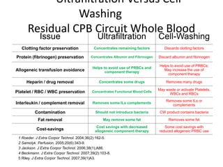
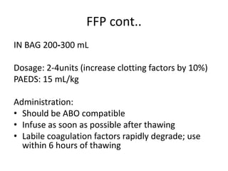
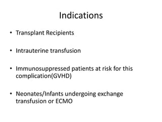
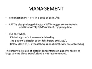
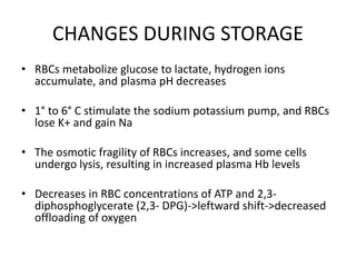
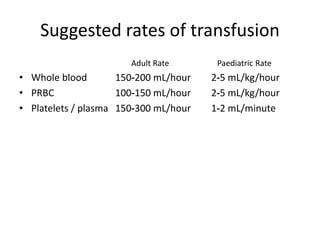
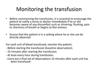
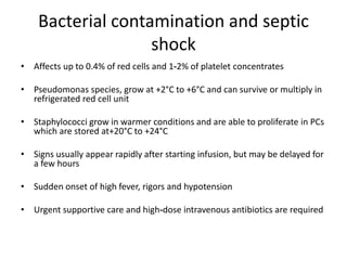
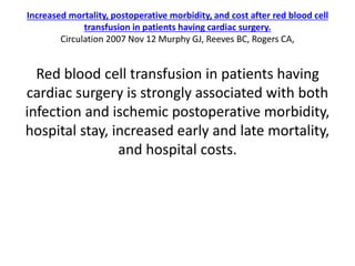
![Processing Residual Circuit Whole
Blood
Three re-infusion methods
– Direct Ann Thorac Surg. 1993;56(4):938-43.
– Cell-Wash JECT. 1996;28(3):134-9.
– Ultrafiltration Perfusion. 2005;20(6):343-9.
Final Infusion Volume Contents
Technique
Volume cc
%
HCT
Plt Cnt
109/L
[Fib]
mg/dL
% Clot
Factors
Direct
700-1800+
17-25 50-140 80-135 15-40
Cell-wash
225-450
40-58 5-25 10-30 2-10
Ultrafiltration
450-1000
45-55 125-325 225-385 85-259](https://image.slidesharecdn.com/bloodpresentationfinal-200916174619/85/Blood-presentation-13-320.jpg)
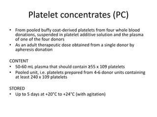
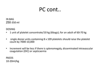
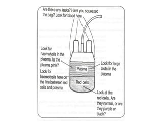


This document discusses red blood cell and component therapy. It covers three pillars of patient blood management: preoperative detection of anemia, intraoperative hemostasis and cell salvage, and postoperative optimization. It then describes the components that make up component therapy, including packed red blood cells (PRBC), platelets, fresh frozen plasma, cryoprecipitate, and leukoreduced and irradiated PRBCs. Indications for transfusion and potential complications are also summarized.


