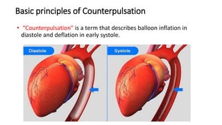
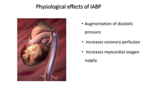
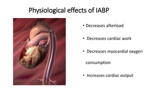
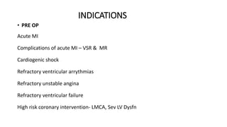
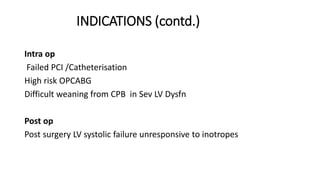
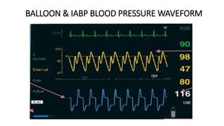
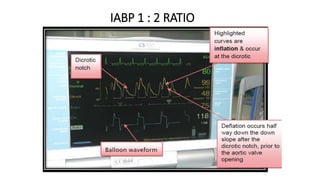
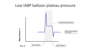
Intra Aortic Balloon
Counterpulsation
Dr. Shamayal Rabbani
TOPICS TO BE COVERED
• HISTORY
• PHYSIOLOGICAL ASPECTS OF IABP
• INDICATIONS & CONTRINDICATIONS
• INSTRUMENTATION & INSERTION
• IABP MONITORING
• NORMAL IAB PRESSURE WAVEFORM
• ABNORMAL IAB PRESSURE WAVEFORM
• WEANING & WITHDRAWL TECHNIQUE
• COMPLICATIONS
History
• Kantrovitz- Idea conceived/Hemidiaphragm wrapping
• Clauss- Systolic blood drainage and diastolic transfusion
• Moulopoulos (1961)-latex tubing with carbon dioxide
• Dennis (1963)- External counterpulsation
• Buckley et al/Mundth et al/Jacobey et al (1970s)
PHYSIOLOGICAL ASPECTS OF IABP
Basic principles of Counterpulsation
• “Counterpulsation” is a term that describes balloon inflation in
diastole and deflation in early systole.
Physiological effects of IABP
• Augmentation of diastolic
pressure
• Increases coronary perfusion
• Increases myocardial oxygen
supply
Physiological effects of IABP
• Decreases afterload
• Decreases cardiac work
• Decreases myocardial oxygen
consumption
• Increases cardiac output
Physiological effects of IABP(CONTD.)
Magnitude of these effects depends upon -:
• Balloon volume - amount of blood displaced is proportional to
volume of balloon
• Heart rate - LV & aortic diastole filling time inversely related to HR,
shorter diastolic time produces lesser balloon augmentation per time
• Aortic compliance - inversely related to diastolic augmentation
IMPORTANT TERMS IN IABP PHYSIOLOGY
• Diastolic Pressure Time Index (DPTI) – Area between LV pressure and
aortic pressure waveform in diastole – Represents pressure and time
available for coronary blood flow
• Tension Time Index (TTI) – Area under the LV pressure waveform in
systole – Represents myocardial work and O2 demand
• Endocardial Viability Ratio ( EVR ) – Ratio of the DPTI to TTI –
Thought to represent the ratio of myocardial O2 supply to demand –
Myocardial ischaemia likely when ratio <0.7
IMPORTANT TERMS IN IABP PHYSIOLOGY(contd.)
Haemodynamic effects of IABP therapy
Aorta ↓systolic pressure, ↑diastolic pressure
Left ventricle
↓systolic pressure, ↓end-diastolic pressure,
↓volume, ↓wall tension
Heart ↓afterload, ↑cardiac output
Blood flow ↑ coronary blood flow
Renal function
• Renal blood flow can increase up to 25%, secondary to increase in
cardiac output.
• Decrease in urine output after insertion of IABP should raise the
suspicion of juxta-renal balloon positioning.
Haematological effects
• The haemoglobin levels and the haematocrit often decrease by up to
5% because of haemolysis from mechanical damage to the red blood
cells.
• Thrombocytopenia can result from mechanical damage to the
platelets, heparin administration, or both.
INDICATIONS
• PRE OP
Acute MI
Complications of acute MI – VSR & MR
Cardiogenic shock
Refractory ventricular arrythmias
Refractory unstable angina
Refractory ventricular failure
High risk coronary intervention- LMCA, Sev LV Dysfn
INDICATIONS (contd.)
Intra op
Failed PCI /Catheterisation
High risk OPCABG
Difficult weaning from CPB in Sev LV Dysfn
Post op
Post surgery LV systolic failure unresponsive to inotropes
CONTRAINDICATIONS
ABSOLUTE
• Significant Aortic regurgitation
• Aortic dissection
• Aortic stents
• Severe aortic and peripheral vascular
atherosclerosis
RELATIVE
• Abdominal aortic aneurysm
• Coagulopathy
• LVOTO
• Uncontrolled sepsis
IABP vs ECMO
IABP instrumentation &
techniques
IABP COMPONENTS
IABP catheter IABP console
IABP CATHETER
Balloon size
IABP Console
IABP Console
IABP CONSOLE
OTHER SITES OF IABP INSERTION
• ASCENDING AORTA
• SUBCLAVIAN ARTERY
• BRACHIAL ARTERY
• AXILLARY ARTERY
POSITIONING OF IABP
• The end of the ballon tip should
be ,∼2 to 3 cm distal to the
origin of the left subclavian
artery (at the level of the
carina),2nd ICS.
• Intraoperatively, balloon
placement can be ascertained
using TEE
MONITORING OF IABP
• CXR – VERIFY POSITION
• Daily Hb & platelet counts – to monitor hemolysis & thrombocytopenia
• Urine output
• Anticoagulation
CARE OF PATIENT WITH IABP
• Should be kept supine in bed (<40 degree elevation)
• Should be evaluated for limb ischemia
• Prophylactic antibiotic are not indicated
• Blood samples should not be obtained from central lumen of IABP
NORMAL IABP WAVEFORM
NORMAL BALLOON PRESSURE WAVEFORM
NORMAL IABP BLOOD PRESSURE WAVEFORM
BALLOON & IABP BLOOD PRESSURE WAVEFORM
IABP 1 : 2 RATIO
ABNORMAL IABP BALLOON
PRESSURE WAVEFORMS
Low IABP balloon plateau pressure
High IABP balloon plateau pressure
High IABP balloon filling pressure baseline
Low (or suddenly decreasing) IABP balloon
filling pressure baseline
IABP TIMING & TRIGGERS
IABP TIMINGS
BALLON PUMP TRIGGERING
ECG PATTERN: Trigger- peak R wave, preset (default) trigger
mode
ARTERIAL PRESSURE: Systolic upstroke - trigger signal.
INTERNAL ASYNCHRONOUS MODE : The balloon inflates and
deflates at a preset rate regardless of the patient’s cardiac
activity. This mode is used in situations where there is no
cardiac output or ECG is unavailable.
A-FIB: Trigger mode of choice in AF
V PACE: Ventricular signal - trigger signal. Mode of
choice in 100% ventricular or AV paced rhythms.
TIMING ERRORS
Scenario 1
Scenario 2
Scenario 3
Scenario 4
TROUBLESHOOTING/
ALARMS
TROUBLESHOOTING
• Loss of trigger:
Check
- ECG trace
- Replace ECG electrodes
- ECG cable
- Choose an alternate ECG lead
• Loss of pressure trace:
Check
- Pressure bag inflated to 300 mm Hg
- Patency of arterial line by withdrawing blood then flushing
- Transducer Level
• Alarms: Leak in IAB circuit / Rapid Gas Loss / IAB disconnected
- Ensure catheter tubing is not leaking
- Check connections along catheter
- Check catheter is not kinked
• Blood detected
- Blood detected in IAB catheter
- Check for blood in tubing if seen stop the pump
- Blood in the inflation catheter could imply rupture of the
balloon.
Complications associated with IABP
• Transient loss of peripheral pulse
• Limb ischaemia
• Thromboembolism
• Compartment syndrome
• Aortic dissection
• Local vascular injury—false
aneurysm, haematoma, bleeding
from the wound
• Infection
• Balloon rupture
• Balloon entrapment
• Haematological changes,
• Malpositioning causing cerebral
or renal compromise
WEANING & WITHDRAWL OF IABP
WEANING OF IABP
• Timing of weaning - Patient should be stable for 12-24 hours
• Minimal inotropic support
• Decrease pump ratio from 1:1 to 1:2 or 1:3
• Decrease augmentation
• Monitor patient closely
• If patient becomes unstable, weaning should be immediately
discontinued
WITHDRAWL OF IABP
Ballon is put on “STAND BY” mode & disconnect
Ballon is deflated, withdraw until resistance met
• Allow backbleed
• Manual pressure proximal to
puncture site
• Manual pressure is then
applied for 30-45 min over
puncture site until adequate
haemostasis achieved
• Compressive dressing
THANK YOU

Iabp presentation

  • 1.
  • 2.
    TOPICS TO BECOVERED • HISTORY • PHYSIOLOGICAL ASPECTS OF IABP • INDICATIONS & CONTRINDICATIONS • INSTRUMENTATION & INSERTION • IABP MONITORING • NORMAL IAB PRESSURE WAVEFORM • ABNORMAL IAB PRESSURE WAVEFORM • WEANING & WITHDRAWL TECHNIQUE • COMPLICATIONS
  • 3.
    History • Kantrovitz- Ideaconceived/Hemidiaphragm wrapping • Clauss- Systolic blood drainage and diastolic transfusion • Moulopoulos (1961)-latex tubing with carbon dioxide • Dennis (1963)- External counterpulsation • Buckley et al/Mundth et al/Jacobey et al (1970s)
  • 4.
  • 5.
    Basic principles ofCounterpulsation • “Counterpulsation” is a term that describes balloon inflation in diastole and deflation in early systole.
  • 6.
    Physiological effects ofIABP • Augmentation of diastolic pressure • Increases coronary perfusion • Increases myocardial oxygen supply
  • 7.
    Physiological effects ofIABP • Decreases afterload • Decreases cardiac work • Decreases myocardial oxygen consumption • Increases cardiac output
  • 8.
    Physiological effects ofIABP(CONTD.) Magnitude of these effects depends upon -: • Balloon volume - amount of blood displaced is proportional to volume of balloon • Heart rate - LV & aortic diastole filling time inversely related to HR, shorter diastolic time produces lesser balloon augmentation per time • Aortic compliance - inversely related to diastolic augmentation
  • 9.
    IMPORTANT TERMS INIABP PHYSIOLOGY • Diastolic Pressure Time Index (DPTI) – Area between LV pressure and aortic pressure waveform in diastole – Represents pressure and time available for coronary blood flow • Tension Time Index (TTI) – Area under the LV pressure waveform in systole – Represents myocardial work and O2 demand • Endocardial Viability Ratio ( EVR ) – Ratio of the DPTI to TTI – Thought to represent the ratio of myocardial O2 supply to demand – Myocardial ischaemia likely when ratio <0.7
  • 10.
    IMPORTANT TERMS INIABP PHYSIOLOGY(contd.)
  • 11.
    Haemodynamic effects ofIABP therapy Aorta ↓systolic pressure, ↑diastolic pressure Left ventricle ↓systolic pressure, ↓end-diastolic pressure, ↓volume, ↓wall tension Heart ↓afterload, ↑cardiac output Blood flow ↑ coronary blood flow
  • 12.
    Renal function • Renalblood flow can increase up to 25%, secondary to increase in cardiac output. • Decrease in urine output after insertion of IABP should raise the suspicion of juxta-renal balloon positioning.
  • 13.
    Haematological effects • Thehaemoglobin levels and the haematocrit often decrease by up to 5% because of haemolysis from mechanical damage to the red blood cells. • Thrombocytopenia can result from mechanical damage to the platelets, heparin administration, or both.
  • 14.
    INDICATIONS • PRE OP AcuteMI Complications of acute MI – VSR & MR Cardiogenic shock Refractory ventricular arrythmias Refractory unstable angina Refractory ventricular failure High risk coronary intervention- LMCA, Sev LV Dysfn
  • 15.
    INDICATIONS (contd.) Intra op FailedPCI /Catheterisation High risk OPCABG Difficult weaning from CPB in Sev LV Dysfn Post op Post surgery LV systolic failure unresponsive to inotropes
  • 16.
    CONTRAINDICATIONS ABSOLUTE • Significant Aorticregurgitation • Aortic dissection • Aortic stents • Severe aortic and peripheral vascular atherosclerosis RELATIVE • Abdominal aortic aneurysm • Coagulopathy • LVOTO • Uncontrolled sepsis
  • 17.
  • 18.
  • 19.
  • 20.
  • 21.
  • 23.
  • 24.
  • 25.
  • 29.
    OTHER SITES OFIABP INSERTION • ASCENDING AORTA • SUBCLAVIAN ARTERY • BRACHIAL ARTERY • AXILLARY ARTERY
  • 30.
    POSITIONING OF IABP •The end of the ballon tip should be ,∼2 to 3 cm distal to the origin of the left subclavian artery (at the level of the carina),2nd ICS. • Intraoperatively, balloon placement can be ascertained using TEE
  • 31.
    MONITORING OF IABP •CXR – VERIFY POSITION • Daily Hb & platelet counts – to monitor hemolysis & thrombocytopenia • Urine output • Anticoagulation CARE OF PATIENT WITH IABP • Should be kept supine in bed (<40 degree elevation) • Should be evaluated for limb ischemia • Prophylactic antibiotic are not indicated • Blood samples should not be obtained from central lumen of IABP
  • 32.
  • 33.
  • 34.
    NORMAL IABP BLOODPRESSURE WAVEFORM
  • 35.
    BALLOON & IABPBLOOD PRESSURE WAVEFORM
  • 36.
    IABP 1 :2 RATIO
  • 37.
  • 38.
    Low IABP balloonplateau pressure
  • 39.
    High IABP balloonplateau pressure
  • 40.
    High IABP balloonfilling pressure baseline
  • 41.
    Low (or suddenlydecreasing) IABP balloon filling pressure baseline
  • 42.
    IABP TIMING &TRIGGERS
  • 43.
  • 44.
    BALLON PUMP TRIGGERING ECGPATTERN: Trigger- peak R wave, preset (default) trigger mode ARTERIAL PRESSURE: Systolic upstroke - trigger signal. INTERNAL ASYNCHRONOUS MODE : The balloon inflates and deflates at a preset rate regardless of the patient’s cardiac activity. This mode is used in situations where there is no cardiac output or ECG is unavailable.
  • 45.
    A-FIB: Trigger modeof choice in AF V PACE: Ventricular signal - trigger signal. Mode of choice in 100% ventricular or AV paced rhythms.
  • 46.
  • 47.
  • 48.
  • 49.
  • 50.
  • 51.
  • 60.
    TROUBLESHOOTING • Loss oftrigger: Check - ECG trace - Replace ECG electrodes - ECG cable - Choose an alternate ECG lead • Loss of pressure trace: Check - Pressure bag inflated to 300 mm Hg - Patency of arterial line by withdrawing blood then flushing - Transducer Level
  • 61.
    • Alarms: Leakin IAB circuit / Rapid Gas Loss / IAB disconnected - Ensure catheter tubing is not leaking - Check connections along catheter - Check catheter is not kinked • Blood detected - Blood detected in IAB catheter - Check for blood in tubing if seen stop the pump - Blood in the inflation catheter could imply rupture of the balloon.
  • 62.
    Complications associated withIABP • Transient loss of peripheral pulse • Limb ischaemia • Thromboembolism • Compartment syndrome • Aortic dissection • Local vascular injury—false aneurysm, haematoma, bleeding from the wound • Infection • Balloon rupture • Balloon entrapment • Haematological changes, • Malpositioning causing cerebral or renal compromise
  • 63.
  • 64.
    WEANING OF IABP •Timing of weaning - Patient should be stable for 12-24 hours • Minimal inotropic support • Decrease pump ratio from 1:1 to 1:2 or 1:3 • Decrease augmentation • Monitor patient closely • If patient becomes unstable, weaning should be immediately discontinued
  • 65.
    WITHDRAWL OF IABP Ballonis put on “STAND BY” mode & disconnect Ballon is deflated, withdraw until resistance met
  • 66.
    • Allow backbleed •Manual pressure proximal to puncture site • Manual pressure is then applied for 30-45 min over puncture site until adequate haemostasis achieved • Compressive dressing
  • 67.

Editor's Notes

  • #7 IABP inflates at onset of diastole , cause diastolic augmentation which increases coronary perfusion and increase myocardial oxygen supply Predominantly associated with enhancement of LV performance and also improves RV function .
  • #9 As aortic compliance increase or SVR decrease the magnitude of diastolic augmentation decreases.
  • #11 • Balloon inflation increases pressure difference between aorta and LV which augments the DPTI - This increases O2 supply Balloon deflation reduces the afterload of the LV which reduces the TTI -- This reduces O2 demand
  • #15 IABP decrease myocardial work and SVR , coronary perfusion increased Severe MR secondary to PAPILLARY MIUSCLE DYSFUNCTION OR rupture after MI can lead to significant instability , initially managed by IABP Life threatening complication of acute MI characterised by low cardiac output ,hypotension unresponsive to fluid administration , elevated filling pressure and tissue hypoperfusion leading to oliguria ,hyperlactemia and altered mental status . Class I indication for management of cardiogenic shock not rapidly reversed by pharmacological therapy. IABP effective in stabilizing patients with refractory ventricular ectopy after MI by increasing coronary perfusion, reducing iuschemia and trans myocardial wall stress and maintain adequate systemic perfusion.
  • #16  IABP is used for stabilization of patients with acute myocardial infarction referred for urgent cardiac surgery. IABP support is often initiated in the cardiac catheterization laboratory and continued through the perioperative period. Elective placement is considered in high-risk patients such as those with significant left main stem disease, severe LV dysfunction (ejection fraction ,30%), congestive heart failure, cardiomyopathy, chronic renal failure, or cerebrovascular disease. Weaning from cardiopulmonary bypass may be difficult in cases where aortic cross-clamping is prolonged, revascularization is only partially achieved, or preexisting myocardial dysfunction is present. Separation from cardiopulmonary bypass may be marked by hypotension and a low cardiac index despite the administration of inotropic drugs. The use of IABP in this setting decreases LV resistance, increases cardiac output, and increases coronary and systemic perfusion, facilitating the patient’s weaning from cardiopulmonary bypass.
  • #17 Essentially, if your aortic valve is incompetent, the diastolic balloon inflation will (instead of nourishing the coronary circulation) send a jet of blood though the aortic valve back into the left ventricle, increasing the preload. The valve was already doing this in diastole (hence the diastolic murmur)- but now the counterpulsation actually augments the regurgitation. The presence of an aortic dissection would make one think twice about inserting a balloon pump. Even if the balloon somehow fails to actually touch the flap, its pulsations will send more blood into the false lumen, expanding the dissection. Any clots forming inside it will be battered around and potentially expelled into the systemic circulation.
  • #21 10-25 cm long polyurethane bladder 30 to 60 cc capacity Optimal 85 % of aorta occlusion (not 100 % ) The shaft of ballon catheter contains 2 lumens- - one allows gas exchange from console to ballon - second lumen for catheter delivery over guide wire & monitoring of central aortic pressure after installation . Blood should not be drawn from central lumen for samples.
  • #24 Mobile console System for helium delivery (Helium is often used because its low density facilitates rapid transfer of gas from console to the balloon. It is also easily absorbed into the blood stream in case of rupture of the balloon ) Computer for inflation & deflation control
  • #32 Patient care should be carried out with 3 primary goals- Evaluation in terms of hemodynamic status , systemic perfusion & relief of cardiac symptoms Observation of early signs of complications including ischaemia , balloon mal-positioning , thrombus formation, bleeding and infection Ensuring proper functioning of IABP , including correct timing, consistent triggering and trouble shout of alarms.
  • #34 The balloon itself has a pressure transducer, and it generates a waveform. Balloon pressure waveforms are also a source of information regarding the behavior of the IABP and its interaction with the cardiovascular physiology of patient. About 40 milliseconds before the dicrotic notch, the IABP balloon inflates. This is timed with the ECG, usually - the end of the T wave is used as a marker that systole has finished. Why the delay? because even the best IABP pistons require a few milliseconds to shoot some helium into the balloon. Balloon deflation (which is also very rapid) is timed with the R wave. The baseline pressure in the helium circuit should be around 10-15mmHg.
  • #35 Balloon inflation causes augmentation of diastolic pressure and a second peak is observed. This peak is referred to as diastolic augmentation. CARDIOVASCULAR CONSEQUENCES OF IABP – Cardiovascular changes in diastolic events – left ventricular diastolic volume is decreased due to systolic unloading. improving left ventricular compliance 2. Cardiovascular changes in systolic pressure – decrease in systolic pressure – decline in SBP by 10 % decrease in end diastolic pressure upto 30 % - indicates systolic unloading decrease in isometric phase of left ventricular contraction decrease in left ventricular wall tensiomn & rate of left ventricular pressure rise increase in cardiac output between 0.5 l & 1.0 litre /min. Factors affecting diastolic augmentation - Patient related factors – Heart rate MAP STROKE VOLUME SVR IAB catheter related – IAB position IAB size Kink in IAB catheter IAB leak Loew helium concenteration IAB PUMP related – Timing
  • #39 The balloon plateau is a function of the pressure inside the helium balloon and the pressure inside the aorta, which relates to the elastic recoil of the aortic walls (and to some extent to the systemic vascular resistance as a whole). Balloon plateau pressure might be low in the following circumstances: - balloon is too small for the patient. - patient is hypotensive, and has low peripheral vascular resistance (eg. septic shock or any other sort of vasoplegia) - patient has a greatly reduced stroke volume (i.e. there is not enough ejected blood in the aorta to displace) balloon is positioned too high or too low in the aorta The consequence of this is a decreased diastolic augmentation, as well as a high aortic end-diastolic pressure. The balloon's low pressure cannot move much blood around the aorta, and the IABP is not being used to its full potential.
  • #40 High balloon plateau pressure, with a square or rounded waveform, reflects either some sort of increase in pressure on the balloon itself, or some impedance to gas flow. Balloon plateau pressure might be high in the following circumstances: balloon catheter is kinked balloon is too big for the patient patient is incredibly hypertensive balloon is positioned too high in the aorta The consequences of this could be severe - the balloon could actually rupture from too much pressure. Or, you could be injuring the aorta. If the catheter is kinked, the balloon cannot empty, and balloon deflation could be delayed, which results in either a failure to improve afterload, or an actual increase in afterload.
  • #41 The baseline pressure in the helium circuit should be around 10-15mmHg. If this pressure rises, there is likely some mechanical fault with the circuit, which limits the normal emptying of the balloon. Balloon filling pressure baseline might be high in the following circumstances: The balloon catheter is kinked and and balloon is not emptying properly The system is overpressurised because the IABP is malfunctioning The consequences of this are balloon could rupture (eventually). Additionally, the increased baseline pressure is transmitted to the aorta, which results in increased afterload and increased myocardial oxygen demand.  Most IABP consoles will begin to alarm with an irritating siren if the baseline pressure climbs over 20mmHg.
  • #42 This could also be a major problem. The filling pressure is adjusted automatically; if this is not happening, there must be either a helium leak somewhere, or a failure of the automated filling mechanism. Alternatively, you have just run out of helium, and the tank needs to be replaced. if the baseline filling pressure dives suddenly, the cause could be either a disconnection of the helium pipe, or (more disturbingly) a balloon rupture.
  • #43 Timing is the process of determining the periods during the cardiac cycle when the balloon should inflate, how long it should remain inflated, and when it should deflate. The beneficial hemodynamic effects of IABPs are critically dependent on timing
  • #44  Inflation of the balloon is triggered by the the beginning of diastole, which correlates with the middle of the T-wave. The balloon is timed to deflate at the very end of diastole. This correlates with the R-wave on the ECG, and this is the most commonly used trigger for balloon deflation.
  • #45 TRIGGER – EVENT THAT PUMP USES TO IDENTIFY THE ONSET OF CARDIAC CYCLE , PUMP MUST HAVE CONSISTENT TRIGGER IN ORDER TO PROVIDE PATIENT ASSIST
  • #48  Early Inflation - Early balloon inflation results in increased afterload -Increased LV oxygen demand, due to increased afterload -Decreased LV oxygen supply, due to decreased diastolic perfusion -Decreased cardiac output, due to decreased stroke volume Physiological effects: potential premature closure of the aortic valve; potential increase in LVEDV and LVEDP or PCWP; increased LV wall stress or afterload; aortic regurgitation; increased MVO2 demand.
  • #49 Waveform characteristics: -inflation of IAB after the dicrotic notch absence of sharp ‘V’ Suboptimal diastolic augmentation Physiological effects: suboptimal coronary artery perfusion
  • #50  Waveform characteristics: - -deflation of IAB is seen as a sharp decrease after diastolic augmentation; -suboptimal diastolic augmentation; -assisted aortic end-diastolic pressure may be equal to or less than the unassisted aortic end-diastolic pressure; - assisted systolic pressure may increase Physiological effects: -suboptimal coronary perfusion; -potential for retrograde coronary and carotid blood flow; -suboptimal afterload reduction; -increased MVO2 demand.
  • #51 Waveform characteristics: -assisted aortic end-diastolic pressure may be equal to the unassisted aortic end-diastolic pressure -rate of increase of assisted systole is prolonged -diastolic augmentation may appear widened. Physiological effects: -afterload reduction is essentially absent increased MVO2 consumption because of the left ventricle ejecting against a greater resistance and a prolonged isovolumetric contraction phase IAB may impede LV ejection and increase the afterload