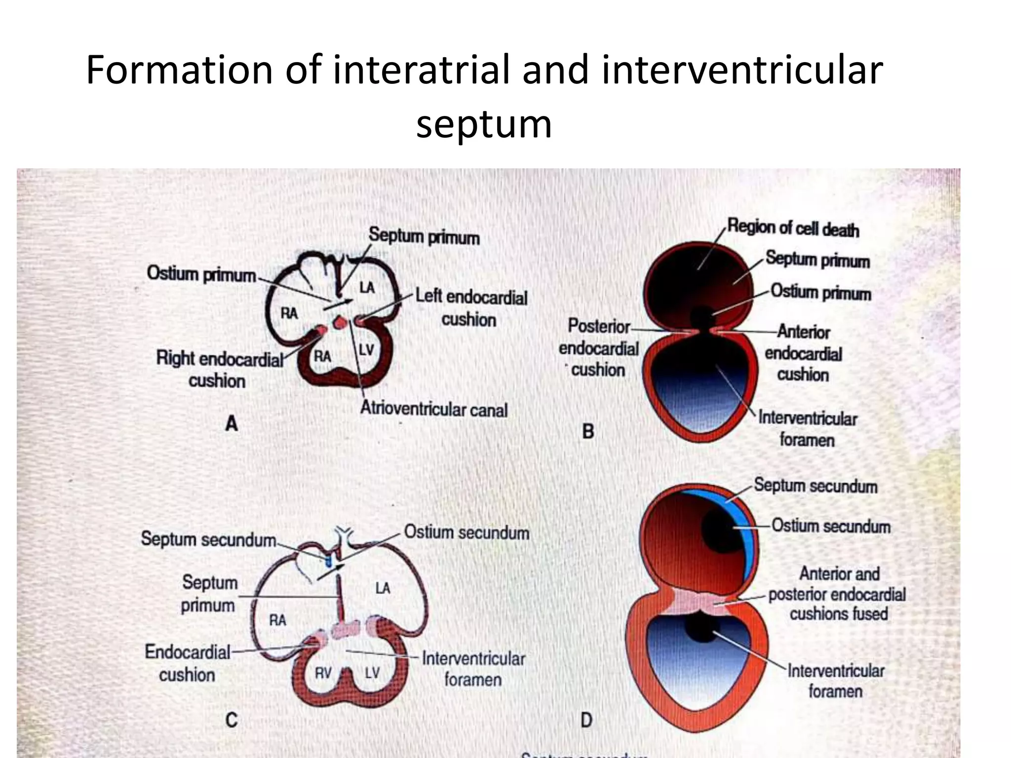
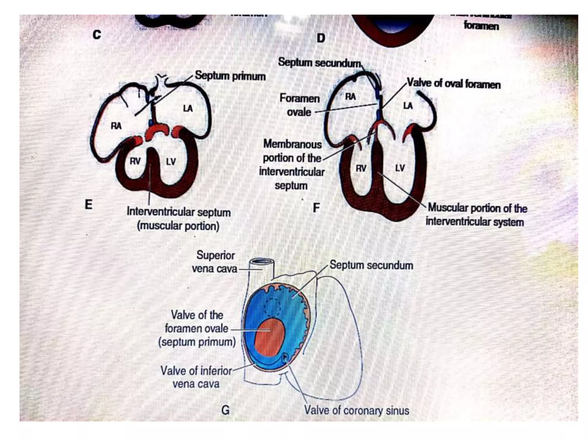
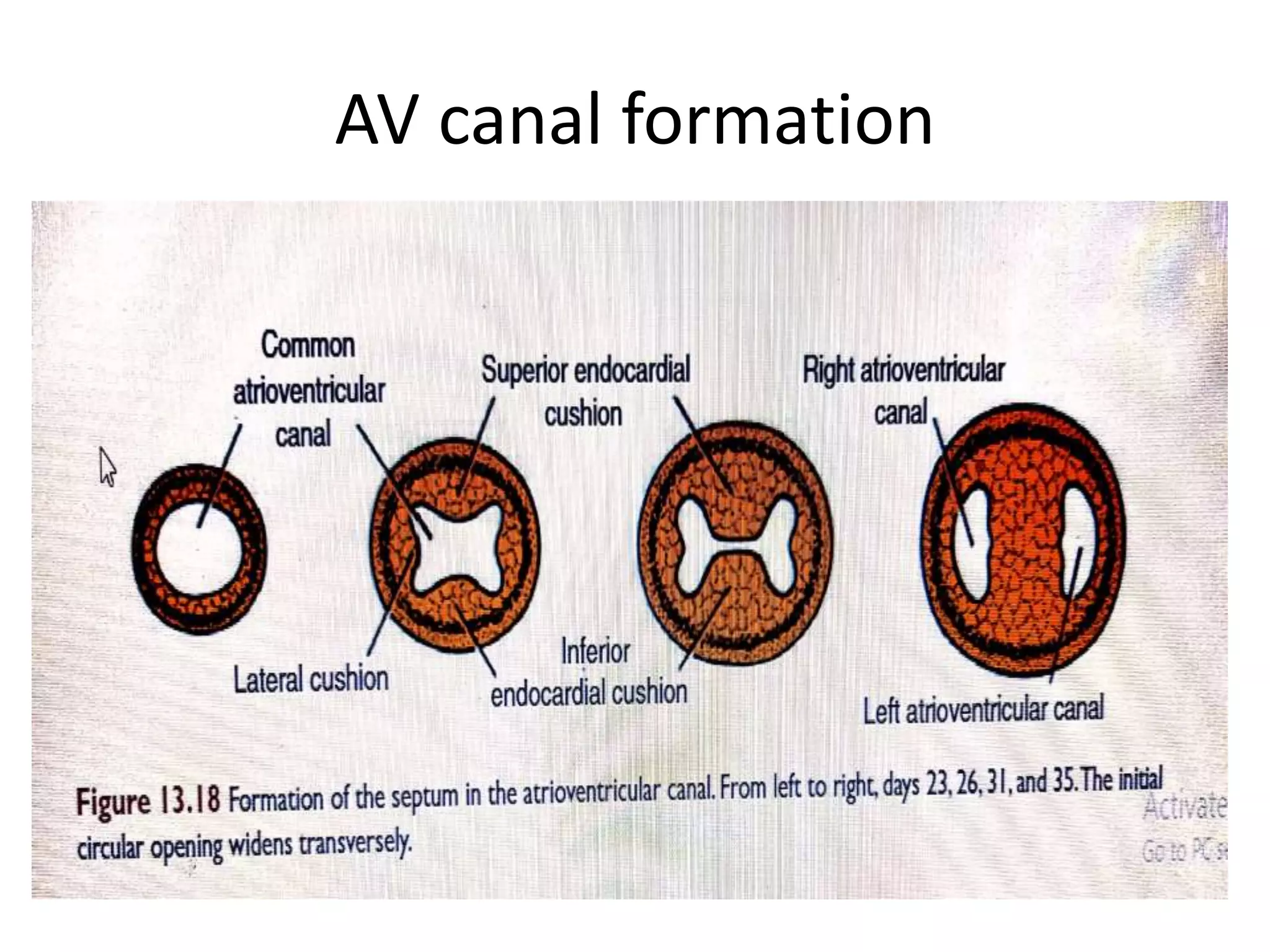
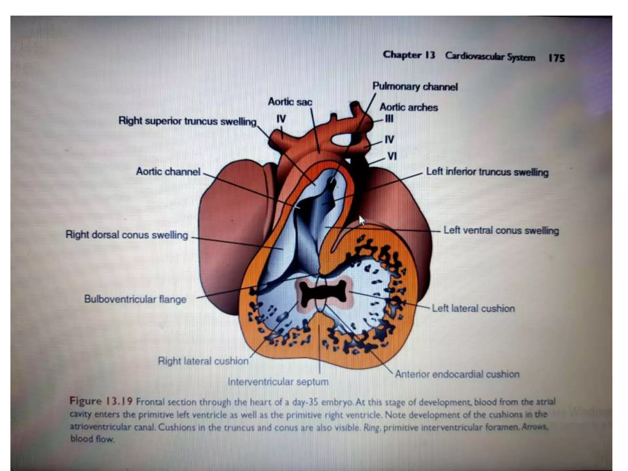
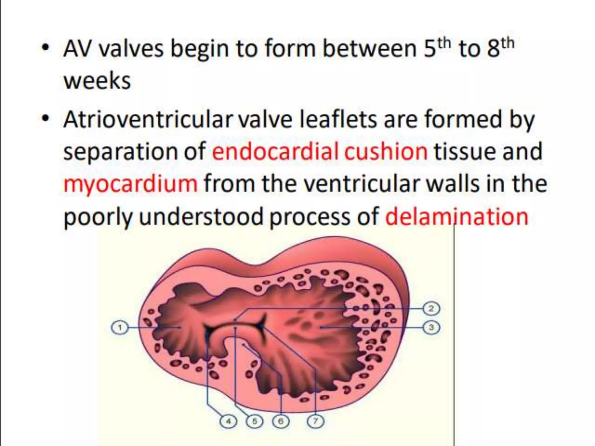
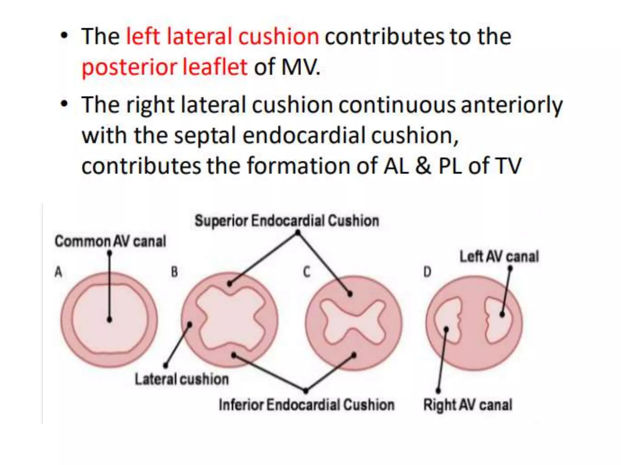
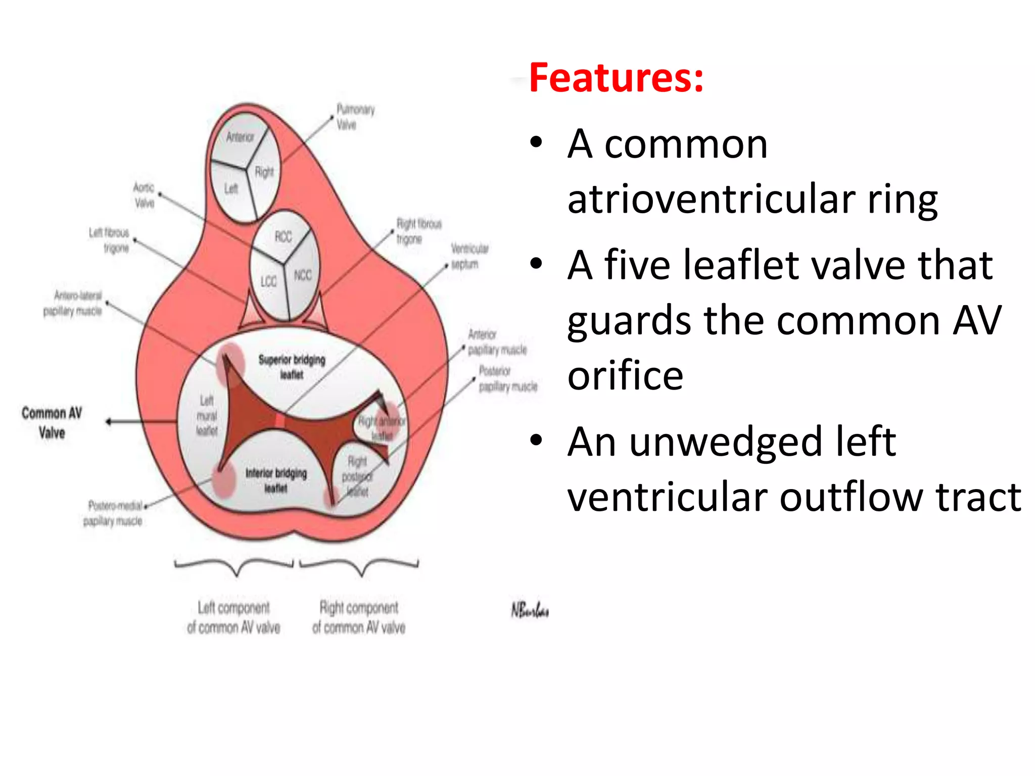
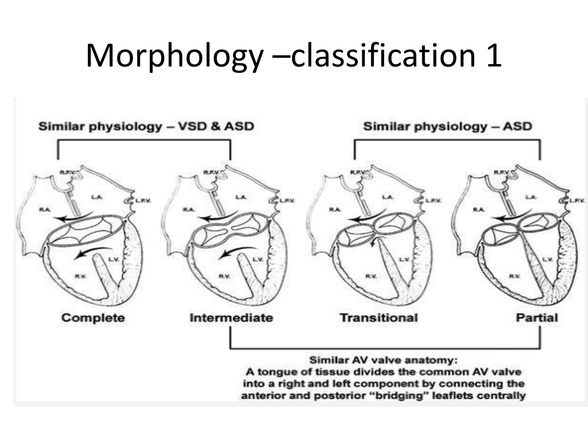
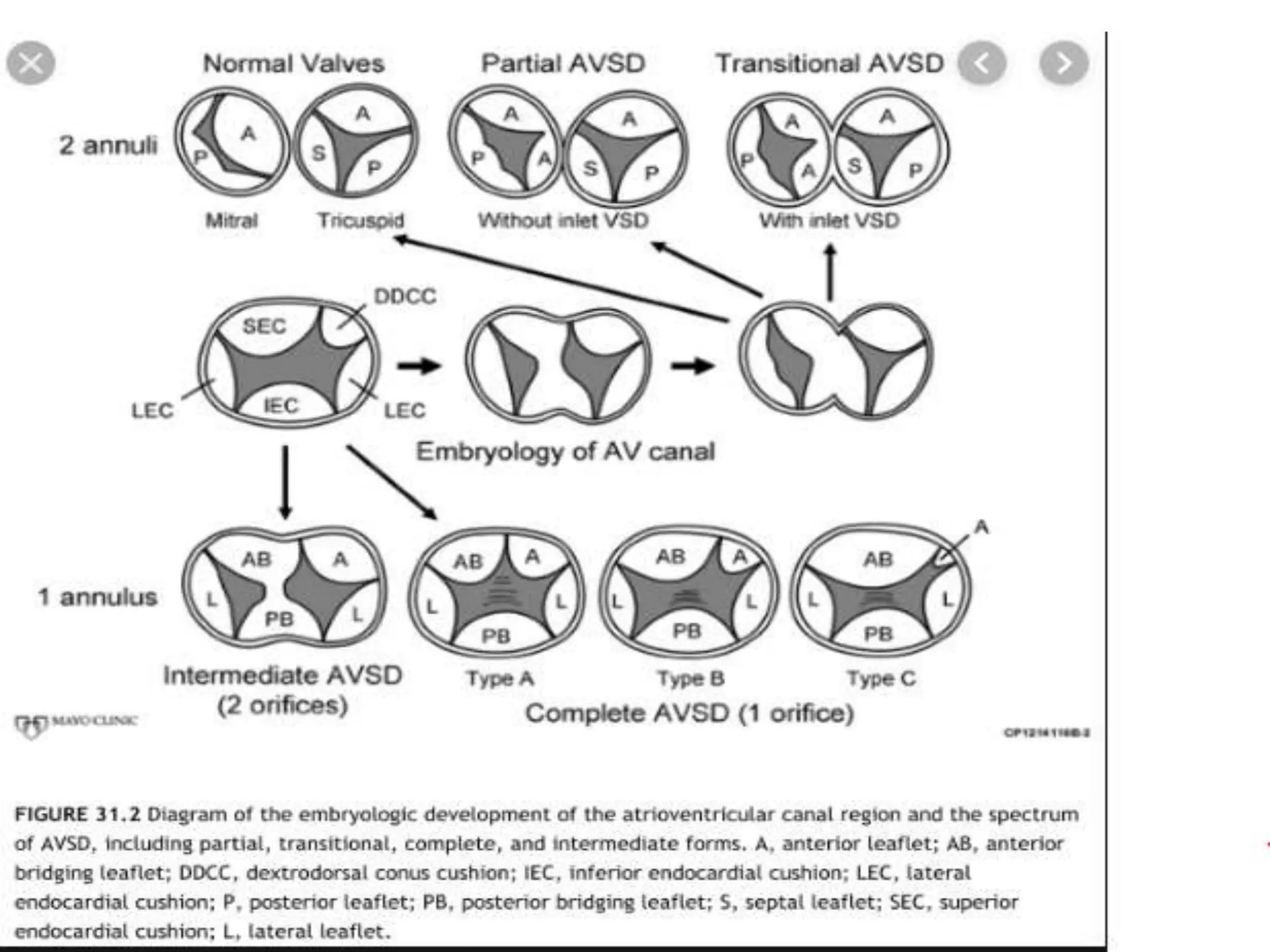
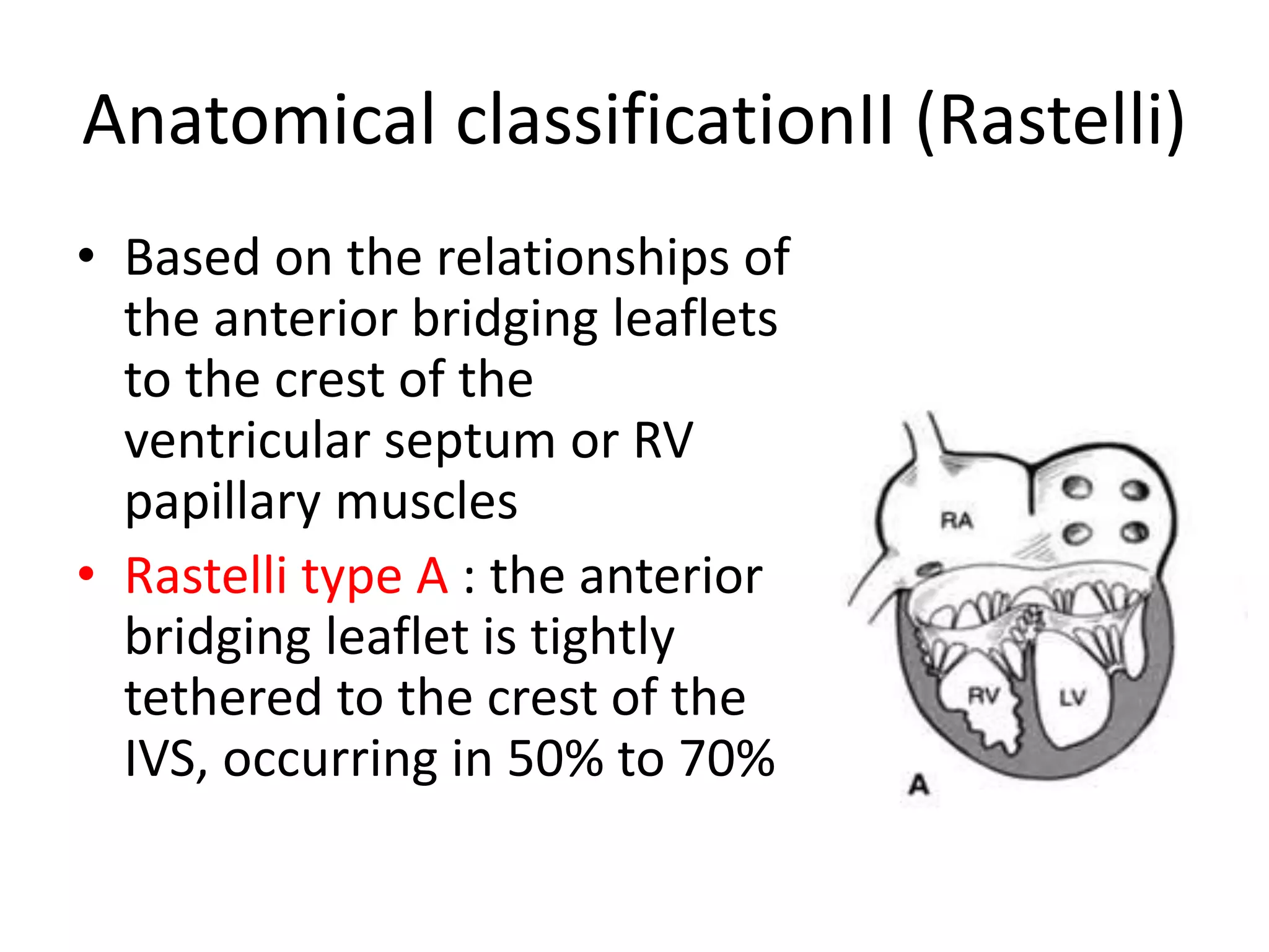
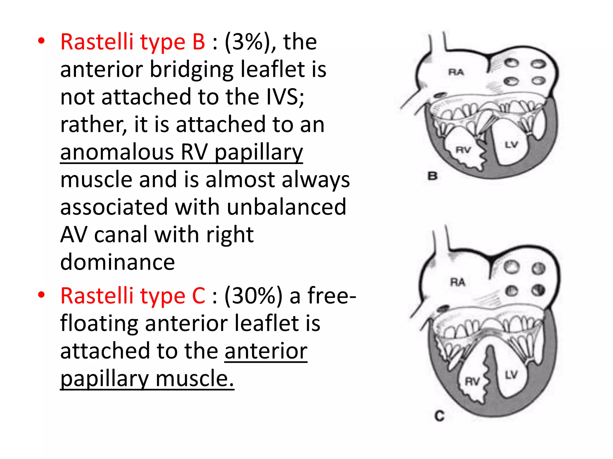
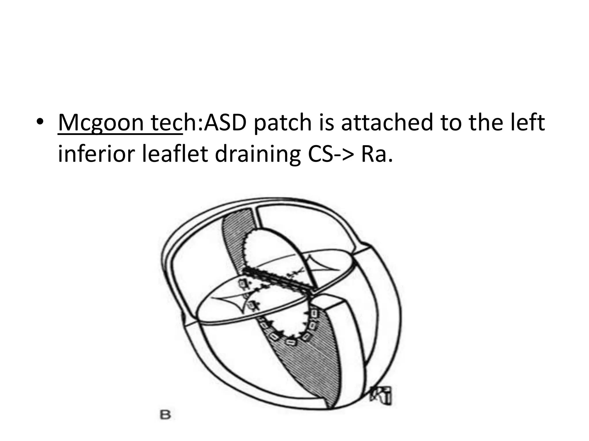
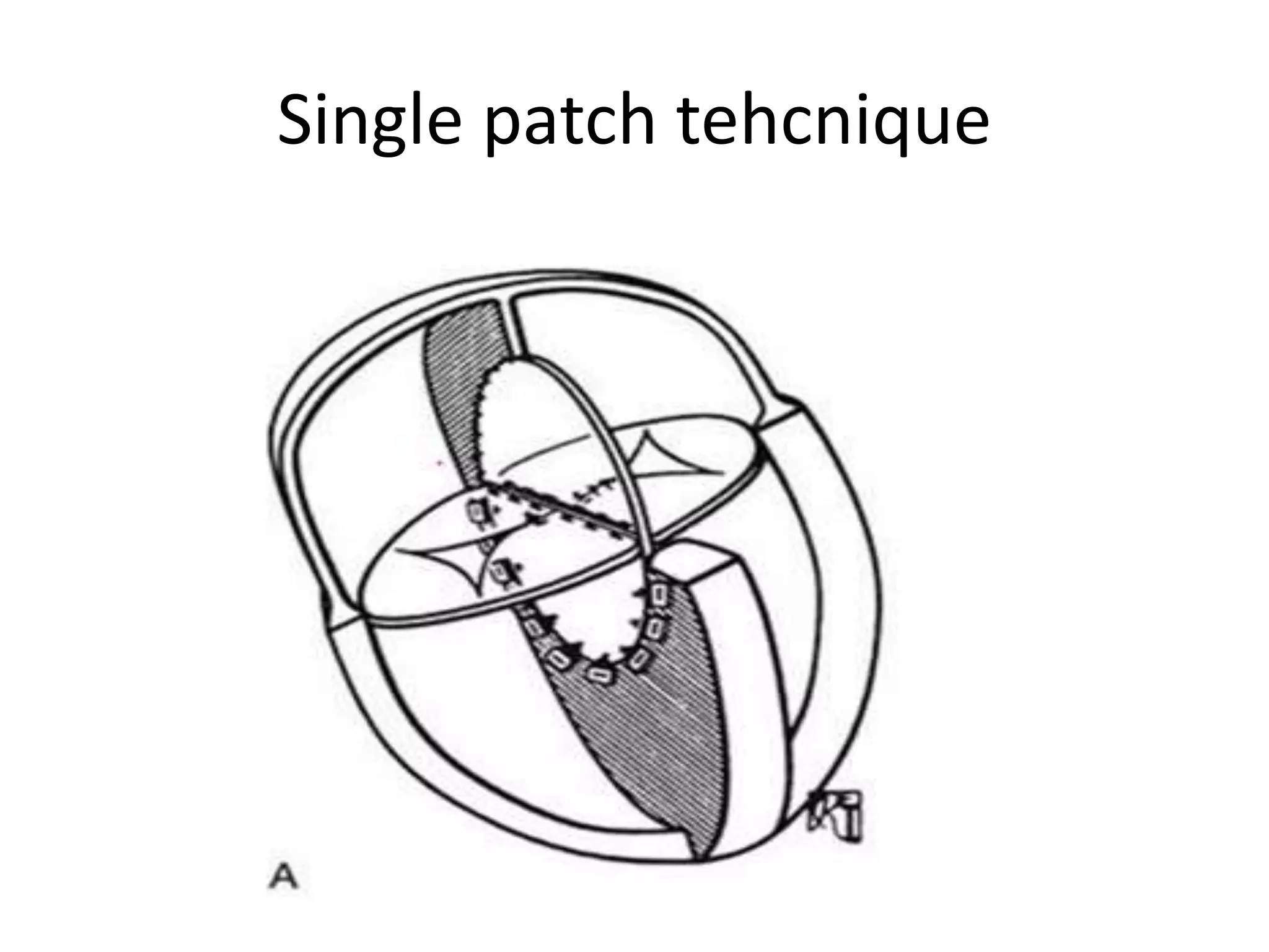
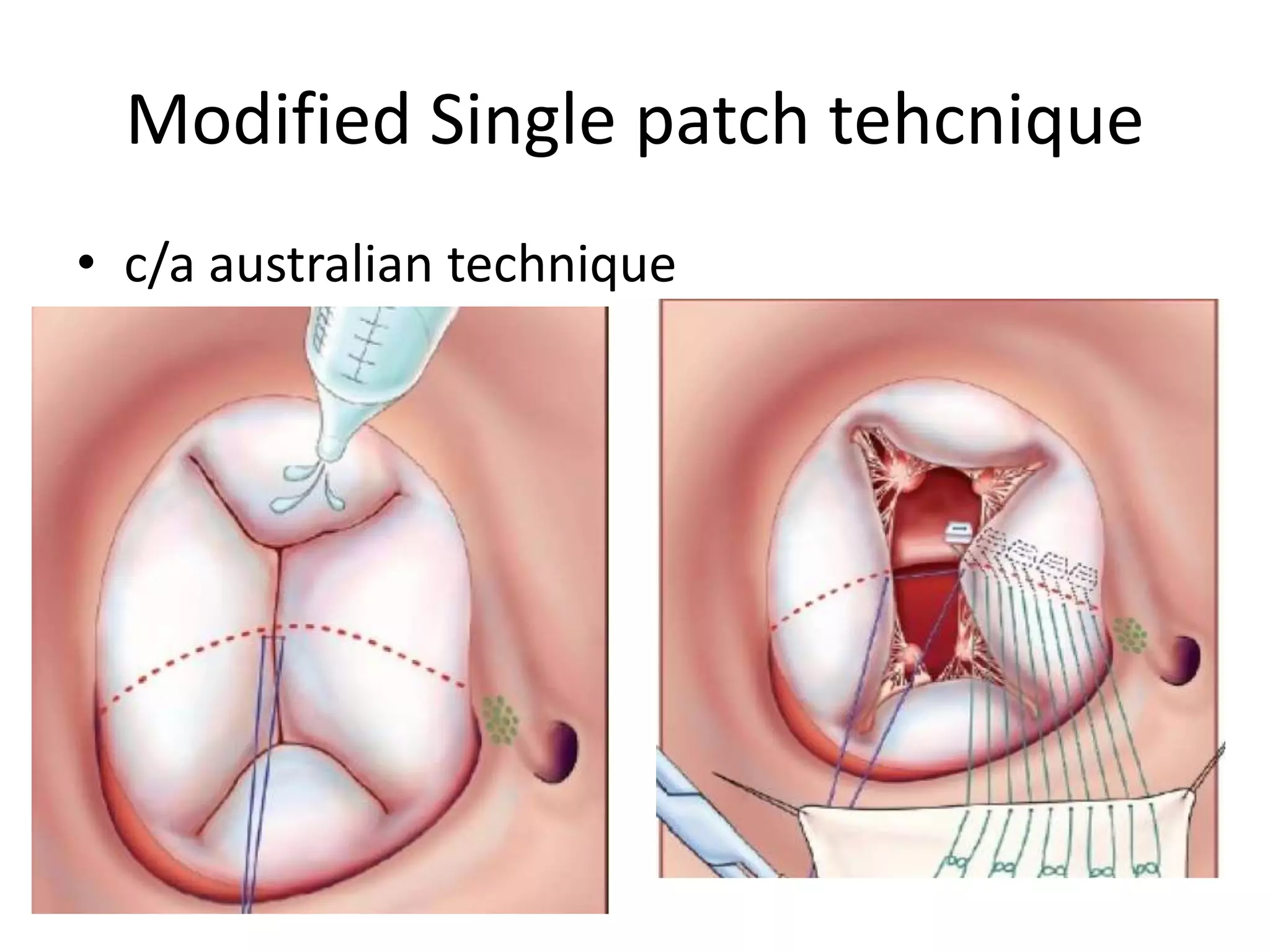
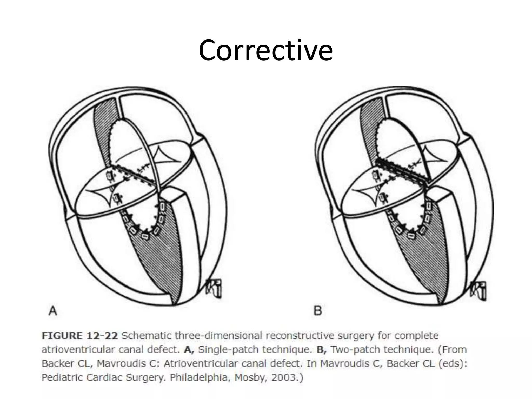
ATRIOVENTRICULAR CANAL
DEFECT
By
Dr VASANTHI
DNB- CARDIAC SURGERY
MADRAS MEDICAL MISSION
Formation of interatrial and interventricular
septum
AV canal formation
AV CANAL DEFECT
• Characterized by complete absence of AV
septum
• Mechanism of development : Abnormal
differentiation and remodeling of endocardial
cushion mesenchyme into valvoseptal tissue
• Also called as
– Endocardial cushion defect,
– Canalis atrioventricularis communis,
– persistent atrioventricular ostium
Features:
• A common
atrioventricular ring
• A five leaflet valve that
guards the common AV
orifice
• An unwedged left
ventricular outflow tract
• Mitral & tricuspid valves achieve the same
septal insertion level because the mitral
annulus is displaced toward the apex
• The distance from mitral annulus to the left
ventricular apex is less than the distance from
the aortic annulus to the apex
• In the normal heart, the aortic valve is wedged
between the mitral and tricuspid annuli. In AVSD the
aortic valve is displaced anteriorly and creates an
elongated, so-called gooseneck deformity of the
LVOT
Incidence
• AVSDs account for 4% to 5% of congenital
heart disease
• Gender distribution is approximately equal or
may show a slight female preponderance
History
• Rogers, Edwards : Recognized morphology of primum
ASD in 1948
• Wakai, Edwards : Term of partial and complete AV
canal defect in 1956
• Bharati& Lev : Term of Intermediate & Transitional in
1980
• Rastelli: Described the of common anterior leaflet in
1966
• Lillehei: 1strepair of AVSD in 1954
• Kirklin, Watkin, Gross: Open repair using oxygenator
Morphology –classification 1
• In partial AVSDs, incomplete fusion of the
superior and inferior endocardial cushions
results in a cleft in the midportion of the AML
,often associated with MR
• Complete AVSD associated with lack of fusion
between the superior and inferior cushions
Anatomical classificationII (Rastelli)
• Based on the relationships of
the anterior bridging leaflets
to the crest of the
ventricular septum or RV
papillary muscles
• Rastelli type A : the anterior
bridging leaflet is tightly
tethered to the crest of the
IVS, occurring in 50% to 70%
• Rastelli type B : (3%), the
anterior bridging leaflet is
not attached to the IVS;
rather, it is attached to an
anomalous RV papillary
muscle and is almost always
associated with unbalanced
AV canal with right
dominance
• Rastelli type C : (30%) a free-
floating anterior leaflet is
attached to the anterior
papillary muscle.
Classificiation III
• Based on level of shunting
– Interatrial and interventricular sunt
– Interatrial shunt only (primum ASD )
– Interventricluar shunt only(av canal VSD)
– AVCD with intact IAS and IVS
Classification IV
• Depending upon the size of ventricular
chambers(univentricular or biventricular)
– Balanced type
– Unbalanced type
• Unbalanced AVCD: 1.ventircular
hypoplasia,2.malalignment of the AV valve
junction. later may affect size of ventricle
development
• Unbalanced Right dominant type : associated
with arch hypoplasia,coarctation of aorta
• Unbalanced with left dominant : associated
with pulmonary stenosis/atresia
Lev description of conduction
• Inferior displacement of AV node and
coronary sinus. Bundle of His is also displaced
inferiorly and coursing at the inferior rim of
scooped out basal portion of IVS
Associated anomalies
Partial AVSD:
• Most common ostium secondum ASD & LSVC to
CS
Complete AVSD
• Type A usually is an isolated defect and is
frequent in patients with Down syndrome.
• Type C –TOF, DORV, TGA and heterotaxy
syndromes
Fetal physiology
• Oxygenated blood may cross the IAS to the RA
• Increase the PO2 of blood in RA, RV, PA
• Slightly higher PO2of blood perfusing the lungs
would decrease pulmonary vasoconstriction and
increase pulmonary blood flow
• It may retard the development of a thick medial
muscle layer, so that a more rapid decrease in
PVR may occur after birth
• An interesting association may develop in some
infants of an obligatory left-to-right shunt
through the AV canal defect simultaneous right-
to-left shunting through the ductus arteriosus
• The increased pulmonary blood flow and PA
pressure interfere with the normal postnatal
maturation of the pulmonary arterioles leads to
early development of Pulmonary vascular
obstructive disease
Clinical manifestation
Partial AVSD
• Patients with primum ASD - usually asymptomatic
during childhood.
• Dyspnea, easy fatigability, recurrent RTI and
growth retardation may be present early in life if
associated with major MR or common atrium
Complete AVSD
• Tachypnea and failure to thrive invariably occur
early in infancy & virtually all patients have
symptoms by 1 year of age because of pulmonary
vascular obstructive disease
Echocardiography
• Primary imaging technique for diagnosing AVSD
• The internal cardiac crux is the most consistent imaging landmark
• Apical four-chamber imaging plane clearly visualizes the internal crux
• The primum ASD is seen as an absence of the lower IAS
Cardiac cath
• Rarely required for diagnosis
• In older patient it may have a role in assessing the
degree of pulmonary vascular obstructive disease or
CAD
• A large Lt to Rt shunt at the atrial level demonstrated
by a significantly higher oxygen saturation sampled
from the RA compared with the blood in the IVC & SVC
• In complete AVSD the PASP is invariably at or near
systemic level, while in partial AVSDs, the PASP is
usually <60% of systemic pressure
• LV angiography -gooseneck deformation of the LVOT
Goose neck deformity
• The left ventricular outflow tract is elongated
and narrowed. The arrows point to the mitral
cleft.
Management
• Elective surgical repair -2 to 4 months of age.
• Early in case of down syndrome because of
their known tendency to develop early
pulmonary vascular obstructive disease
• Palliative:
– Unbalanced AV canal (PA banding )
Corrective surgeries
• Single patch technique
• Double patch technique
• Modified single patch technique
Double patch technique
• Separate patch for both ASD and VSD closure
• VSD –dacron and ASD – pericardial patch to avoid
hemolysis in case of AVVR
• Mitral valve cleft is closed by coaptation of SBL
and IBL using horizontal mattress
• Kirklin tech : attaching ASD patch to the right of
AV node invloves suturing the right inferior leaflet
.leaving CS ->LA
– Advantage : far from conductive tissue
– Diaadvantgae : some desaturation becoz of CS->LA
• Mcgoon tech:ASD patch is attached to the left
inferior leaflet draining CS-> Ra.
Single patch tehcnique
Modified Single patch tehcnique
• c/a australian technique
• Disadvantage : development of lvoto because
of VSD is closed primarily witout patch
material
Corrective
Atrioventricular canal defect

Atrioventricular canal defect

  • 1.
    ATRIOVENTRICULAR CANAL DEFECT By Dr VASANTHI DNB-CARDIAC SURGERY MADRAS MEDICAL MISSION
  • 2.
    Formation of interatrialand interventricular septum
  • 4.
  • 11.
    AV CANAL DEFECT •Characterized by complete absence of AV septum • Mechanism of development : Abnormal differentiation and remodeling of endocardial cushion mesenchyme into valvoseptal tissue
  • 12.
    • Also calledas – Endocardial cushion defect, – Canalis atrioventricularis communis, – persistent atrioventricular ostium
  • 13.
    Features: • A common atrioventricularring • A five leaflet valve that guards the common AV orifice • An unwedged left ventricular outflow tract
  • 14.
    • Mitral &tricuspid valves achieve the same septal insertion level because the mitral annulus is displaced toward the apex • The distance from mitral annulus to the left ventricular apex is less than the distance from the aortic annulus to the apex
  • 15.
    • In thenormal heart, the aortic valve is wedged between the mitral and tricuspid annuli. In AVSD the aortic valve is displaced anteriorly and creates an elongated, so-called gooseneck deformity of the LVOT
  • 16.
    Incidence • AVSDs accountfor 4% to 5% of congenital heart disease • Gender distribution is approximately equal or may show a slight female preponderance
  • 17.
    History • Rogers, Edwards: Recognized morphology of primum ASD in 1948 • Wakai, Edwards : Term of partial and complete AV canal defect in 1956 • Bharati& Lev : Term of Intermediate & Transitional in 1980 • Rastelli: Described the of common anterior leaflet in 1966 • Lillehei: 1strepair of AVSD in 1954 • Kirklin, Watkin, Gross: Open repair using oxygenator
  • 18.
  • 19.
    • In partialAVSDs, incomplete fusion of the superior and inferior endocardial cushions results in a cleft in the midportion of the AML ,often associated with MR • Complete AVSD associated with lack of fusion between the superior and inferior cushions
  • 21.
    Anatomical classificationII (Rastelli) •Based on the relationships of the anterior bridging leaflets to the crest of the ventricular septum or RV papillary muscles • Rastelli type A : the anterior bridging leaflet is tightly tethered to the crest of the IVS, occurring in 50% to 70%
  • 22.
    • Rastelli typeB : (3%), the anterior bridging leaflet is not attached to the IVS; rather, it is attached to an anomalous RV papillary muscle and is almost always associated with unbalanced AV canal with right dominance • Rastelli type C : (30%) a free- floating anterior leaflet is attached to the anterior papillary muscle.
  • 23.
    Classificiation III • Basedon level of shunting – Interatrial and interventricular sunt – Interatrial shunt only (primum ASD ) – Interventricluar shunt only(av canal VSD) – AVCD with intact IAS and IVS
  • 24.
    Classification IV • Dependingupon the size of ventricular chambers(univentricular or biventricular) – Balanced type – Unbalanced type
  • 25.
    • Unbalanced AVCD:1.ventircular hypoplasia,2.malalignment of the AV valve junction. later may affect size of ventricle development • Unbalanced Right dominant type : associated with arch hypoplasia,coarctation of aorta • Unbalanced with left dominant : associated with pulmonary stenosis/atresia
  • 26.
    Lev description ofconduction • Inferior displacement of AV node and coronary sinus. Bundle of His is also displaced inferiorly and coursing at the inferior rim of scooped out basal portion of IVS
  • 27.
    Associated anomalies Partial AVSD: •Most common ostium secondum ASD & LSVC to CS Complete AVSD • Type A usually is an isolated defect and is frequent in patients with Down syndrome. • Type C –TOF, DORV, TGA and heterotaxy syndromes
  • 28.
    Fetal physiology • Oxygenatedblood may cross the IAS to the RA • Increase the PO2 of blood in RA, RV, PA • Slightly higher PO2of blood perfusing the lungs would decrease pulmonary vasoconstriction and increase pulmonary blood flow • It may retard the development of a thick medial muscle layer, so that a more rapid decrease in PVR may occur after birth
  • 29.
    • An interestingassociation may develop in some infants of an obligatory left-to-right shunt through the AV canal defect simultaneous right- to-left shunting through the ductus arteriosus • The increased pulmonary blood flow and PA pressure interfere with the normal postnatal maturation of the pulmonary arterioles leads to early development of Pulmonary vascular obstructive disease
  • 30.
    Clinical manifestation Partial AVSD •Patients with primum ASD - usually asymptomatic during childhood. • Dyspnea, easy fatigability, recurrent RTI and growth retardation may be present early in life if associated with major MR or common atrium Complete AVSD • Tachypnea and failure to thrive invariably occur early in infancy & virtually all patients have symptoms by 1 year of age because of pulmonary vascular obstructive disease
  • 31.
    Echocardiography • Primary imagingtechnique for diagnosing AVSD • The internal cardiac crux is the most consistent imaging landmark • Apical four-chamber imaging plane clearly visualizes the internal crux • The primum ASD is seen as an absence of the lower IAS
  • 33.
    Cardiac cath • Rarelyrequired for diagnosis • In older patient it may have a role in assessing the degree of pulmonary vascular obstructive disease or CAD • A large Lt to Rt shunt at the atrial level demonstrated by a significantly higher oxygen saturation sampled from the RA compared with the blood in the IVC & SVC • In complete AVSD the PASP is invariably at or near systemic level, while in partial AVSDs, the PASP is usually <60% of systemic pressure • LV angiography -gooseneck deformation of the LVOT
  • 34.
    Goose neck deformity •The left ventricular outflow tract is elongated and narrowed. The arrows point to the mitral cleft.
  • 35.
    Management • Elective surgicalrepair -2 to 4 months of age. • Early in case of down syndrome because of their known tendency to develop early pulmonary vascular obstructive disease • Palliative: – Unbalanced AV canal (PA banding )
  • 36.
    Corrective surgeries • Singlepatch technique • Double patch technique • Modified single patch technique
  • 37.
    Double patch technique •Separate patch for both ASD and VSD closure • VSD –dacron and ASD – pericardial patch to avoid hemolysis in case of AVVR • Mitral valve cleft is closed by coaptation of SBL and IBL using horizontal mattress • Kirklin tech : attaching ASD patch to the right of AV node invloves suturing the right inferior leaflet .leaving CS ->LA – Advantage : far from conductive tissue – Diaadvantgae : some desaturation becoz of CS->LA
  • 38.
    • Mcgoon tech:ASDpatch is attached to the left inferior leaflet draining CS-> Ra.
  • 39.
  • 40.
    Modified Single patchtehcnique • c/a australian technique
  • 41.
    • Disadvantage :development of lvoto because of VSD is closed primarily witout patch material
  • 42.