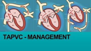
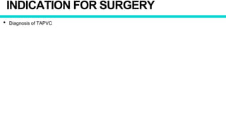
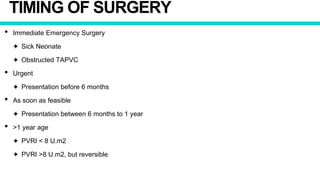
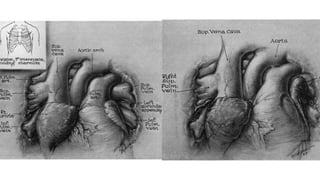
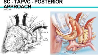
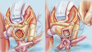
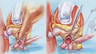
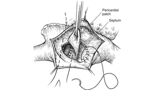
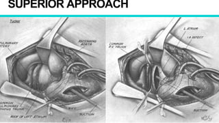
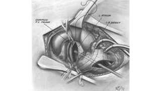
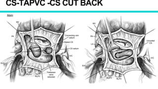
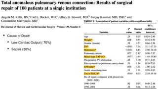
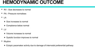
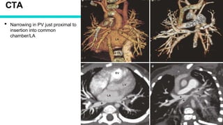
TAPVC - MANAGEMENT
TYPES
DIAGNOSIS
DIAGNOSIS
• Clinical Presentation
• ECG
• Imaging
✦ CXR
✦ Echocardiography
✦ Contrast CT
✦ Catheter Angiography
ECG
• Obstructed TAPVC
✦ Right ventricular hypertrophy
★ qR complex or only R wavein right chest leads
✦ Right atrial enlargement is infrequent.
★ Only seen in cardiac TAPVC with obstruction at ASD level
• Non-obstructed TAPVC
✦ Resembles that of an OS-ASD
✦ Right axis deviation
✦ Right atrial and ventricular enlargement
✦ Incomplete right bundle branch block pattern.
CXR
• Non Obstructed
✦ Cardiomegaly
✦ RV Apex
✦ RA and RV Enlargement
✦ Enlarged PA Segment
✦ Pulmonary Plethora
CXR
• Obstruction
✦ Vertical Vein - Normal Cardiac Size
✦ ASD - Cardiomegaly
✦ White out / Ground Glass Opacity lung field
★ DDX
• Meconium Aspiration Syndrome
• Hyaline Membrane Disease
✦ Enlarged PA Segment
SC-TAPVC - SNOWMAN
OBSTRUCTED TAPVC
• CXR
✦ Plethoric Lung Fields / Ground
Glass appearance
✦ Absence of cardiomegaly
✦ Indistinct cardiac silhouette
• DDX
✦ Meconium Aspiration
✦ Hyaline Membrane Disease
ECHOCARDIOGRAPHY
• Diagnostic and Investigation of Choice
• Important Observation
✦ PV not draining into LA
✦ Type of TAPVC
★ Cardiac - Dilated CS
★ Supracardiac - Dilated innominate vein and SVC
★ Infracardiac - Dilated IVC
✦ Insertion of Vertical Vein
✦ Narrowing of Vertical Vein
✦ Adequacy of ASD
✦ RA and RV Dilatation
✦ TR
✦ PAH
✦ RV Function
✦ Small LA
✦ LV size and Function
✦ PV Narrowing
✦ Size
✦ Doppler - Velocity > 2m/s
✦ Coexisting Congenital Defect
✦ VSD
✦ PDA
CARDIAC CATHERISATION
• Always Diagnostic
• Indication
✦ Measure PVRI / Reversibility in older children
✦ Diagnosis not clear on Echocardiography
✦ Intervention - BAS, PV dilatation, Vertical Vein Dilatation
• PA/PV Angiogram
✦ PA injection with venous follow through preferred over direct PV injection
✦ Anatomy of TAPVC
✦ Type
✦ Drainage
✦ Site of Obstruction
CARDIAC CATHERISATION
• Oximetry
✦ Similar saturation in all chamber
• Drawbacks
✦ Delays management
✦ Aggravate pulmonary edema
✦ Contrast related
SC-TAPVC CATHETER ANGIO
IC-TAPVC CATHTER ANGIO
CROSS SECTIONAL IMAGING
• CT and MRI
• Indication
✦ Complex pulmonary venous drainage pattern, not clearly defined on Echo
✦ Heterotaxy syndromes with TAPVC
✦ Post operative Pulmonary vein Stenosis
• Drawbacks
✦ Contrast related
✦ Requires sedation
SC- TAPVC CTA
CARDIAC TAPVC
• CTA
✦ Common chamber opening into
CS
✦ Small LA
✦ Dilated RA
✦ ASD
IC-TAPVC CTA
DECISION MAKING
FACTORS TO CONSIDER
• Age of Patient
• Weight of the patient
• Type of TAPVC
• Obstructed or Non Obstructed
• Clinical Status of Patient
✦ Ventilator
✦ Inotropes
✦ Acidosis
• LV and RV function
• PAH
• Presence of PVOD
INDICATION FOR SURGERY
• Diagnosis of TAPVC
TIMING OF SURGERY
• Immediate Emergency Surgery
✦ Sick Neonate
✦ Obstructed TAPVC
• Urgent
✦ Presentation before 6 months
• As soon as feasible
✦ Presentation between 6 months to 1 year
• >1 year age
✦ PVRI < 8 U.m2
✦ PVRI >8 U.m2, but reversible
MEDICAL TREATMENT
MEDICAL THERAPY
• Only supportive in managing TAPVC
• Intended to stabilise the patient before surgery
• Goals
✦ Correct acidosis and hypotension
✦ Fluid management
✦ Mechanical ventilation
• Prostaglandin
✦ Helpful in keeping ductus venosus open in infra-diaphragmatic TAPVC
✦ Other forms - Increases right to left shunting and systemic desaturation
CATHETER BASED INTERVENTION
BAS
• Short term palliation
• Indication
✦ Sick neonate with TAPVC
✦ Obstruction at ASD level
• Goal
✦ Improve mixing
✦ Improve cardiac output
✦ Relief systemic and pulmonary congestion
• Technically challenging
✦ Small left atrium
✦ Echocardiographic guidance is often helpful
• Success of a BAS
✦ Drop in pulmonary artery pressure
✦ Increase in systemic pressure
✦ Drop in systemic saturation
BALLON DILATION/ STENTING VV
• Occasionally performed to relieve obstruction
• Indication
✦ Sick neonates presenting with obstructed TAPVC
• Technically challenging
SURGICAL TREATMENT
PRINCIPLES
• Principle
✦ On CPB or Circulatory arrest
✦ Identify 4 PV, common chamber and dissect and loop vertical vein
✦ Achieve wide tension free non obstructing anastomosis between common chamber and LA
✦ Avoiding purse stringing of suture line
✦ Divert all pulmonary venous blood to LA
✦ Close intra-atrial communication
★ Patch Material
• Pericardium
• Dacron ( Polyethylene terephthalate )
✦ Ligate vertical vein close to insertion into systemic vein
CLOSED APPROACH
William H Muller
SC - TAPVC - POSTERIOR
APPROACHPaul Ebert
SC-TAPVC RIGHT BIATRIAL APPROACH
Shumacker and King
RIGHT SIDED APPROACH - LA
Kirklin
TRANS RA TRANS LA
Cooley
SUPERIOR APPROACH
Tucker
CS-TAPVC -CS CUT BACK
Malm
CS-TAPVC
Van Prragh
IC- TAPVC - ORIENTATION OF COMMON CHAMBER
SUTURELESS TECHNIQUE
Lacour Gayet
CONTROVERSIES
PRIMARY SUTURELESS VS CONVENTIONAL REPAIR
PVO AND REOPERATIONPostOpPVOReopPVO
OVERALL AND EARLY MORTALITYOverallMortalityEarlyMortality
VERTICAL VEIN LIGATION
• Proponents of Ligation
✦ Large left to right shunt with RV volume overload
✦ Requires additional procedure for closure
✦ No survival benefit
✦ Ligating the vertical vein close to its insertion into the systemic vein, the vertical vein adds to the
reservoir of the LA
• Proponents of Leaving the vertical vein open
✦ Serves as a pop off for the non compliant left heart
✦ Spontaneously closes over time
✦ Less turbulent immediate postoperative course, especially in obstructed TAPVC
Surviours - 12
Not ligated vertical vein - 10
Median Follow up - 4.7 years
Spontaneous closure - 5
Required Ligation - 4
Circulation. 2010;122:2718-2726
LA ENLARGMENT
• Proponents
✦ Increase in reservoir capacity
✦ Better LV Filling and Cardiac Output
✦ Decreased incidence of Pulmonary venous hypertension and reactive PAH
• Oponents
✦ Incorporation of common venous sinus enlarges LA adequately
✦ Patch material is non contractile
• Literature
✦ No significant difference in survival with LA enlargement
★ Katz NM, Kirklin JW, Pacifico AD. Concepts and practices in surgery for total anomalous
pulmonary venous connection. Ann Thorac Sure 1977
PFO
• Benefits
✦ Popoff for RV in patients with severe PAH
✦ Maintain cardio output at the cost of desaturation
• Drawbacks
✦ Risk of paradoxical embolism
✦ Significant right to left requiring additional procedure for closure
POST OPERATIVE MANAGEMENT
PHYSIOLOGY
• Preoperative
✦ Small LA
✦ Underfilled LV
✦ RA/RV Volume Overload
✦ Increased Qp
• Postoperative
✦ Offloading of Right Heart
✦ Volume loading in not compliant LV
ANTICIPATED PROBLEMS
• Non Compliant LV
✦ > Acute Dilatation > Septal Dysfunction > LV Dysfunction > Increase in LA Pressure
• Non Compliant LA > Surges in LA pressure >
✦ PVH > PAH Crisis
✦ Pulmonary Hemorrhage > Stiff Lungs
• Post Op PAH > RV Dysfunction > LCOS
• LV Dysfunction > LCOS
COMPLICATIONS OF SURGERY
• Bleeding
• LV diastolic dysfunction (Low Compliance)
• RV dysfunction
• Pulmonary hypertension and PAH crisis
• Gradient across anastomosis
• Pulmonary vein stenosis
STRATEGIES
• Avoid surges is LA pressure
✦ Fluid Restriction
✦ Avoid Fluid Bolus
• Inodilators
✦ Dobutamine/Milrinone
• Afterload Reduction - Vasodilators
✦ NTG/SNP
• Avoid Triggers of PAH
• Refractory PAH
✦ Echo - R/o Anastamotic stenosis/ Pulmonary vein stenosis
✦ NO Ventilator
✦ Mechanical Support
ISSUES
• LV dysfunction
✦ Inodilators
✦ Monitor LA pressure
✦ Positive pressure ventilation
✦ Prevent fluid overloading ( Low Threshold for PD)
✦ ECMO
ISSUES
• PAH
✦ Adequate sedation in the first 24-48 hours
✦ Avoid
★ Hypercardbia
★ Hypoxia
★ Acidosis
✦ Adequate lung recruitment
✦ Pulmonary dilatation
★ SNP, Milrinone
✦ Avoid triggers of PAH
★ Suctioning
✦ Refractory Cases - NO Ventilation
PEARLS
• Avoid Fluid Bolus
• Accept low BP if no Lactic Acidosis
• Avoiding Weaning when LA pressure high
• Low threshold for Peritoneal dialysis
• Avoid triggers of PAH
• Elective CPAP after extubation helps with LV dysfunction
RESULTS
MORTALITY
• Early Mortality
✦ STS - 10-30%
✦ Individual Centers - 2-20%
• Late Mortality
✦ Very few late mortality
✦ Survival depends on early survival
MODES OF DEATH
• Cardiac Failure
• PAH Crisis
• Sepsis
• Pathophyiological correlates to mortality
✦ Preoperative cardiopulmonary instability
★ Acidosis
★ Ventilator Requirement
✦ Post op PAH Crisis
★ Preop Lung Injury
★ CPB related
★ Postop Acidosis
✦ Progression of Pulmonary Vein Stenosis
• Cause of Death
• Low Cardiac Output ( 70%)
• Sepsis (30%)
RISK FACTORS FOR EARLY DEATH
• Low birth weight
• Infracardiac TAPVC
• Mixed TAPVC
• Obstructed TAPVC
• Poor preoperative physiological state
• Postop Pulmonary Venous Obstruction
• Increased PVRI
• Single ventricle physiology
Over all mortality
<2 Kg
Mixed vs Others
Postop PVO
Reoperation PVO
Reoperation Mixed
TAPVC
HEMODYNAMIC OUTCOME
• RV - Size decreases to normal
• PA - Pressure normalises
• LA
✦ Size increases to normal
✦ Compliance below normal
• LV
✦ Volume increases to normal
✦ Systolic function improves to normal
• Rhythm
✦ Ectopic pacemaker activity due to damage of internodal preferential pathway
FUNCTIONAL STATUS
• Most surviving patients are NYHA 1
ary and musculoskeletal exercise performance after repair for total anomalous pulmonary venous connection during infancy. J Thorac Cardiovasc Surg 2007;133:1533-9.
AIIMS RESULT
• Patients - 248 (168 boys, 80 girls)
• Age - 1 day to 24 years (median 8 months) ; 145 infants
• Weight - 2 to 52 kg (median 5 kg). 70% were less than the 50th percentile of predicted weight for age and
sex.
• Type
✦ Supracardiac - 134 (54%)
✦ Cardiac - 80 (32.2%)
✦ Infracardiac - 9 (3.6%)
✦ Mixed - 25 (10.1%)
• Obstructed TAPVC - 50 (20.2%)
• Mod-Sev PAH - 76 patients (30.2%)
Choudhary SK, Bhan A, Sharma R, et al. Total anomalous pulmonary venous connection: surgical experience in Indians. Indian Heart J. 20
• Emergency surgery - 45 patients (18.1%)
• Circulatory Arrest - 114
• Early Mortality - 45 (19.1%)
✦ Pulmonary arterial hypertensive crisis - 19
✦ Low cardiac output syndrome - 17
• Risk Factors
✦ Age < 1 year (OR 2.16; 95% CI: 1.22-3.82, p=0.008)
✦ Severe pulmonary arterial hypertension (AR 5.86; 95% CI: 2-17, p=0.001),
✦ Emergency surgery (OR 3.65; 95% CI: 1.59-8.38, p=0.002)
• Late Mortality - 4
• Follow-up ranged from 1 to 180 months (median 48 months).
• Actuarial survival at 12 years - 92.6% +/- 2.8%.
Choudhary SK, Bhan A, Sharma R, et al. Total anomalous pulmonary venous connection: surgical experience in Indians. Indian Heart J. 20
REOPERATION
ANASTAMOTIC STENOSIS
• Incidence - 10%
• Occurs early postoperative period
✦ Later anastomosis grows and child grows
• Factors which decrease risk
✦ Open anastomosis
✦ Wide anastomosis
✦ Absorbable monofilament sutures
✦ No difference between continuous and interrupted suture technique
• Reoperation
✦ Risk of restenosis
✦ Poor outcome
PULMONARY VEIN STENOSIS
• Incidence - 5- 15%
• Usually occurs within the first 6 months
• Pathophysiology
✦ Diffuse fibrotic thickening of PV
✦ Localised narrowing at PV-LA junction
✦ May or may not be associate with anastomotic stenosis
• Presentation
✦ Progressive Dyspnea
• Investigation
✦ Echocardiography
★ PV Flow velocity > 2m/s
✦ Cross Sectional Imaging
Circulation. 2010;122:2718-2726
CTA
• Narrowing in PV just proximal to
insertion into common
chamber/LA
PULMONARY VEIN STENOSIS
• Treatment
✦ Steroids or Chemotherapy to minimise fibrosis
✦ Balloon dilatation/stenting
✦ Ostial endarterectomy of intimal hyperplasia IRevising the common pulmonary vein to left atrium
anastomosis with or without patch enlargement
✦ Sutureless repair
• Developed PVO - 71/406 (17.5%)
✦ Cardiac - 9/67 (13%)
✦ Supracardiac -25/205 (12%)
✦ Infracardiac - 25/110 (23%)
✦ Mixed - 11/37 (30%)
• Median time to diagnosis from surgery - 49days ( 0 - 5.9 yrs)
✦ Diagnosed within 6 months of surgery - 59/71 (83%)
✦ Echo - 30% , Angiography - 54% and CT/MRI - 16%
• Intervention
✦ 60 patients required intervention
✦ 56 received intervention
★ 50% required more than 1 intervention
THANK YOU

Management of tapvc

  • 1.
  • 2.
  • 6.
  • 7.
    DIAGNOSIS • Clinical Presentation •ECG • Imaging ✦ CXR ✦ Echocardiography ✦ Contrast CT ✦ Catheter Angiography
  • 8.
    ECG • Obstructed TAPVC ✦Right ventricular hypertrophy ★ qR complex or only R wavein right chest leads ✦ Right atrial enlargement is infrequent. ★ Only seen in cardiac TAPVC with obstruction at ASD level • Non-obstructed TAPVC ✦ Resembles that of an OS-ASD ✦ Right axis deviation ✦ Right atrial and ventricular enlargement ✦ Incomplete right bundle branch block pattern.
  • 9.
    CXR • Non Obstructed ✦Cardiomegaly ✦ RV Apex ✦ RA and RV Enlargement ✦ Enlarged PA Segment ✦ Pulmonary Plethora
  • 10.
    CXR • Obstruction ✦ VerticalVein - Normal Cardiac Size ✦ ASD - Cardiomegaly ✦ White out / Ground Glass Opacity lung field ★ DDX • Meconium Aspiration Syndrome • Hyaline Membrane Disease ✦ Enlarged PA Segment
  • 11.
  • 12.
    OBSTRUCTED TAPVC • CXR ✦Plethoric Lung Fields / Ground Glass appearance ✦ Absence of cardiomegaly ✦ Indistinct cardiac silhouette • DDX ✦ Meconium Aspiration ✦ Hyaline Membrane Disease
  • 13.
    ECHOCARDIOGRAPHY • Diagnostic andInvestigation of Choice • Important Observation ✦ PV not draining into LA ✦ Type of TAPVC ★ Cardiac - Dilated CS ★ Supracardiac - Dilated innominate vein and SVC ★ Infracardiac - Dilated IVC ✦ Insertion of Vertical Vein ✦ Narrowing of Vertical Vein ✦ Adequacy of ASD
  • 14.
    ✦ RA andRV Dilatation ✦ TR ✦ PAH ✦ RV Function ✦ Small LA ✦ LV size and Function ✦ PV Narrowing ✦ Size ✦ Doppler - Velocity > 2m/s ✦ Coexisting Congenital Defect ✦ VSD ✦ PDA
  • 17.
    CARDIAC CATHERISATION • AlwaysDiagnostic • Indication ✦ Measure PVRI / Reversibility in older children ✦ Diagnosis not clear on Echocardiography ✦ Intervention - BAS, PV dilatation, Vertical Vein Dilatation • PA/PV Angiogram ✦ PA injection with venous follow through preferred over direct PV injection ✦ Anatomy of TAPVC ✦ Type ✦ Drainage ✦ Site of Obstruction
  • 18.
    CARDIAC CATHERISATION • Oximetry ✦Similar saturation in all chamber • Drawbacks ✦ Delays management ✦ Aggravate pulmonary edema ✦ Contrast related
  • 20.
  • 21.
  • 22.
    CROSS SECTIONAL IMAGING •CT and MRI • Indication ✦ Complex pulmonary venous drainage pattern, not clearly defined on Echo ✦ Heterotaxy syndromes with TAPVC ✦ Post operative Pulmonary vein Stenosis • Drawbacks ✦ Contrast related ✦ Requires sedation
  • 23.
  • 24.
    CARDIAC TAPVC • CTA ✦Common chamber opening into CS ✦ Small LA ✦ Dilated RA ✦ ASD
  • 25.
  • 26.
  • 27.
    FACTORS TO CONSIDER •Age of Patient • Weight of the patient • Type of TAPVC • Obstructed or Non Obstructed • Clinical Status of Patient ✦ Ventilator ✦ Inotropes ✦ Acidosis • LV and RV function • PAH • Presence of PVOD
  • 28.
    INDICATION FOR SURGERY •Diagnosis of TAPVC
  • 29.
    TIMING OF SURGERY •Immediate Emergency Surgery ✦ Sick Neonate ✦ Obstructed TAPVC • Urgent ✦ Presentation before 6 months • As soon as feasible ✦ Presentation between 6 months to 1 year • >1 year age ✦ PVRI < 8 U.m2 ✦ PVRI >8 U.m2, but reversible
  • 31.
  • 32.
    MEDICAL THERAPY • Onlysupportive in managing TAPVC • Intended to stabilise the patient before surgery • Goals ✦ Correct acidosis and hypotension ✦ Fluid management ✦ Mechanical ventilation • Prostaglandin ✦ Helpful in keeping ductus venosus open in infra-diaphragmatic TAPVC ✦ Other forms - Increases right to left shunting and systemic desaturation
  • 33.
  • 34.
    BAS • Short termpalliation • Indication ✦ Sick neonate with TAPVC ✦ Obstruction at ASD level • Goal ✦ Improve mixing ✦ Improve cardiac output ✦ Relief systemic and pulmonary congestion • Technically challenging ✦ Small left atrium ✦ Echocardiographic guidance is often helpful • Success of a BAS ✦ Drop in pulmonary artery pressure ✦ Increase in systemic pressure ✦ Drop in systemic saturation
  • 35.
    BALLON DILATION/ STENTINGVV • Occasionally performed to relieve obstruction • Indication ✦ Sick neonates presenting with obstructed TAPVC • Technically challenging
  • 38.
  • 39.
    PRINCIPLES • Principle ✦ OnCPB or Circulatory arrest ✦ Identify 4 PV, common chamber and dissect and loop vertical vein ✦ Achieve wide tension free non obstructing anastomosis between common chamber and LA ✦ Avoiding purse stringing of suture line ✦ Divert all pulmonary venous blood to LA ✦ Close intra-atrial communication ★ Patch Material • Pericardium • Dacron ( Polyethylene terephthalate ) ✦ Ligate vertical vein close to insertion into systemic vein
  • 40.
  • 42.
    SC - TAPVC- POSTERIOR APPROACHPaul Ebert
  • 46.
    SC-TAPVC RIGHT BIATRIALAPPROACH Shumacker and King
  • 48.
  • 51.
    TRANS RA TRANSLA Cooley
  • 54.
  • 56.
  • 58.
  • 60.
    IC- TAPVC -ORIENTATION OF COMMON CHAMBER
  • 63.
  • 66.
  • 67.
    PRIMARY SUTURELESS VSCONVENTIONAL REPAIR
  • 68.
  • 69.
    OVERALL AND EARLYMORTALITYOverallMortalityEarlyMortality
  • 70.
    VERTICAL VEIN LIGATION •Proponents of Ligation ✦ Large left to right shunt with RV volume overload ✦ Requires additional procedure for closure ✦ No survival benefit ✦ Ligating the vertical vein close to its insertion into the systemic vein, the vertical vein adds to the reservoir of the LA • Proponents of Leaving the vertical vein open ✦ Serves as a pop off for the non compliant left heart ✦ Spontaneously closes over time ✦ Less turbulent immediate postoperative course, especially in obstructed TAPVC
  • 74.
    Surviours - 12 Notligated vertical vein - 10 Median Follow up - 4.7 years Spontaneous closure - 5 Required Ligation - 4
  • 75.
  • 76.
    LA ENLARGMENT • Proponents ✦Increase in reservoir capacity ✦ Better LV Filling and Cardiac Output ✦ Decreased incidence of Pulmonary venous hypertension and reactive PAH • Oponents ✦ Incorporation of common venous sinus enlarges LA adequately ✦ Patch material is non contractile • Literature ✦ No significant difference in survival with LA enlargement ★ Katz NM, Kirklin JW, Pacifico AD. Concepts and practices in surgery for total anomalous pulmonary venous connection. Ann Thorac Sure 1977
  • 77.
    PFO • Benefits ✦ Popofffor RV in patients with severe PAH ✦ Maintain cardio output at the cost of desaturation • Drawbacks ✦ Risk of paradoxical embolism ✦ Significant right to left requiring additional procedure for closure
  • 78.
  • 79.
    PHYSIOLOGY • Preoperative ✦ SmallLA ✦ Underfilled LV ✦ RA/RV Volume Overload ✦ Increased Qp • Postoperative ✦ Offloading of Right Heart ✦ Volume loading in not compliant LV
  • 80.
    ANTICIPATED PROBLEMS • NonCompliant LV ✦ > Acute Dilatation > Septal Dysfunction > LV Dysfunction > Increase in LA Pressure • Non Compliant LA > Surges in LA pressure > ✦ PVH > PAH Crisis ✦ Pulmonary Hemorrhage > Stiff Lungs • Post Op PAH > RV Dysfunction > LCOS • LV Dysfunction > LCOS
  • 81.
    COMPLICATIONS OF SURGERY •Bleeding • LV diastolic dysfunction (Low Compliance) • RV dysfunction • Pulmonary hypertension and PAH crisis • Gradient across anastomosis • Pulmonary vein stenosis
  • 82.
    STRATEGIES • Avoid surgesis LA pressure ✦ Fluid Restriction ✦ Avoid Fluid Bolus • Inodilators ✦ Dobutamine/Milrinone • Afterload Reduction - Vasodilators ✦ NTG/SNP • Avoid Triggers of PAH • Refractory PAH ✦ Echo - R/o Anastamotic stenosis/ Pulmonary vein stenosis ✦ NO Ventilator ✦ Mechanical Support
  • 83.
    ISSUES • LV dysfunction ✦Inodilators ✦ Monitor LA pressure ✦ Positive pressure ventilation ✦ Prevent fluid overloading ( Low Threshold for PD) ✦ ECMO
  • 84.
    ISSUES • PAH ✦ Adequatesedation in the first 24-48 hours ✦ Avoid ★ Hypercardbia ★ Hypoxia ★ Acidosis ✦ Adequate lung recruitment ✦ Pulmonary dilatation ★ SNP, Milrinone ✦ Avoid triggers of PAH ★ Suctioning ✦ Refractory Cases - NO Ventilation
  • 85.
    PEARLS • Avoid FluidBolus • Accept low BP if no Lactic Acidosis • Avoiding Weaning when LA pressure high • Low threshold for Peritoneal dialysis • Avoid triggers of PAH • Elective CPAP after extubation helps with LV dysfunction
  • 86.
  • 87.
    MORTALITY • Early Mortality ✦STS - 10-30% ✦ Individual Centers - 2-20% • Late Mortality ✦ Very few late mortality ✦ Survival depends on early survival
  • 88.
    MODES OF DEATH •Cardiac Failure • PAH Crisis • Sepsis • Pathophyiological correlates to mortality ✦ Preoperative cardiopulmonary instability ★ Acidosis ★ Ventilator Requirement ✦ Post op PAH Crisis ★ Preop Lung Injury ★ CPB related ★ Postop Acidosis ✦ Progression of Pulmonary Vein Stenosis
  • 89.
    • Cause ofDeath • Low Cardiac Output ( 70%) • Sepsis (30%)
  • 90.
    RISK FACTORS FOREARLY DEATH • Low birth weight • Infracardiac TAPVC • Mixed TAPVC • Obstructed TAPVC • Poor preoperative physiological state • Postop Pulmonary Venous Obstruction • Increased PVRI • Single ventricle physiology
  • 92.
    Over all mortality <2Kg Mixed vs Others Postop PVO Reoperation PVO Reoperation Mixed TAPVC
  • 94.
    HEMODYNAMIC OUTCOME • RV- Size decreases to normal • PA - Pressure normalises • LA ✦ Size increases to normal ✦ Compliance below normal • LV ✦ Volume increases to normal ✦ Systolic function improves to normal • Rhythm ✦ Ectopic pacemaker activity due to damage of internodal preferential pathway
  • 95.
    FUNCTIONAL STATUS • Mostsurviving patients are NYHA 1 ary and musculoskeletal exercise performance after repair for total anomalous pulmonary venous connection during infancy. J Thorac Cardiovasc Surg 2007;133:1533-9.
  • 99.
    AIIMS RESULT • Patients- 248 (168 boys, 80 girls) • Age - 1 day to 24 years (median 8 months) ; 145 infants • Weight - 2 to 52 kg (median 5 kg). 70% were less than the 50th percentile of predicted weight for age and sex. • Type ✦ Supracardiac - 134 (54%) ✦ Cardiac - 80 (32.2%) ✦ Infracardiac - 9 (3.6%) ✦ Mixed - 25 (10.1%) • Obstructed TAPVC - 50 (20.2%) • Mod-Sev PAH - 76 patients (30.2%) Choudhary SK, Bhan A, Sharma R, et al. Total anomalous pulmonary venous connection: surgical experience in Indians. Indian Heart J. 20
  • 100.
    • Emergency surgery- 45 patients (18.1%) • Circulatory Arrest - 114 • Early Mortality - 45 (19.1%) ✦ Pulmonary arterial hypertensive crisis - 19 ✦ Low cardiac output syndrome - 17 • Risk Factors ✦ Age < 1 year (OR 2.16; 95% CI: 1.22-3.82, p=0.008) ✦ Severe pulmonary arterial hypertension (AR 5.86; 95% CI: 2-17, p=0.001), ✦ Emergency surgery (OR 3.65; 95% CI: 1.59-8.38, p=0.002) • Late Mortality - 4 • Follow-up ranged from 1 to 180 months (median 48 months). • Actuarial survival at 12 years - 92.6% +/- 2.8%. Choudhary SK, Bhan A, Sharma R, et al. Total anomalous pulmonary venous connection: surgical experience in Indians. Indian Heart J. 20
  • 101.
  • 102.
    ANASTAMOTIC STENOSIS • Incidence- 10% • Occurs early postoperative period ✦ Later anastomosis grows and child grows • Factors which decrease risk ✦ Open anastomosis ✦ Wide anastomosis ✦ Absorbable monofilament sutures ✦ No difference between continuous and interrupted suture technique • Reoperation ✦ Risk of restenosis ✦ Poor outcome
  • 103.
    PULMONARY VEIN STENOSIS •Incidence - 5- 15% • Usually occurs within the first 6 months • Pathophysiology ✦ Diffuse fibrotic thickening of PV ✦ Localised narrowing at PV-LA junction ✦ May or may not be associate with anastomotic stenosis • Presentation ✦ Progressive Dyspnea • Investigation ✦ Echocardiography ★ PV Flow velocity > 2m/s ✦ Cross Sectional Imaging
  • 104.
  • 108.
    CTA • Narrowing inPV just proximal to insertion into common chamber/LA
  • 109.
    PULMONARY VEIN STENOSIS •Treatment ✦ Steroids or Chemotherapy to minimise fibrosis ✦ Balloon dilatation/stenting ✦ Ostial endarterectomy of intimal hyperplasia IRevising the common pulmonary vein to left atrium anastomosis with or without patch enlargement ✦ Sutureless repair
  • 115.
    • Developed PVO- 71/406 (17.5%) ✦ Cardiac - 9/67 (13%) ✦ Supracardiac -25/205 (12%) ✦ Infracardiac - 25/110 (23%) ✦ Mixed - 11/37 (30%) • Median time to diagnosis from surgery - 49days ( 0 - 5.9 yrs) ✦ Diagnosed within 6 months of surgery - 59/71 (83%) ✦ Echo - 30% , Angiography - 54% and CT/MRI - 16% • Intervention ✦ 60 patients required intervention ✦ 56 received intervention ★ 50% required more than 1 intervention
  • 118.

Editor's Notes

  • #73 Vertical vein was looped and snared in all patients. Loosened in all patients with obstructed TAPVC
  • #76 Group 1 - No VV ligation Group 2 - VV ligation