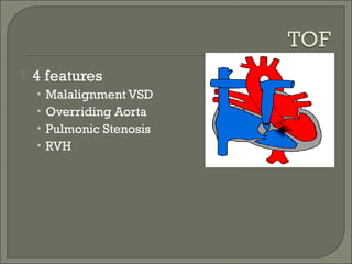
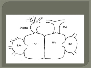
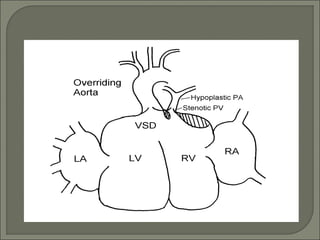
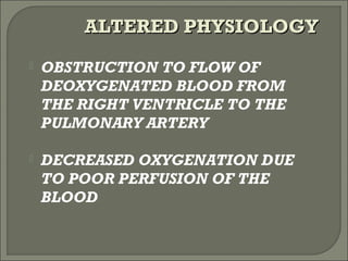
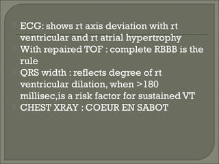
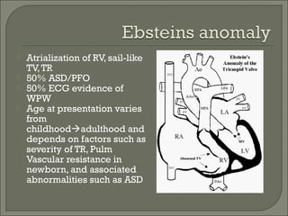
CHAIRPERSON: DR B.L
BHARDWAJ
 US: 1,000,000 adults with congenital heart dz
 20,000 more patients reach adolescents yearly
 Incidence increasing due to increased survival
of children with CHD
 Genetic : as in downs , noonans , holt-
oram, williams etc
 Environmental : maternal rubella , drugs
like thalidomide , isotretinoin
 Hyperviscosity syndrome : headache ,
dizziness , nausea , paresthesias due to
increased erythrocytosis bcoz of
cyanosis
 Bleeding tendencies
 Paradoxical embolisation , brain abscess
 Proteinuria , hyperuricemia
 Hypertrophic osteoarthropathy causing
arthralgias and bone pain
 Atrial Septal Defect
 Ventricular septal defect
 PDA
 TOF
 Eisenmenger
 Ebstein anomaly
 Pulm. Stenosis with intact ventr. septum
 Aortic stenosis
 COA
.
 4 possibilities:
 1..if ecg reveals LAD of >30 deg :
suggests ostium primum defect
 2..pt may hav a floppy mitral valve
 3..ostium secundum defect assoc with
rheumatic MR
 4..secundum ASD rarely assoc with cleft
mitral valve
• Effort dyspnea is seen in 30% of patients
by 3rd
decade and over 75% of patients by
the fifth decade
• SVT (atrial fibrillation or flutter) and right-
sided heart failure develop by age 40 in
10-20% of patients
• The chest radiograph shows large pulmonary arteries, increased
pulmonary vascularity, an enlarged RA and RV, and a small aortic
knob with all pre-tricuspid cardiac left-to-right shunts.
 Percutaneous Closure
• only for secundum (contra in others)
• adequate superior/inferior rim around ASD
 Surgical Closure
• Good prognosis:
 closure age < 25, PA pressure <40
 If >25 or PA>40, decreased survival due to CHF,
stroke, and afib
 First described by roger in 1879
 Term” maladie de roger” : refers to small
asymptomatic VSD
 Most common CHD in children (25%)
 Isolated VSD found in only 10% of adults
with CHD
 75-80% of small VSD’s close spontaneously
by late childhood
 10-15% of large VSD’s close spontaneously
 60% of defects close before age 3, and 90%
before age 8
 Risk factors for decreased survival for
unoperated patients include:
• Cardiomegaly on CXR, Elevated PASP (>50
mmHg), and CV symptoms
 Perimembranous defect (70-80%)
• Less likely to be associated with other defects
• Highest rate of spontaneous closure
 Muscular or apical defects (5-20%)
• Typically occur in isolation
• High spontaneous closure rates unless multiple defects r present
 AV-Canal type (5-8%)
• Rarely close spontaneously, commonly seen in Trisomy 21
• Usually large & associated with abnormal AV valve
 Supracristal or subaortic defects (5-7%)
• Often small but need closure due to associated AR
 Arterial pulse is often normal
 There may be a systolic thrill on palpation of the
precordium (maximal in lt 3rd
or 4th
ICS)
 Holosystolic, high frequency murmur (grade 4-
6/6) with small VSD and normal PAP
 Once PAP increases above the systemic
pressures the holosystolic murmur disappears
 Increase flow across pulmonary valve causes a
SEM
 A loud P2 component is heard in this setting
 May be normal but often shows LVH and LAE
 Presence of RAD represents elevated RVP and PAP
 Postoperative RBBB is common
 Cardiomegaly with LAE and LVE will be seen with large L to R shunts
 A large defect associated with a small heart and oligemic lung fields
should raise the suspicion of pulmonary vascular disease
 Hemodynamic severity grading of isolated VSDs in
adults:
• Small: Qp:Qs <1.4, and pulmonary to aortic systolic pressure
<0.3
• Moderate: Qp:Qs = 1.4-2.2, and systolic pressure ratio >0.3
• Large: Qp:Qs >2.2, and systolic pressure ratio >0.3
• Eisenmenger: Qp:Qs <1.5 and systolic pressure ratio >0.9
 Clinical severity grading:
• Small: Causes negligible hemodynamic changes. LV size normal
w/o PHTN
• Moderate: Causes LV and LA enlargment, and usually some
PHTN (reversible)
• Large: Results in pulmonary vascular obstructive disease and
Eisenmenger physiology unless there is coexistent RVOTO
 When repair is performed in the first two years of life,
asymptomatic adult survival with normal growth and
development can be anticipated
 When surgery is undertaken in older children, a late
postopeartive increase in LV chamber size, together with
decreased systolic function is seen
 Development of late postoperative PHTN is largely
determined by the age at surgery and preoperative PVR
 Risk of SBE persists and requires prophylaxis
 Indications for intervention: Geade C, Level IV
• Presence of a significant VSD (symptomatic QP/QS = 2/1, PASP
> 50 mmHg), deteriorating ventricular fx due to volume (LV) or
pressure (RV) overload
• Significant RVOTO (pk to pk gradient of > 50 mmHg, or
instantaneous gradient >70 mmHg)
• Perimembranous or doubly committed VSD with more than mild
AR
• Hx of endocarditis especially if recurrent
 Successful closure is associated with excellent survival if
ventricular fx is normal. Elevated PAP preop may
progress, regress, or remain the same postop
 A. fib may occur, especially if there has been
longstanding volume overload of the left heart
 Pregnancy is well tolerated in women with small or
moderate VSD and in women with repaired VSD
 Pregnancy is contraindicated in women with
Eisenmenger syndrome due to both high maternal
(>50%) and fetal (~60%) mortality
 4 features
• Malalignment VSD
• Overriding Aorta
• Pulmonic Stenosis
• RVH
 OBSTRUCTION TO FLOW OF
DEOXYGENATED BLOOD FROM
THE RIGHT VENTRICLE TO THE
PULMONARY ARTERY
 DECREASED OXYGENATION DUE
TO POOR PERFUSION OF THE
BLOOD
ALTERED PHYSIOLOGYALTERED PHYSIOLOGY
 Rt-lt shunting across vsd
 Degree of cyanosis reflects the severity
of RVOTO and level of SVR
 Severity of cyanosis is directly
propotional to severity of pulmonic
stenosis
 MORE SHUNTING ACROSS THE VSD
– MORE DESATURATION OF
SYSTEMIC BLOOD – PERIPHERAL
ACIDOSIS – FUTHER SYSTEMIC
VASODILATATION – FURTHER
DECREASE IN SVR – VICIOUS CYCLE
 Variable cyanosis
 Rt ventricular impulse and systolic thrill
along left sternal border
 Repaired
• RVOT obstruction
• Pulmonary or tricuspid regurgitation
• LV/RV dysfunction
• Atrial/ventricular arrhythmias
 Unrepaired
• Significant morbidity
• Consider later repair
 Since rt ventricle is effectively
decompressed by VSD , CCF never
occurs....exceptions to this rule are:
 1.anemia
 2.infective endocarditis
 3.syst HTN
 4. aortic or pulm.Valve regurgitation
 ECG: shows rt axis deviation with rt
ventricular and rt atrial hypertrophy
 With repaired TOF : complete RBBB is the
rule
 QRS width : reflects degree of rt
ventricular dilation, when >180
millisec,is a risk factor for sustained VT
 CHEST XRAY : COEUR EN SABOT
 ONLY SURGICAL
• PALLIATIVE SURGERY
• DEFINITIVE SURGERY
TREATMENT OPTIONSTREATMENT OPTIONS
 Atrialization of RV, sail-like
TV,TR
 50% ASD/PFO
 50% ECG evidence of
WPW
 Age at presentation varies
from
childhoodadulthood and
depends on factors such as
severity of TR, Pulm
Vascular resistance in
newborn, and associated
abnormalities such as ASD
 Commonly associated with:
• ASD or PFO (90%)
• VSD, AV canal defect
• Pulmonary stenosis/atresia (20-25%)
• Wolff-Parkinson-White
 Syndromes:
• Down, Marfan, Noonan, Cornelia de Lange
 Heart sounds
• First heart sound widely split with loud tricuspid
component
• Second heart sound usually is normal but may
be widely split due to RBBB
• Third and fourth heart sounds commonly present
 Murmurs
• Holosystolic murmur of tricuspid regurgitation
 Cyanosis
• Due to R  L shunt at atrial level
 Fatigue and dyspnea
• Secondary to RV failure and decreased LV
ejection fraction
 Palpitations and sudden cardiac death
 Incidental murmur
 Paradoxic embolism
 Due to right atrial enlargement and high
prevalence of accessory pathways
 30-50% have evidence of WPW
secondary to the atrialized RV tissue
 Mapping and ablation are difficult
• Atrial dilation disrupts anatomic landmarks
• Accessory pathways are often multiple
 Low voltage
 Peaked p waves in lead 2 and v1: reflect
rt atrial enlargement
 Prolonged PR interval
 RBBB common in adults
 Pediatric
• murmur
 Adult (unrepaired with ASD)
• atrial arrhythmias
• murmur
• cyanosis
 RL shunt
• exercise intolerance
 Narrowing in proximal descending aorta
 May be long/tubular but most commonly
discrete ridge
 Natural hx: poor prognosis if unrepaired
• Aortic Aneurysm/dissection
• CHF
• Premature CADz
Pathophysiology
 Narrowed aorta produces increased left
ventricular afterload and wall stress, left
ventricular hypertrophy, and congestive
heart failure.
 Systemic perfusion is dependent on the
ductal flow and collateralization in severe
coarctation
 Associated pathology
 1. Collateral circulation
 * Inflow : primary from branches of both subclavian arteries
 . internal mammary artery . vertebral artery
 . costocervical trunk . thyrocervical trunk
 * Outflow : into descending aorta, two pairs of intercostal
arteries
 2. Aneurysm formation of intercostal arteries
 * 3rd, & 4th rib notching * rare before 10 years of age
 3. Coronary artery dilatation and tortuosity
 * due to LVH
 4. Aortic valve
 * bicuspid (27-45%) * stenosis ( 6 - 7%)
 5. Intracranial aneurysm
 * berry type intracranial aneurysm in some patients
 6. Associated cardiac anomaly
 * 85% of neonates presenting COA
 Most repaired, but adult presentation may be:
• Upper limb HTN
• murmur (continuous or systolic murmur heard
in back or SEM/ejection click of bicuspid AV)
 weak/delayed LE pulses
 Interscapular systolic murmur and widespread
crescendo-decrescendo murmurs due to
collaterals
 Rib notching on CXR pathognomonic
 ECG : shows RV hypertrophy, rt axis deviation
 Despite surgery, patients still have significant
morbidity/mortality with average age 38
 Up to 70% of repaired patients still go on to
develop HTN, pathology not well understood
 Recurrence in 8-54% of repairs, can undergo
repeat surgery or balloon angioplasty
 Aortic Aneurysm/ruputure may occur despite
successful repair and correction of HTN (freq
around anastomosis site on patch repair – 30%
in one study)

Asd and vsd

  • 1.
  • 2.
     US: 1,000,000adults with congenital heart dz  20,000 more patients reach adolescents yearly  Incidence increasing due to increased survival of children with CHD
  • 3.
     Genetic :as in downs , noonans , holt- oram, williams etc  Environmental : maternal rubella , drugs like thalidomide , isotretinoin
  • 4.
     Hyperviscosity syndrome: headache , dizziness , nausea , paresthesias due to increased erythrocytosis bcoz of cyanosis  Bleeding tendencies  Paradoxical embolisation , brain abscess  Proteinuria , hyperuricemia  Hypertrophic osteoarthropathy causing arthralgias and bone pain
  • 5.
     Atrial SeptalDefect  Ventricular septal defect  PDA  TOF  Eisenmenger  Ebstein anomaly  Pulm. Stenosis with intact ventr. septum  Aortic stenosis  COA .
  • 6.
     4 possibilities: 1..if ecg reveals LAD of >30 deg : suggests ostium primum defect  2..pt may hav a floppy mitral valve  3..ostium secundum defect assoc with rheumatic MR  4..secundum ASD rarely assoc with cleft mitral valve
  • 7.
    • Effort dyspneais seen in 30% of patients by 3rd decade and over 75% of patients by the fifth decade • SVT (atrial fibrillation or flutter) and right- sided heart failure develop by age 40 in 10-20% of patients
  • 8.
    • The chestradiograph shows large pulmonary arteries, increased pulmonary vascularity, an enlarged RA and RV, and a small aortic knob with all pre-tricuspid cardiac left-to-right shunts.
  • 9.
     Percutaneous Closure •only for secundum (contra in others) • adequate superior/inferior rim around ASD  Surgical Closure • Good prognosis:  closure age < 25, PA pressure <40  If >25 or PA>40, decreased survival due to CHF, stroke, and afib
  • 10.
     First describedby roger in 1879  Term” maladie de roger” : refers to small asymptomatic VSD
  • 11.
     Most commonCHD in children (25%)  Isolated VSD found in only 10% of adults with CHD  75-80% of small VSD’s close spontaneously by late childhood  10-15% of large VSD’s close spontaneously  60% of defects close before age 3, and 90% before age 8  Risk factors for decreased survival for unoperated patients include: • Cardiomegaly on CXR, Elevated PASP (>50 mmHg), and CV symptoms
  • 12.
     Perimembranous defect(70-80%) • Less likely to be associated with other defects • Highest rate of spontaneous closure  Muscular or apical defects (5-20%) • Typically occur in isolation • High spontaneous closure rates unless multiple defects r present  AV-Canal type (5-8%) • Rarely close spontaneously, commonly seen in Trisomy 21 • Usually large & associated with abnormal AV valve  Supracristal or subaortic defects (5-7%) • Often small but need closure due to associated AR
  • 13.
     Arterial pulseis often normal  There may be a systolic thrill on palpation of the precordium (maximal in lt 3rd or 4th ICS)  Holosystolic, high frequency murmur (grade 4- 6/6) with small VSD and normal PAP  Once PAP increases above the systemic pressures the holosystolic murmur disappears  Increase flow across pulmonary valve causes a SEM  A loud P2 component is heard in this setting
  • 14.
     May benormal but often shows LVH and LAE  Presence of RAD represents elevated RVP and PAP  Postoperative RBBB is common
  • 15.
     Cardiomegaly withLAE and LVE will be seen with large L to R shunts  A large defect associated with a small heart and oligemic lung fields should raise the suspicion of pulmonary vascular disease
  • 16.
     Hemodynamic severitygrading of isolated VSDs in adults: • Small: Qp:Qs <1.4, and pulmonary to aortic systolic pressure <0.3 • Moderate: Qp:Qs = 1.4-2.2, and systolic pressure ratio >0.3 • Large: Qp:Qs >2.2, and systolic pressure ratio >0.3 • Eisenmenger: Qp:Qs <1.5 and systolic pressure ratio >0.9
  • 17.
     Clinical severitygrading: • Small: Causes negligible hemodynamic changes. LV size normal w/o PHTN • Moderate: Causes LV and LA enlargment, and usually some PHTN (reversible) • Large: Results in pulmonary vascular obstructive disease and Eisenmenger physiology unless there is coexistent RVOTO
  • 18.
     When repairis performed in the first two years of life, asymptomatic adult survival with normal growth and development can be anticipated  When surgery is undertaken in older children, a late postopeartive increase in LV chamber size, together with decreased systolic function is seen  Development of late postoperative PHTN is largely determined by the age at surgery and preoperative PVR  Risk of SBE persists and requires prophylaxis
  • 19.
     Indications forintervention: Geade C, Level IV • Presence of a significant VSD (symptomatic QP/QS = 2/1, PASP > 50 mmHg), deteriorating ventricular fx due to volume (LV) or pressure (RV) overload • Significant RVOTO (pk to pk gradient of > 50 mmHg, or instantaneous gradient >70 mmHg) • Perimembranous or doubly committed VSD with more than mild AR • Hx of endocarditis especially if recurrent
  • 20.
     Successful closureis associated with excellent survival if ventricular fx is normal. Elevated PAP preop may progress, regress, or remain the same postop  A. fib may occur, especially if there has been longstanding volume overload of the left heart  Pregnancy is well tolerated in women with small or moderate VSD and in women with repaired VSD  Pregnancy is contraindicated in women with Eisenmenger syndrome due to both high maternal (>50%) and fetal (~60%) mortality
  • 21.
     4 features •Malalignment VSD • Overriding Aorta • Pulmonic Stenosis • RVH
  • 24.
     OBSTRUCTION TOFLOW OF DEOXYGENATED BLOOD FROM THE RIGHT VENTRICLE TO THE PULMONARY ARTERY  DECREASED OXYGENATION DUE TO POOR PERFUSION OF THE BLOOD ALTERED PHYSIOLOGYALTERED PHYSIOLOGY
  • 25.
     Rt-lt shuntingacross vsd  Degree of cyanosis reflects the severity of RVOTO and level of SVR  Severity of cyanosis is directly propotional to severity of pulmonic stenosis
  • 26.
     MORE SHUNTINGACROSS THE VSD – MORE DESATURATION OF SYSTEMIC BLOOD – PERIPHERAL ACIDOSIS – FUTHER SYSTEMIC VASODILATATION – FURTHER DECREASE IN SVR – VICIOUS CYCLE
  • 27.
     Variable cyanosis Rt ventricular impulse and systolic thrill along left sternal border  Repaired • RVOT obstruction • Pulmonary or tricuspid regurgitation • LV/RV dysfunction • Atrial/ventricular arrhythmias  Unrepaired • Significant morbidity • Consider later repair
  • 28.
     Since rtventricle is effectively decompressed by VSD , CCF never occurs....exceptions to this rule are:  1.anemia  2.infective endocarditis  3.syst HTN  4. aortic or pulm.Valve regurgitation
  • 29.
     ECG: showsrt axis deviation with rt ventricular and rt atrial hypertrophy  With repaired TOF : complete RBBB is the rule  QRS width : reflects degree of rt ventricular dilation, when >180 millisec,is a risk factor for sustained VT  CHEST XRAY : COEUR EN SABOT
  • 30.
     ONLY SURGICAL •PALLIATIVE SURGERY • DEFINITIVE SURGERY TREATMENT OPTIONSTREATMENT OPTIONS
  • 32.
     Atrialization ofRV, sail-like TV,TR  50% ASD/PFO  50% ECG evidence of WPW  Age at presentation varies from childhoodadulthood and depends on factors such as severity of TR, Pulm Vascular resistance in newborn, and associated abnormalities such as ASD
  • 35.
     Commonly associatedwith: • ASD or PFO (90%) • VSD, AV canal defect • Pulmonary stenosis/atresia (20-25%) • Wolff-Parkinson-White  Syndromes: • Down, Marfan, Noonan, Cornelia de Lange
  • 36.
     Heart sounds •First heart sound widely split with loud tricuspid component • Second heart sound usually is normal but may be widely split due to RBBB • Third and fourth heart sounds commonly present  Murmurs • Holosystolic murmur of tricuspid regurgitation
  • 37.
     Cyanosis • Dueto R  L shunt at atrial level  Fatigue and dyspnea • Secondary to RV failure and decreased LV ejection fraction  Palpitations and sudden cardiac death  Incidental murmur  Paradoxic embolism
  • 38.
     Due toright atrial enlargement and high prevalence of accessory pathways  30-50% have evidence of WPW secondary to the atrialized RV tissue  Mapping and ablation are difficult • Atrial dilation disrupts anatomic landmarks • Accessory pathways are often multiple
  • 40.
     Low voltage Peaked p waves in lead 2 and v1: reflect rt atrial enlargement  Prolonged PR interval  RBBB common in adults
  • 41.
     Pediatric • murmur Adult (unrepaired with ASD) • atrial arrhythmias • murmur • cyanosis  RL shunt • exercise intolerance
  • 42.
     Narrowing inproximal descending aorta  May be long/tubular but most commonly discrete ridge  Natural hx: poor prognosis if unrepaired • Aortic Aneurysm/dissection • CHF • Premature CADz
  • 43.
    Pathophysiology  Narrowed aortaproduces increased left ventricular afterload and wall stress, left ventricular hypertrophy, and congestive heart failure.  Systemic perfusion is dependent on the ductal flow and collateralization in severe coarctation
  • 44.
     Associated pathology 1. Collateral circulation  * Inflow : primary from branches of both subclavian arteries  . internal mammary artery . vertebral artery  . costocervical trunk . thyrocervical trunk  * Outflow : into descending aorta, two pairs of intercostal arteries  2. Aneurysm formation of intercostal arteries  * 3rd, & 4th rib notching * rare before 10 years of age  3. Coronary artery dilatation and tortuosity  * due to LVH  4. Aortic valve  * bicuspid (27-45%) * stenosis ( 6 - 7%)  5. Intracranial aneurysm  * berry type intracranial aneurysm in some patients  6. Associated cardiac anomaly  * 85% of neonates presenting COA
  • 46.
     Most repaired,but adult presentation may be: • Upper limb HTN • murmur (continuous or systolic murmur heard in back or SEM/ejection click of bicuspid AV)  weak/delayed LE pulses  Interscapular systolic murmur and widespread crescendo-decrescendo murmurs due to collaterals  Rib notching on CXR pathognomonic  ECG : shows RV hypertrophy, rt axis deviation
  • 48.
     Despite surgery,patients still have significant morbidity/mortality with average age 38  Up to 70% of repaired patients still go on to develop HTN, pathology not well understood  Recurrence in 8-54% of repairs, can undergo repeat surgery or balloon angioplasty  Aortic Aneurysm/ruputure may occur despite successful repair and correction of HTN (freq around anastomosis site on patch repair – 30% in one study)