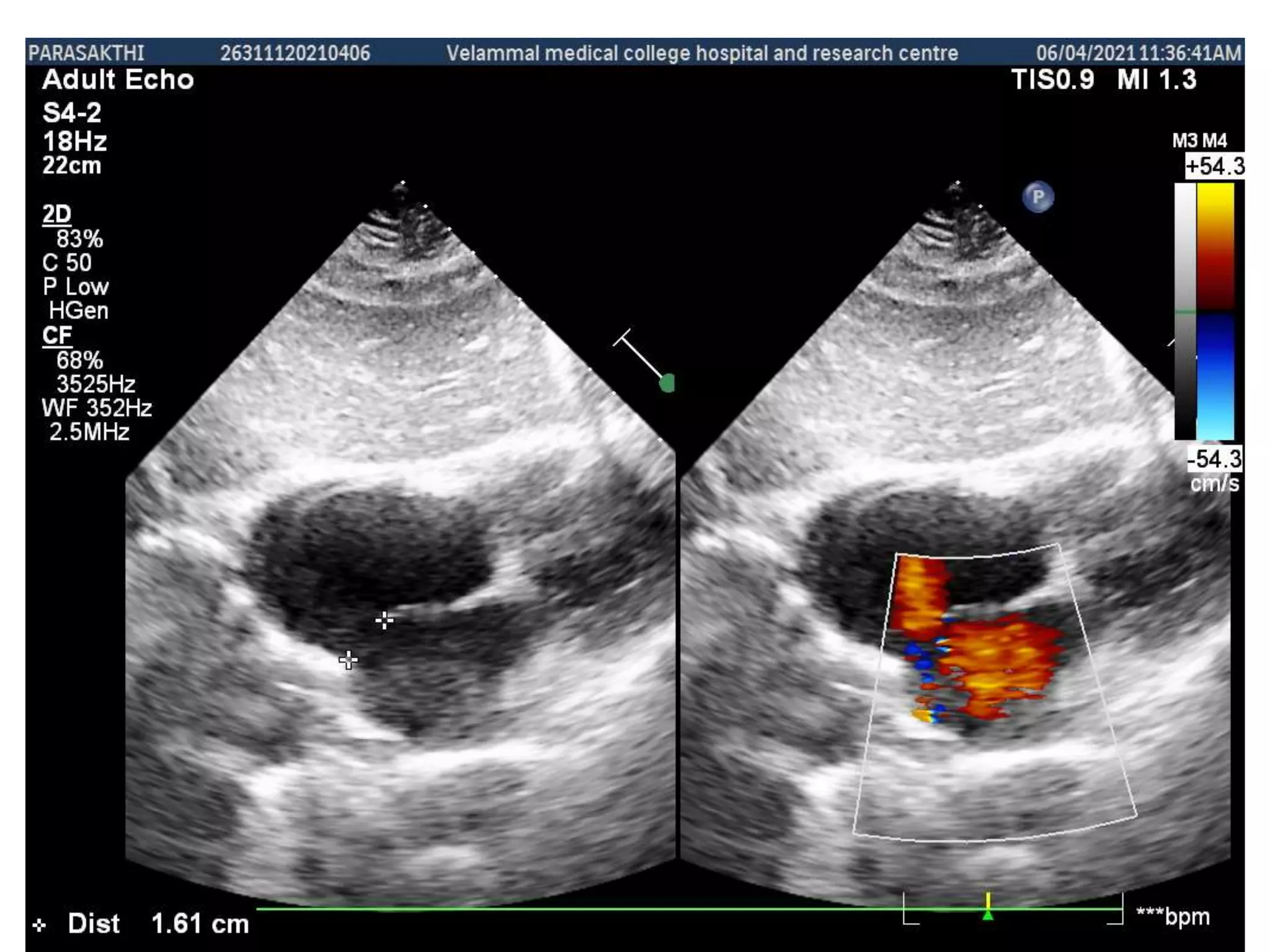
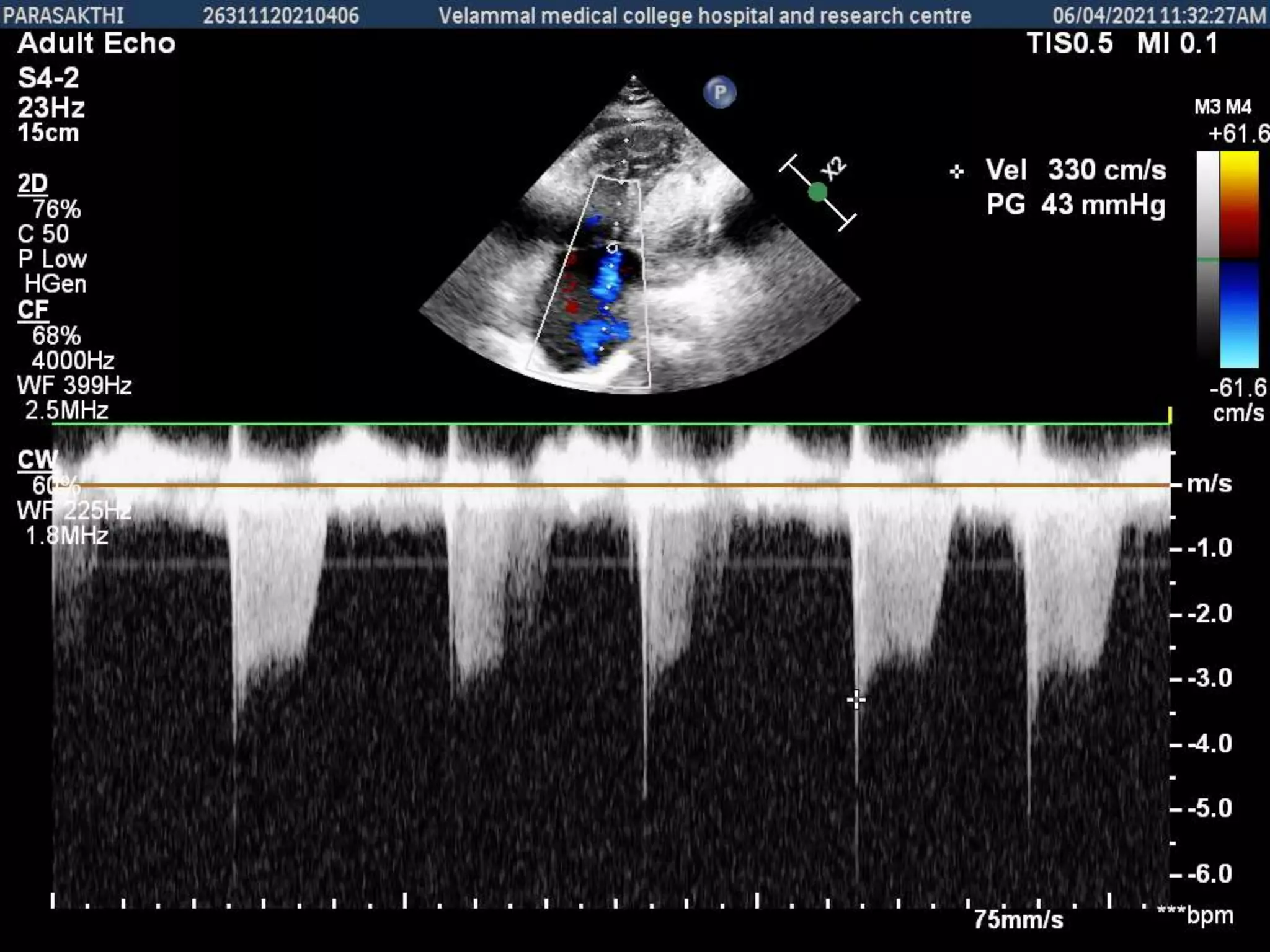
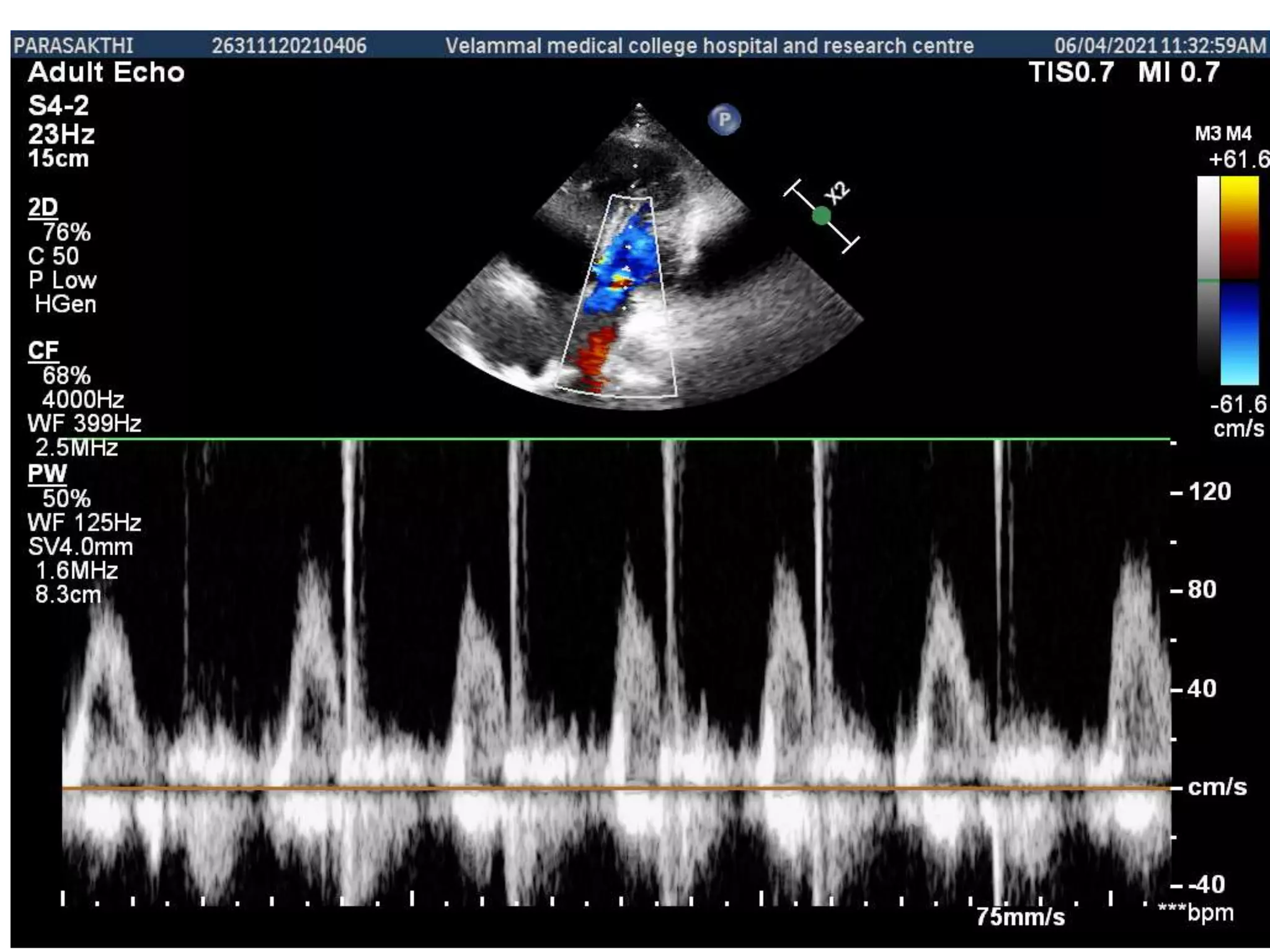
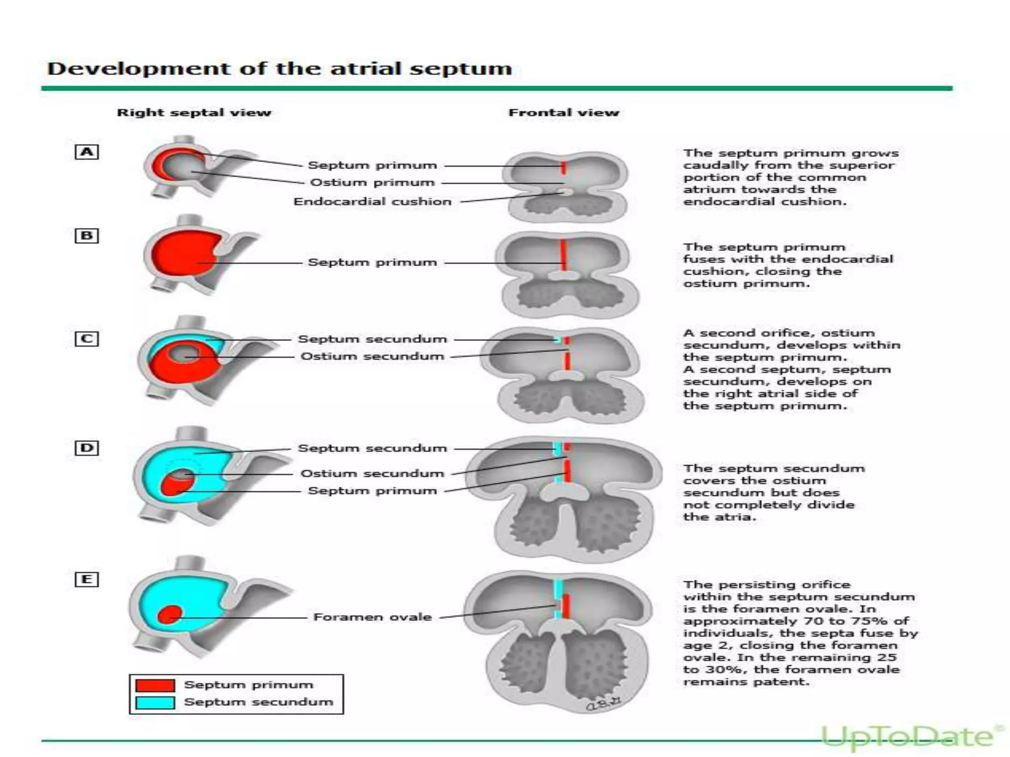
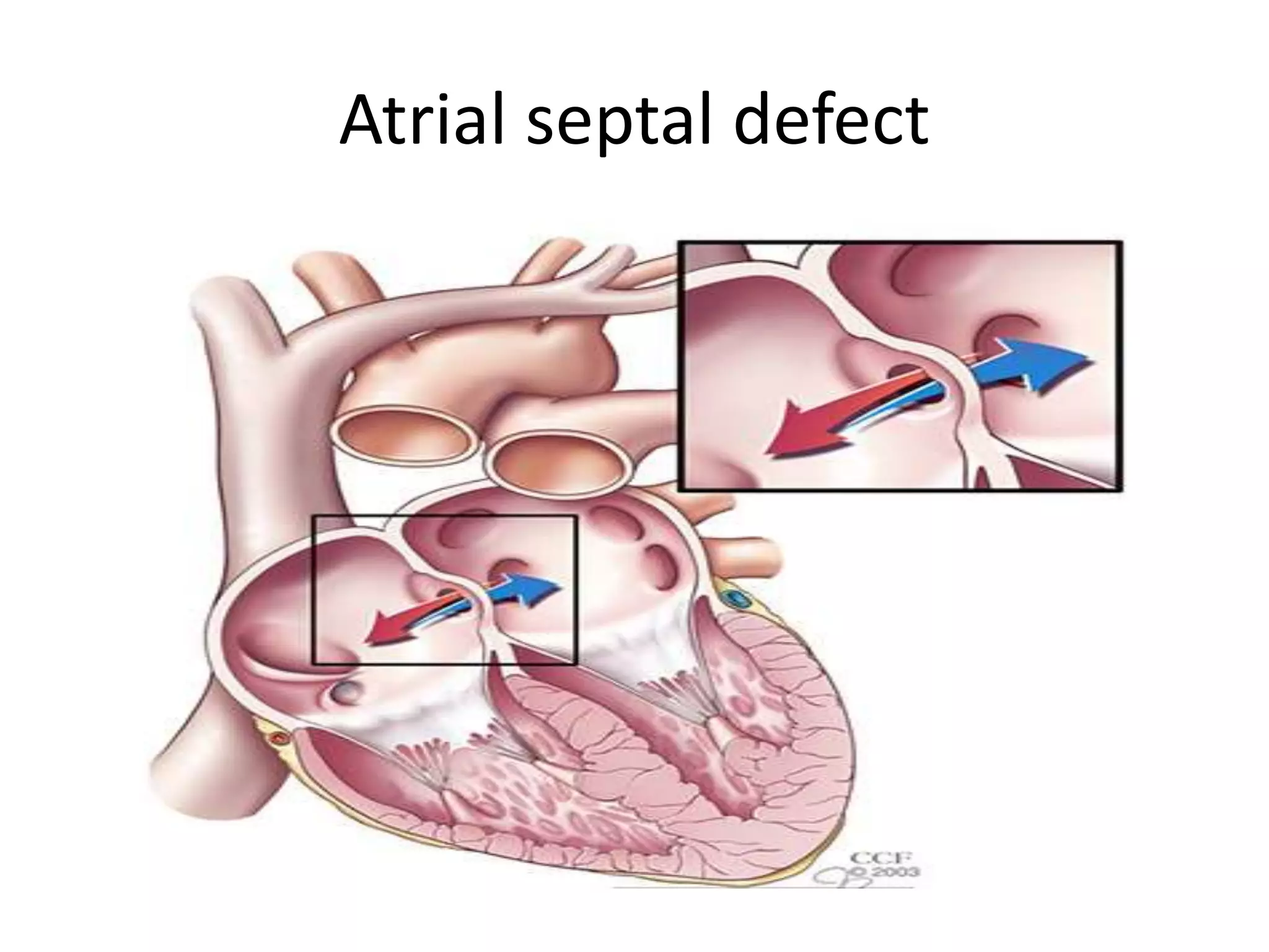
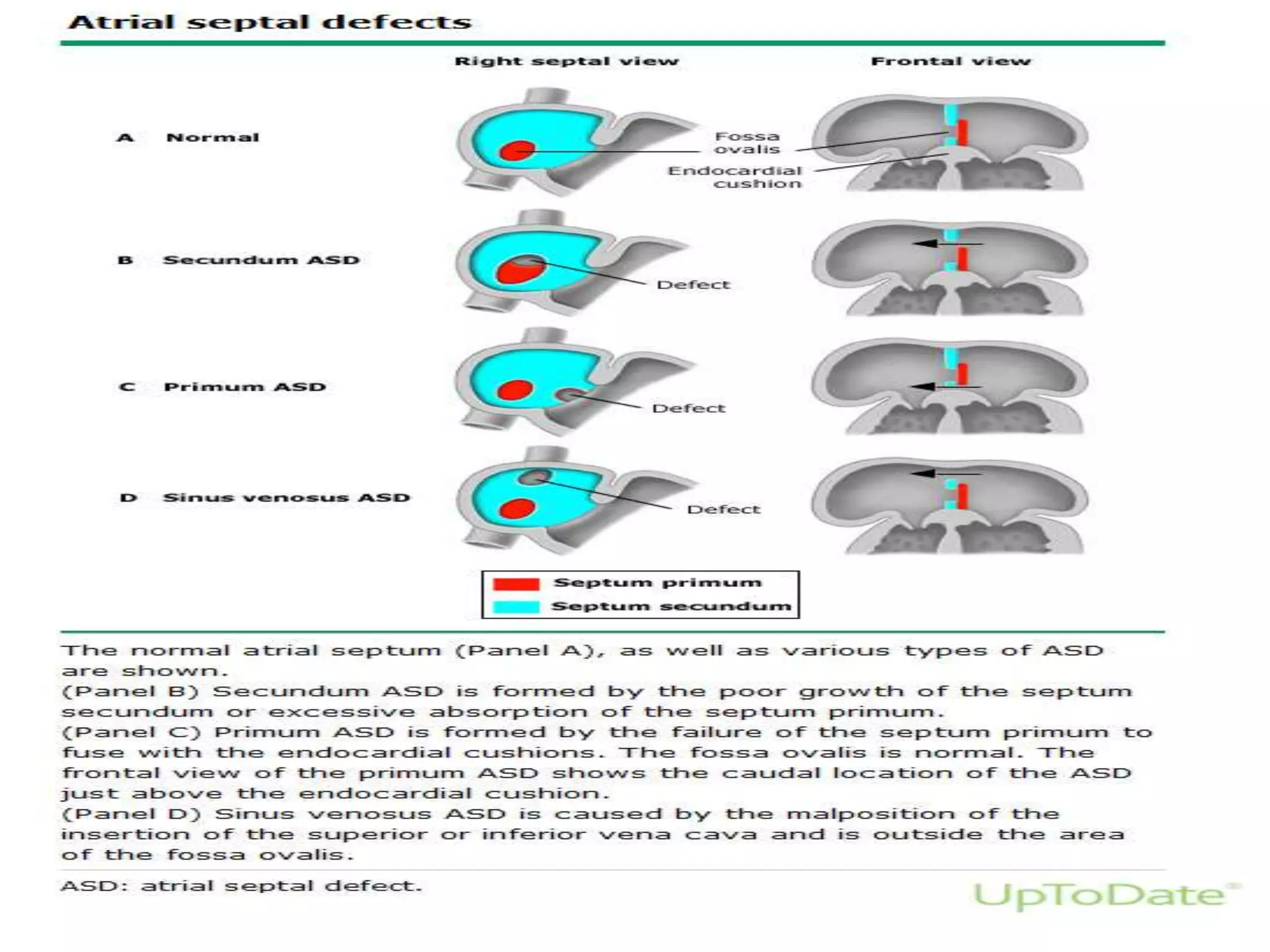
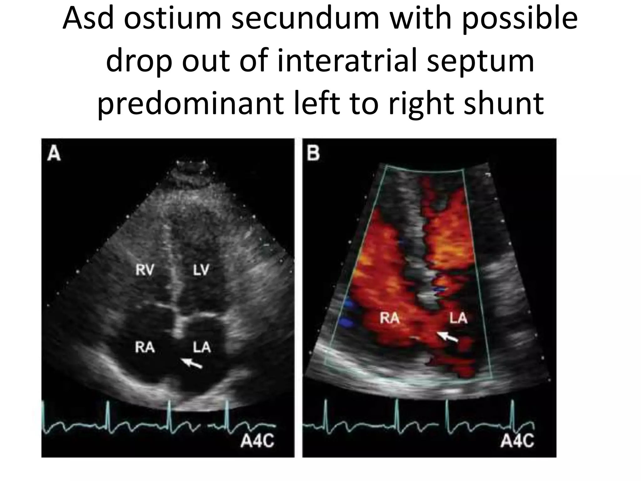
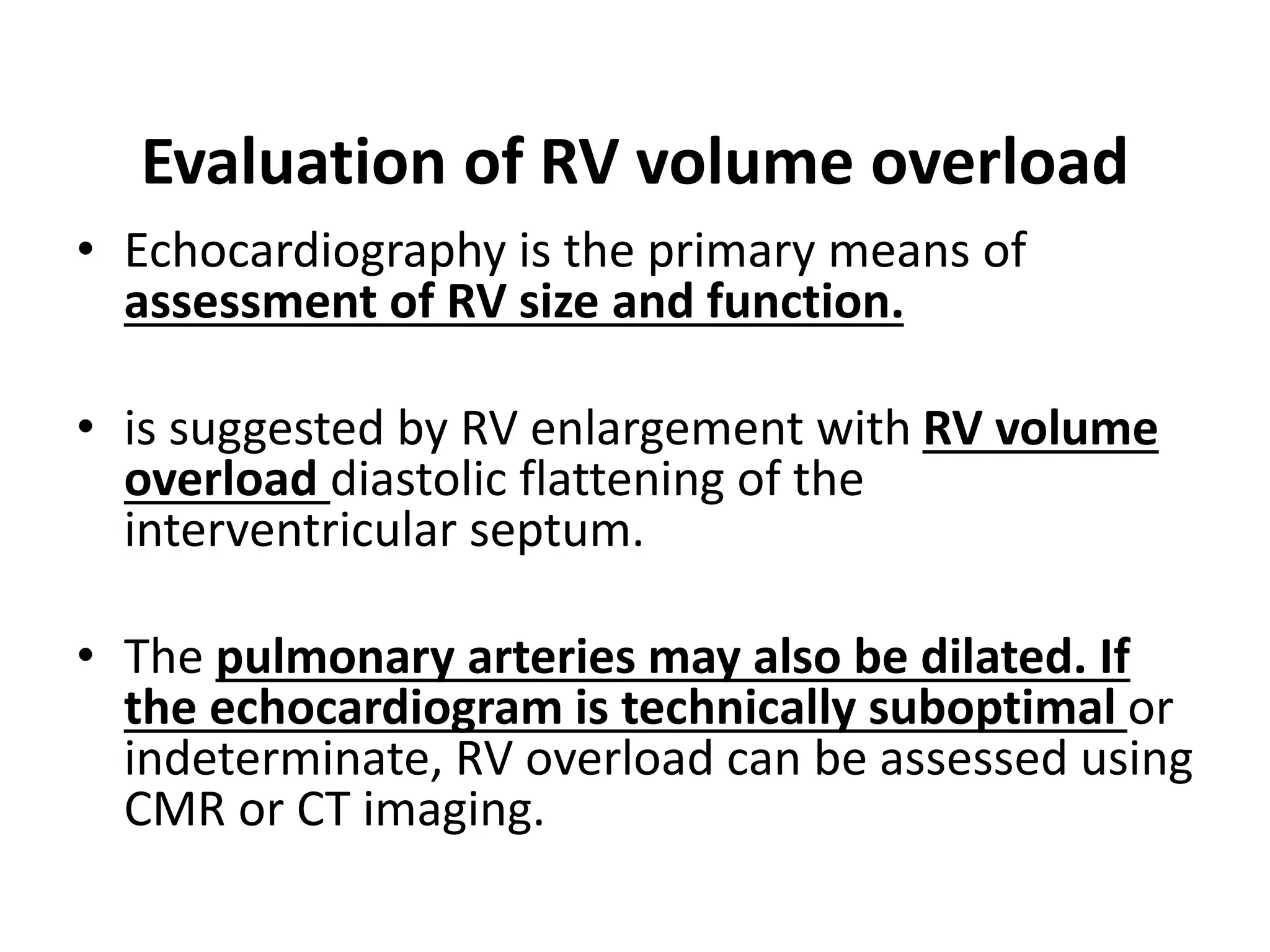
SlideShare a Scribd company logo
1 of 77
A 45 year old woman Parasakthi
came with complaints of shortness
of breath which increased while
climbing stairs and on exertion.
There was no associated clubbing or
cyanosis. The following are her
echocardiographic images. What is
your probable diagnosis ??
Atrial septal defect
Dr.S.R.Sruthi
Meenaxshi,MBBS,MD,PDF
• Atrial septal defect (ASD) is the most common
congenital heart lesion in adults and is often
asymptomatic until adulthood.
• Diagnosis is important, as timely ASD repair
improves outcomes
EMBRYOLOGY AND CLASSIFICATION
• ASDs result from lack of sufficient tissue to
completely septate the atria and are classified
according to their location in the atrial
septum.
• The location of the defect in relation to
adjacent cardiac structures defines the
anomalies associated with the ASD and
impacts the natural history and requirements
for repair.
Atrial septal defect
Types of atrial septal defect
Asd ostium secundum with possible
drop out of interatrial septum
predominant left to right shunt
• The location of the defect in relation to
adjacent cardiac structures defines the
anomalies associated with the ASD and
impacts the natural history and requirements
for repair.
• Atrial septation begins as early as the fifth
week of gestation.
• The septum primum arises from the superior
portion of the common atrium and grows
caudally to the endocardial cushions located
between the atria and ventricles, eventually
closing the orifice (ostium primum) between
the atria
• A second orifice (the ostium secundum) develops in
the septum primum; this orifice is covered by another
septum (the septum secundum) that arises on the right
atrial side of the septum primum.
• The septum secundum grows caudally and covers the
ostium secundum.
• However, the septum secundum does not completely
divide the atria, but leaves an oval orifice (the foramen
ovale) that is covered but not sealed on the left side by
the flexible flap of the septum primum
• Flow through the foramen ovale is essential
for fetal circulation.
• The foramen ovale closes spontaneously
within the first two years of life in 70 percent
of children.
• However, in a significant proportion (20 to 30
percent) of the population, the septea do not
fuse, leading to a patent foramen ovale
Secundum ASD
• Secundum ASD accounts for 70 to 75 percent of all
ASDs.
• Secundum ASD is a defect in the septum primum
resulting from poor growth of the secundum septum or
excessive absorption of the septum
• Although most secundum ASDs are isolated defects,
familial forms exist, some of which are associated with
other congenital cardiac and extracardiac
abnormalities.
• Other genes linked to familial isolated secundum ASD
include GATA 4,MYH6, NKX2-5. These syndromes
typically present in childhood or adolescence
The genetic disorder associated with secundum ASD is the HOLT
ORAM SYNDROME (also known as heart-hand syndrome) which
is caused by various mutations, most commonly mutations in
the TBX5 gene
Holt oram syndrome
ASD with rheumatic mitral stenosis –
LUTEMBACHER SYNDROME
• Secundum ASDs are occasionally associated
with partial anomalous pulmonary venous
connection and/or pulmonary stenosis.
• The rare combination of an ASD with
rheumatic mitral stenosis is known as
Lutembacher syndrome.
Primum ASD
• Primum ASD accounts for 15 to 20 percent of ASDs.
• A primum ASD is a defect in the septum secundum
caused by failure of the primum septum to fuse with
the endocardial cushions at the base of the interatrial
septum
• This results from maldevelopment/malalignment of
the ventricular septum due to malformation of the
endocardial cushions rather than a decrease in atrial
septal tissue
• Primum ASDs are nearly always associated with
anomalies of the atrioventricular (AV) valves,
particularly a cleft in the anterior mitral valve leaflet,
with or without a contiguous defect in the inlet
ventricular septum.
• When the combination of the primum ASD, cleft
mitral valve, and an inlet ventricular septal defect are
seen, this is called a partial AV septal defect (AVSD).
• The most severe form of AVSD (or endocardial cushion
defect) is the complete AV septal (or canal) defect, in
which a primum ASD and inlet ventricular septal defect
are present along with a common AV valve
Complete av canal defect
Av canal defect
• Discrete subaortic stenosis as well as
elongation (often referred to as a "goose-neck
deformity") of the left ventricular outflow
tract are often seen in association with
endocardial cushion defects.
• Endocardial cushion defects are often noted in
patients with Trisomy 21 down syndrome
Sinus venosus defect
• Sinus venosus defects account for 5 to 10 percent of ASDs and are located
in the venoatrial portion of the atrial septum.
• Sinus venosus defects represent an abnormality in the insertion of the
superior or inferior vena cava, which overrides the interatrial septum;
the interatrial communication is then formed within the mouth of the
overriding vein and is outside the area of the fossa ovalis .
• Thus, sinus venosus defects are technically not ASDs since the defect is
within the sinus venosus septum.
• An anomalous connection involving one or more pulmonary veins is
present in most patients with sinus venosus ASD
• Sinus venosus defects are of two types
• Superior sinus venosus defects are located
immediately below the orifice of the superior
vena cava.
• The right upper lobe and middle lobe
pulmonary veins often connect to the junction
of the superior vena cava and right atrium or
on the superior vena cava, resulting in a
partial anomalous pulmonary venous
connection
Inferior sinus venosus defects
• Inferior sinus venosus defects, also known as
inferior vena caval defects, are much less
common.
• They are located immediately above the
orifice of the inferior vena cava.
• These defects are also often associated with
partial anomalous connection of the right
pulmonary veins to the junction of the right
atrium and inferior vena cava.
Unroofed coronary sinus
• Unroofed coronary sinus (also known as
coronary sinus defect) is caused by absence of
part or all of the common wall between the
coronary sinus and the left atrium.
• This defect accounts for less than 1 percent of
ASDs and is commonly associated with a
persistent left superior vena cava.
PATHOPHYSIOLOGY
• ASDs in adults are associated with left-to-right shunt causing
volume overload of the right heart chambers
• The severity of the shunt is determined by the size of the defect
and atrial and ventricular compliance and pressure.
• The left-to-right shunting occurs primarily in late ventricular
systole and early diastole, with some augmentation during atrial
systole.
• The shunt flow due to an ASD moves from the left to the right
atrium, right ventricle (RV), pulmonary circulation, back to the left
atrium, and through the defect back to the right atrium.
• This leads to volume overload of the right heart chambers and
pulmonary arteries with possible late development of progressive
pulmonary vascular obstructive disease and pulmonary
hypertension when the degree of shunting is substantial and, more
commonly, in sinus venosus defects and primum ASDs.
• In addition, there is transient right-to-left
shunting at the onset of ventricular contraction,
particularly under conditions of bradycardia
and/or decreased intrathoracic pressure
• This explains the possibility of paradoxical
embolism in the setting of ASD.
• Significant right-to-left shunting can develop
later in life if severe pulmonary hypertension or
tricuspid regurgitation develops.
• Left atrial enlargement is also seen in adults
with ASDs, particularly
• in patients older than 50 years
• with atrial fibrillation
• with diastolic dysfunction with elevated left
heart filling pressure
• with a primum defect associated with cleft
mitral valve and mitral regurgitation.
NATURAL HISTORY
• The natural course of isolated ASDs varies from
spontaneous closure in secundum ASDs to
asymptomatic right ventricular enlargement and to
increasing symptoms with age.
• Spontaneous closure of ASDs, noted in approximately
40 percent of secundum ASDs, mostly occurs when
ASDs are small, usually less than 8 mm in diameter,
and in childhood. Secundum ASDs ≥8 mm in diameter
and those in adults do not typically close
spontaneously.
• Primum ASDs, sinus venosus defects, and coronary
sinus defects do not close spontaneously.
• Electrocardiogram — An electrocardiogram
(ECG) is routinely performed in patients with a
suspected ASD. The ECG may be normal with
an uncomplicated small ASD.
• Most individuals with an ASD have normal
sinus rhythm, but atrial arrhythmias often
occur in adults.
• The frontal plane QRS axis often ranges from
+95 to +135° (right axis deviation) with a
clockwise loop.
• A northwest (right superior) QRS axis (an axis
from -90 to ±180°) usually suggests the
presence of an AV canal defect.
• P waves are typically normal with secundum
ASDs.
• In comparison, sinus venosus defects are
often associated with a leftward frontal plane
P-wave axis (ie, negative in leads III and aVF
and positive in lead aVL)
• This leftward shift is caused by an ectopic
pacemaker resulting from an ASD located near
the sinus node.
• First-degree AV block can occur in any type of
ASD but is classically present in ostium primum
defects in association with complete right
bundle branch block and left anterior fascicular
block.
• The rim of the ostium primum defect is in close
spatial relationship to the His bundle, accounting
for abnormalities of impulse conduction through
this area.
• The QRS complex is often slightly prolonged and has a
characteristic rSr' or rsR' pattern that is thought to
result from disproportionate thickening of the right
ventricular outflow tract, which is the last portion of
the ventricle to depolarize.
• Patients with increasing severity of pulmonary
hypertension tend to lose the rSr' pattern in V1 and
develop a tall monophasic R wave with a deeply
inverted T wave as right ventricular hypertrophy
develops.
• A notch on the R wave in the inferior leads (a
pattern called "crochetage") has also been
suggested as a sensitive and specific ECG sign
of secundum ASD
notched r waves in inferior leads
echocardiography
• TTE with Doppler is generally the initial test
for diagnosis and evaluation of ASDs, as it
identifies most secundum and primum ASDs
• may also identify unroofed coronary sinus
and
• some variants of partial anomalous
pulmonary venous connection (PAPVC)
contrast echocardiography –
• If comprehensive TTE is not conclusive for ASD,
echocardiography with agitated saline contrast
with maneuvers (Valsalva and cough) may be
helpful to identify an intracardiac shunt
• Agitated saline contrast in a left upper extremity
vein is also helpful for identifying persistent left
superior vena cava (which commonly
accompanies unroofed coronary sinus.
• Transesophageal echocardiography (TEE) is suggested if TTE is technically
suboptimal or fails to show an ASD in a patient with suspected ASD.
• TEE is more sensitive than TTE in detection of ASDs, enables diagnosis of
sinus venosus defects (of superior vena cava or inferior vena type), and
aids in the sizing of secundum ASDs (as well as determination of
suitability for transcatheter device closure)
• TEE is also helpful in identification of the most common forms of PAPVC.
• The TEE procedure should be performed by an experienced examiner or in
conjunction with a congenital heart specialist since identification of the
ASD and anomalous pulmonary veins can be challenging.
Evaluation of RV volume overload
• Echocardiography is the primary means of
assessment of RV size and function.
• is suggested by RV enlargement with RV volume
overload diastolic flattening of the
interventricular septum.
• The pulmonary arteries may also be dilated. If
the echocardiogram is technically suboptimal or
indeterminate, RV overload can be assessed using
CMR or CT imaging.
Evaluation of pulmonary artery
pressure
• Pulmonary pressures should be assessed in all patients with
ASDs.
• RV and pulmonary artery systolic pressures are estimated
using Doppler echocardiography by obtaining the peak
tricuspid regurgitation continuous-wave Doppler signal,
using the modified Bernoulli equation, and adding the
estimated right atrial pressure
• RV volume overload is suggested by diastolic flattening of
the interventricular septum
• RV pressure overload is suggested by systolic (or systolic
and diastolic) flattening of the interventricular septum
• Estimation of Qp:Qs - Estimation of the
pulmonary blood flow to systemic blood flow
(Qp:Qs) ratio is helpful in cases in which the
cause of right atrial and RV chamber enlargement
is uncertain and in cases in which the degree of
shunt may help determine whether intervention
would be beneficial.
• •The Qp:Qs ratio can generally be estimated
noninvasively using CMR imaging; thus, cardiac
catheterization is generally not required to
determine the shunt flow ratio
Etimation of systemic flow across the
lvot
Pulmonary flow across the rvot
Color flow showing interartrial septal
defect
• The Qp:Qs estimated by Doppler
echocardiography has also been described,
but this technique has limited reliability.
• •When required, a formal "shunt run" at
cardiac catheterization measures the oxygen
content in the blood at multiple sites, and the
Fick equation is then used to calculate the
Qp:Qs ratio
Echocardiography
• Echocardiography is the imaging modality of
choice for the diagnosis of ASDs, as it generally
identifies and characterizes the ASD as well as
associated abnormalities and complications.
• Since the sensitivity of echocardiography varies
with technology, acoustic windows, and
operator/patient factors, negative suboptimal or
noncomprehensive echocardiograms do not
exclude an ASD.
ASD and associated flow
• TTE is usually diagnostic for secundum and primum ASDs when a
complete examination (including multiple precordial windows) is
performed by a trained sonographer/imager, unless the shunt is
very small or images are technically suboptimal.
• Clues to the presence of a secundum or primum defect include
abrupt discontinuity or drop out of the interatrial septum.
• Hypermobility of the septum, particularly in association with an
abrupt discontinuity, is also suggestive of secundum defect.
• TTE may also detect unroofed coronary sinus defects.
• The interatrial septum is often best visualized in
the subcostal view in which the ultrasound beam
is generally nearly perpendicular to the atrial
septum
• However, this view is suboptimal in some
individuals, particularly in obese subjects.
• Off-axis apical, parasternal short axis, and other
nonstandard views are frequently necessary to
interrogate the whole interatrial septum.
• Contrast echocardiography using peripheral
vein injection of agitated saline, when
performed, identified all defects missed by
two-dimensional TTE.
• The interatrial septum may also be visualized
in the apical four-chamber view, but this view
should generally not be relied upon given the
risk of artifactual echo dropout (low signal) as
the ultrasound beam may be parallel to the
atrial septum in this view.
• The size of an ASD on two-dimensional TTE does not
correlate well with shunt flow measured at catheterization.
ASD size is better assessed with color flow and pulsed-
wave Doppler or three-dimensional TTE or TEE imaging.
• The addition of color flow Doppler imaging can help
identify or confirm the presence of an ASD and indicate the
overall direction of the flow across the atrial septum
• Reducing the Nyquist limit (the upper limit of velocity that
can be detected with a given Doppler pulse frequency) may
enable detection of the turbulent shunt flow.
• There are several limitations to color flow
Doppler echocardiography in the diagnosis of
ASD:
• ●Ghosting of color across the interatrial septum
sometimes gives the false impression of shunt
flow (in particular with apical imaging).
• ●An ASD can be missed by TTE when there is
associated severe pulmonary hypertension as the
latter reduces the shunt flow across the ASD.
• In this setting, agitated saline injection or TEE
should be considered.
Associated findings
Since initial imaging of the interatrial septum may
be inconclusive, other evidence of ASD such as right
atrial and ventricular enlargement due to volume
overload (ie, diastolic interventricular septal
flattening and increased pulmonary artery
velocities without anatomic stenosis) and
pulmonary artery dilatation should be sought.
Continuous-wave Doppler echocardiography is
used to estimate the RV and (thus indirectly)
pulmonary artery systolic pressures.
Associated findings
• in addition, associated congenital lesions should be sought:
• ●Primum ASDs are generally accompanied by cleft
anterior mitral valve leaflet and, less commonly, tricuspid
valve, ventricular septal, and left ventricular outflow tract
abnormalities.
• ●Unroofed coronary sinus is commonly accompanied by
persistent left superior vena cava.
• ●Partial anomalous pulmonary venous drainage (primarily
of the right upper and middle pulmonary veins) frequently
accompanies superior sinus venosus defects and less
frequently occurs with secundum ASDs
• •PAPVC with right-sided pulmonary veins
connecting into the inferior vena cava
(Scimitar) or left-sided pulmonary veins
connecting into the innominate vein (the most
common type) are better seen by TTE than
TEE
• •TEE is more helpful in identifying PAPVC to
the superior vena cava.
Agitated saline contrast
• If comprehensive TTE with two-dimensional and color Doppler
imaging is not conclusive for ASD, imaging following
agitated saline contrast injection in a peripheral vein at rest and
with one or more maneuvers (Valsalva or cough) is helpful to
confirm the diagnosis
• Agitated saline contrast can be performed with TTE or TEE imaging
(movie 3).
• Contrast injection can be via any peripheral vein, but a left upper
extremity injection site is required to assess persistent left superior
vena cava (commonly associated with unroofed coronary sinus).
• The presence of unroofed coronary sinus with persistent left
superior vena cava is identified after intravenous
agitated saline injection in the left arm by detection of contrast in
the left atrium before or simultaneous to the right atrium.
Agitated saline contrast in asd
• An adequate contrast injection causes opacification of the right
atrium and ventricle. If there is a right-to-left interatrial shunt
(transient or net), early appearance of contrast can be seen in the
left atrium".)
• A right-to-left interatrial shunt can be detected by contrast
echocardiography in three circumstances:
• ●Patent foramen ovale, with no or net left-to-right shunt, with
transient elevation in right atrial pressure above left atrial pressure
(generally timed during the end of the T wave)
• ●With an uncomplicated ASD with net left-to-right shunt, when
flow is temporarily reversed with transient increases in right atrial
pressure relative to left atrial pressure (eg, with a Valsalva
maneuver or coughing) or briefly during the onset of left ventricular
contraction
• Agitated saline contrast echocardiography may also be
used in the detection of a left-to-right shunt.
• However, negative contrast in the right atrium is an
insensitive and nonspecific sign of left-to-right
interatrial shunting as flow from the contrast-free left
atrium produces areas in the right atrium in which
contrast is not seen.
• Care must be taken to distinguish this finding from lack
of contrast opacification of the right atrium due to
streaming of blood from vena cava inflow.
Stages of closure with amplatzer
Atrial septal defect Echocardiography

More Related Content

What's hot

Ventricular Septal defects Echocardiography
Ventricular Septal defects EchocardiographyVentricular Septal defects Echocardiography
Ventricular Septal defects EchocardiographySruthi Meenaxshi
 
Tte and tee assessment for asd closure 2
Tte and tee assessment for asd closure 2Tte and tee assessment for asd closure 2
Tte and tee assessment for asd closure 2Rahul Chalwade
 
Aortic stenosis - Echocardiography
Aortic stenosis - EchocardiographyAortic stenosis - Echocardiography
Aortic stenosis - EchocardiographyAnkur Gupta
 
ECHOCARDIOGRAPHIC EVALUATION OF AORTIC REGURGITATION
ECHOCARDIOGRAPHIC EVALUATION OF AORTIC REGURGITATIONECHOCARDIOGRAPHIC EVALUATION OF AORTIC REGURGITATION
ECHOCARDIOGRAPHIC EVALUATION OF AORTIC REGURGITATIONPraveen Nagula
 
Echocardiographic Evaluation of LV Diastolic Function
Echocardiographic Evaluation of LV Diastolic FunctionEchocardiographic Evaluation of LV Diastolic Function
Echocardiographic Evaluation of LV Diastolic FunctionJunhao Koh
 
Echo in restrictive cardiomyopathy
Echo in restrictive cardiomyopathyEcho in restrictive cardiomyopathy
Echo in restrictive cardiomyopathysruthiMeenaxshiSR
 
Echo Mitral Stenosis
Echo Mitral StenosisEcho Mitral Stenosis
Echo Mitral StenosisMashiul Alam
 
Aorto pulmonary window may 2021
Aorto   pulmonary window  may 2021Aorto   pulmonary window  may 2021
Aorto pulmonary window may 2021rajasthan govt
 
Percutaneous Pulmonary Valve Interventions
Percutaneous Pulmonary Valve InterventionsPercutaneous Pulmonary Valve Interventions
Percutaneous Pulmonary Valve InterventionsPraveen Nagula
 
Right heart catheters
Right heart cathetersRight heart catheters
Right heart cathetersRohitWalse2
 
Calculation of bidirectional shunts
Calculation of bidirectional shuntsCalculation of bidirectional shunts
Calculation of bidirectional shuntsRamachandra Barik
 
FFR(fractional flow reserve)
FFR(fractional flow reserve)FFR(fractional flow reserve)
FFR(fractional flow reserve)DIPAK PATADE
 
Echo assessment of cardiomyopathy pdf
Echo assessment of cardiomyopathy pdfEcho assessment of cardiomyopathy pdf
Echo assessment of cardiomyopathy pdfNizam Uddin
 
Echo assessment of aortic stenosis
Echo assessment of aortic stenosisEcho assessment of aortic stenosis
Echo assessment of aortic stenosisNizam Uddin
 

What's hot (20)

Echo assessment of RV function
Echo assessment of RV functionEcho assessment of RV function
Echo assessment of RV function
 
Ventricular Septal defects Echocardiography
Ventricular Septal defects EchocardiographyVentricular Septal defects Echocardiography
Ventricular Septal defects Echocardiography
 
Tte and tee assessment for asd closure 2
Tte and tee assessment for asd closure 2Tte and tee assessment for asd closure 2
Tte and tee assessment for asd closure 2
 
Aortic stenosis - Echocardiography
Aortic stenosis - EchocardiographyAortic stenosis - Echocardiography
Aortic stenosis - Echocardiography
 
ECHOCARDIOGRAPHIC EVALUATION OF AORTIC REGURGITATION
ECHOCARDIOGRAPHIC EVALUATION OF AORTIC REGURGITATIONECHOCARDIOGRAPHIC EVALUATION OF AORTIC REGURGITATION
ECHOCARDIOGRAPHIC EVALUATION OF AORTIC REGURGITATION
 
Echocardiographic Evaluation of LV Diastolic Function
Echocardiographic Evaluation of LV Diastolic FunctionEchocardiographic Evaluation of LV Diastolic Function
Echocardiographic Evaluation of LV Diastolic Function
 
Echo in restrictive cardiomyopathy
Echo in restrictive cardiomyopathyEcho in restrictive cardiomyopathy
Echo in restrictive cardiomyopathy
 
ECHO ASSESSMENT OF ASD FOR DEVICE CLOSURE
ECHO ASSESSMENT OF ASD FOR DEVICE CLOSURE ECHO ASSESSMENT OF ASD FOR DEVICE CLOSURE
ECHO ASSESSMENT OF ASD FOR DEVICE CLOSURE
 
Echo Mitral Stenosis
Echo Mitral StenosisEcho Mitral Stenosis
Echo Mitral Stenosis
 
Aorto pulmonary window may 2021
Aorto   pulmonary window  may 2021Aorto   pulmonary window  may 2021
Aorto pulmonary window may 2021
 
SEGMENTAL ANALYSIS OF CONGENITAL HEART DISEASE
SEGMENTAL ANALYSIS OF CONGENITAL HEART DISEASE SEGMENTAL ANALYSIS OF CONGENITAL HEART DISEASE
SEGMENTAL ANALYSIS OF CONGENITAL HEART DISEASE
 
Percutaneous Pulmonary Valve Interventions
Percutaneous Pulmonary Valve InterventionsPercutaneous Pulmonary Valve Interventions
Percutaneous Pulmonary Valve Interventions
 
Right heart catheters
Right heart cathetersRight heart catheters
Right heart catheters
 
Calculation of bidirectional shunts
Calculation of bidirectional shuntsCalculation of bidirectional shunts
Calculation of bidirectional shunts
 
Lv systolic function
Lv systolic functionLv systolic function
Lv systolic function
 
L-TGA or CCTGA
L-TGA or CCTGA L-TGA or CCTGA
L-TGA or CCTGA
 
FRACTIONAL FLOW RESERVE
FRACTIONAL FLOW RESERVEFRACTIONAL FLOW RESERVE
FRACTIONAL FLOW RESERVE
 
FFR(fractional flow reserve)
FFR(fractional flow reserve)FFR(fractional flow reserve)
FFR(fractional flow reserve)
 
Echo assessment of cardiomyopathy pdf
Echo assessment of cardiomyopathy pdfEcho assessment of cardiomyopathy pdf
Echo assessment of cardiomyopathy pdf
 
Echo assessment of aortic stenosis
Echo assessment of aortic stenosisEcho assessment of aortic stenosis
Echo assessment of aortic stenosis
 

Similar to Atrial septal defect Echocardiography

Atrial Septal Defects.pptx
Atrial Septal Defects.pptxAtrial Septal Defects.pptx
Atrial Septal Defects.pptxVannalaRaju2
 
Atrial septal defects 16 3-15
Atrial septal defects 16 3-15Atrial septal defects 16 3-15
Atrial septal defects 16 3-15Dr. Harshil Joshi
 
Atrial septal defect DR NIKUNJ R SHEKHADA (MBBS,MS GEN SURG,DNB CTS SR
Atrial septal defect DR NIKUNJ R SHEKHADA (MBBS,MS GEN SURG,DNB CTS SRAtrial septal defect DR NIKUNJ R SHEKHADA (MBBS,MS GEN SURG,DNB CTS SR
Atrial septal defect DR NIKUNJ R SHEKHADA (MBBS,MS GEN SURG,DNB CTS SRDR NIKUNJ SHEKHADA
 
Av canal defect
Av canal defectAv canal defect
Av canal defectdrsrb
 
Atrioventricular septal defects
Atrioventricular septal defectsAtrioventricular septal defects
Atrioventricular septal defectsIndia CTVS
 
Congenital heart diseases (acyanotic)
Congenital heart diseases (acyanotic)Congenital heart diseases (acyanotic)
Congenital heart diseases (acyanotic)Ashish Mankar
 
Atrioventricular canal defect, Firas Aljanadi,MD
Atrioventricular canal defect, Firas Aljanadi,MDAtrioventricular canal defect, Firas Aljanadi,MD
Atrioventricular canal defect, Firas Aljanadi,MDFIRAS ALJANADI
 
ASD by Dr Kuntal Surana
ASD by Dr Kuntal SuranaASD by Dr Kuntal Surana
ASD by Dr Kuntal SuranaKuntal Surana
 
cyanotic and acyanotic Congenital heart disease for undergraduated student uo...
cyanotic and acyanotic Congenital heart disease for undergraduated student uo...cyanotic and acyanotic Congenital heart disease for undergraduated student uo...
cyanotic and acyanotic Congenital heart disease for undergraduated student uo...Azad Haleem
 
Electrocardiography in Adult Congenital Heart Diseases
Electrocardiography in Adult Congenital Heart DiseasesElectrocardiography in Adult Congenital Heart Diseases
Electrocardiography in Adult Congenital Heart DiseasesSaleh AL-Hatem
 
Atrioventricular canal defect
Atrioventricular canal defectAtrioventricular canal defect
Atrioventricular canal defectDrvasanthi
 

Similar to Atrial septal defect Echocardiography (20)

Atrial septal defect
Atrial septal defect Atrial septal defect
Atrial septal defect
 
Atrial Septal Defects.pptx
Atrial Septal Defects.pptxAtrial Septal Defects.pptx
Atrial Septal Defects.pptx
 
Atrial Septal Defect
Atrial Septal DefectAtrial Septal Defect
Atrial Septal Defect
 
atio ventricular septal defects
atio ventricular septal defectsatio ventricular septal defects
atio ventricular septal defects
 
Atrial septal defects 16 3-15
Atrial septal defects 16 3-15Atrial septal defects 16 3-15
Atrial septal defects 16 3-15
 
Asd new
Asd newAsd new
Asd new
 
Atrial septal defect DR NIKUNJ R SHEKHADA (MBBS,MS GEN SURG,DNB CTS SR
Atrial septal defect DR NIKUNJ R SHEKHADA (MBBS,MS GEN SURG,DNB CTS SRAtrial septal defect DR NIKUNJ R SHEKHADA (MBBS,MS GEN SURG,DNB CTS SR
Atrial septal defect DR NIKUNJ R SHEKHADA (MBBS,MS GEN SURG,DNB CTS SR
 
Av canal defect
Av canal defectAv canal defect
Av canal defect
 
An approach to a patient with Atrial septal defect (ASD)
An approach to a patient with Atrial  septal defect (ASD)An approach to a patient with Atrial  septal defect (ASD)
An approach to a patient with Atrial septal defect (ASD)
 
Cogenital heart ds.
Cogenital heart ds.Cogenital heart ds.
Cogenital heart ds.
 
CHD.pptx
CHD.pptxCHD.pptx
CHD.pptx
 
Atrioventricular septal defects
Atrioventricular septal defectsAtrioventricular septal defects
Atrioventricular septal defects
 
Atrial septal defect
Atrial septal defectAtrial septal defect
Atrial septal defect
 
Congenital heart diseases (acyanotic)
Congenital heart diseases (acyanotic)Congenital heart diseases (acyanotic)
Congenital heart diseases (acyanotic)
 
Atrioventricular canal defect, Firas Aljanadi,MD
Atrioventricular canal defect, Firas Aljanadi,MDAtrioventricular canal defect, Firas Aljanadi,MD
Atrioventricular canal defect, Firas Aljanadi,MD
 
ASD by Dr Kuntal Surana
ASD by Dr Kuntal SuranaASD by Dr Kuntal Surana
ASD by Dr Kuntal Surana
 
Cc tga
Cc tgaCc tga
Cc tga
 
cyanotic and acyanotic Congenital heart disease for undergraduated student uo...
cyanotic and acyanotic Congenital heart disease for undergraduated student uo...cyanotic and acyanotic Congenital heart disease for undergraduated student uo...
cyanotic and acyanotic Congenital heart disease for undergraduated student uo...
 
Electrocardiography in Adult Congenital Heart Diseases
Electrocardiography in Adult Congenital Heart DiseasesElectrocardiography in Adult Congenital Heart Diseases
Electrocardiography in Adult Congenital Heart Diseases
 
Atrioventricular canal defect
Atrioventricular canal defectAtrioventricular canal defect
Atrioventricular canal defect
 

More from Sruthi Meenaxshi

MEN SYNDROMESAND GENETIC BASIS.pptx
MEN SYNDROMESAND GENETIC BASIS.pptxMEN SYNDROMESAND GENETIC BASIS.pptx
MEN SYNDROMESAND GENETIC BASIS.pptxSruthi Meenaxshi
 
ADULT NEURODEGENERATIVE DISORDERS .pptx
ADULT NEURODEGENERATIVE DISORDERS .pptxADULT NEURODEGENERATIVE DISORDERS .pptx
ADULT NEURODEGENERATIVE DISORDERS .pptxSruthi Meenaxshi
 
DRUG INDUCED LIVER INJURY.pptx
DRUG INDUCED LIVER INJURY.pptxDRUG INDUCED LIVER INJURY.pptx
DRUG INDUCED LIVER INJURY.pptxSruthi Meenaxshi
 
acute hiv inffection and cdc criteria.pptx
acute hiv inffection and cdc criteria.pptxacute hiv inffection and cdc criteria.pptx
acute hiv inffection and cdc criteria.pptxSruthi Meenaxshi
 
head ache dizziness and sphincter disturbance s.pptx
head ache dizziness and sphincter disturbance s.pptxhead ache dizziness and sphincter disturbance s.pptx
head ache dizziness and sphincter disturbance s.pptxSruthi Meenaxshi
 
inflammatory bowel disease.pptx
inflammatory bowel disease.pptxinflammatory bowel disease.pptx
inflammatory bowel disease.pptxSruthi Meenaxshi
 
Arrythmogenic rv dysplasia (ARVD)
Arrythmogenic rv dysplasia (ARVD)Arrythmogenic rv dysplasia (ARVD)
Arrythmogenic rv dysplasia (ARVD)Sruthi Meenaxshi
 
Left ventricular noncompaction
Left ventricular noncompactionLeft ventricular noncompaction
Left ventricular noncompactionSruthi Meenaxshi
 
Vector borne diseases recent concepts in management and elimination targets...
Vector borne diseases   recent concepts in management and elimination targets...Vector borne diseases   recent concepts in management and elimination targets...
Vector borne diseases recent concepts in management and elimination targets...Sruthi Meenaxshi
 
Echo in prosthetic valve evaluation
Echo in prosthetic valve evaluationEcho in prosthetic valve evaluation
Echo in prosthetic valve evaluationSruthi Meenaxshi
 
Infective endocarditis Echocardiography
Infective endocarditis EchocardiographyInfective endocarditis Echocardiography
Infective endocarditis EchocardiographySruthi Meenaxshi
 
Conduction abnormalities part 2
Conduction abnormalities part 2Conduction abnormalities part 2
Conduction abnormalities part 2Sruthi Meenaxshi
 
Conduction abnormalities part I
Conduction abnormalities part IConduction abnormalities part I
Conduction abnormalities part ISruthi Meenaxshi
 

More from Sruthi Meenaxshi (20)

MEN SYNDROMESAND GENETIC BASIS.pptx
MEN SYNDROMESAND GENETIC BASIS.pptxMEN SYNDROMESAND GENETIC BASIS.pptx
MEN SYNDROMESAND GENETIC BASIS.pptx
 
ADULT NEURODEGENERATIVE DISORDERS .pptx
ADULT NEURODEGENERATIVE DISORDERS .pptxADULT NEURODEGENERATIVE DISORDERS .pptx
ADULT NEURODEGENERATIVE DISORDERS .pptx
 
lipid guidelines.pptx
lipid guidelines.pptxlipid guidelines.pptx
lipid guidelines.pptx
 
DRUG INDUCED LIVER INJURY.pptx
DRUG INDUCED LIVER INJURY.pptxDRUG INDUCED LIVER INJURY.pptx
DRUG INDUCED LIVER INJURY.pptx
 
AORTIC STENOSIS.pptx
AORTIC STENOSIS.pptxAORTIC STENOSIS.pptx
AORTIC STENOSIS.pptx
 
drugoverdose.pptx
drugoverdose.pptxdrugoverdose.pptx
drugoverdose.pptx
 
scorpion envenomation.pptx
scorpion envenomation.pptxscorpion envenomation.pptx
scorpion envenomation.pptx
 
acute hiv inffection and cdc criteria.pptx
acute hiv inffection and cdc criteria.pptxacute hiv inffection and cdc criteria.pptx
acute hiv inffection and cdc criteria.pptx
 
head ache dizziness and sphincter disturbance s.pptx
head ache dizziness and sphincter disturbance s.pptxhead ache dizziness and sphincter disturbance s.pptx
head ache dizziness and sphincter disturbance s.pptx
 
inflammatory bowel disease.pptx
inflammatory bowel disease.pptxinflammatory bowel disease.pptx
inflammatory bowel disease.pptx
 
Ventricular arrythmia
Ventricular arrythmiaVentricular arrythmia
Ventricular arrythmia
 
Digoxin toxicity
Digoxin toxicityDigoxin toxicity
Digoxin toxicity
 
Arrythmogenic rv dysplasia (ARVD)
Arrythmogenic rv dysplasia (ARVD)Arrythmogenic rv dysplasia (ARVD)
Arrythmogenic rv dysplasia (ARVD)
 
Left ventricular noncompaction
Left ventricular noncompactionLeft ventricular noncompaction
Left ventricular noncompaction
 
Vector borne diseases recent concepts in management and elimination targets...
Vector borne diseases   recent concepts in management and elimination targets...Vector borne diseases   recent concepts in management and elimination targets...
Vector borne diseases recent concepts in management and elimination targets...
 
Asd device closure
Asd device closureAsd device closure
Asd device closure
 
Echo in prosthetic valve evaluation
Echo in prosthetic valve evaluationEcho in prosthetic valve evaluation
Echo in prosthetic valve evaluation
 
Infective endocarditis Echocardiography
Infective endocarditis EchocardiographyInfective endocarditis Echocardiography
Infective endocarditis Echocardiography
 
Conduction abnormalities part 2
Conduction abnormalities part 2Conduction abnormalities part 2
Conduction abnormalities part 2
 
Conduction abnormalities part I
Conduction abnormalities part IConduction abnormalities part I
Conduction abnormalities part I
 

Recently uploaded

College Call Girls Pune Mira 9907093804 Short 1500 Night 6000 Best call girls...
College Call Girls Pune Mira 9907093804 Short 1500 Night 6000 Best call girls...College Call Girls Pune Mira 9907093804 Short 1500 Night 6000 Best call girls...
College Call Girls Pune Mira 9907093804 Short 1500 Night 6000 Best call girls...Miss joya
 
CALL ON ➥9907093804 🔝 Call Girls Hadapsar ( Pune) Girls Service
CALL ON ➥9907093804 🔝 Call Girls Hadapsar ( Pune)  Girls ServiceCALL ON ➥9907093804 🔝 Call Girls Hadapsar ( Pune)  Girls Service
CALL ON ➥9907093804 🔝 Call Girls Hadapsar ( Pune) Girls ServiceMiss joya
 
Call Girls Service Pune Vaishnavi 9907093804 Short 1500 Night 6000 Best call ...
Call Girls Service Pune Vaishnavi 9907093804 Short 1500 Night 6000 Best call ...Call Girls Service Pune Vaishnavi 9907093804 Short 1500 Night 6000 Best call ...
Call Girls Service Pune Vaishnavi 9907093804 Short 1500 Night 6000 Best call ...Miss joya
 
Call Girls Service Navi Mumbai Samaira 8617697112 Independent Escort Service ...
Call Girls Service Navi Mumbai Samaira 8617697112 Independent Escort Service ...Call Girls Service Navi Mumbai Samaira 8617697112 Independent Escort Service ...
Call Girls Service Navi Mumbai Samaira 8617697112 Independent Escort Service ...Call girls in Ahmedabad High profile
 
Book Paid Powai Call Girls Mumbai 𖠋 9930245274 𖠋Low Budget Full Independent H...
Book Paid Powai Call Girls Mumbai 𖠋 9930245274 𖠋Low Budget Full Independent H...Book Paid Powai Call Girls Mumbai 𖠋 9930245274 𖠋Low Budget Full Independent H...
Book Paid Powai Call Girls Mumbai 𖠋 9930245274 𖠋Low Budget Full Independent H...Call Girls in Nagpur High Profile
 
Call Girl Number in Panvel Mumbai📲 9833363713 💞 Full Night Enjoy
Call Girl Number in Panvel Mumbai📲 9833363713 💞 Full Night EnjoyCall Girl Number in Panvel Mumbai📲 9833363713 💞 Full Night Enjoy
Call Girl Number in Panvel Mumbai📲 9833363713 💞 Full Night Enjoybabeytanya
 
Bangalore Call Girls Marathahalli 📞 9907093804 High Profile Service 100% Safe
Bangalore Call Girls Marathahalli 📞 9907093804 High Profile Service 100% SafeBangalore Call Girls Marathahalli 📞 9907093804 High Profile Service 100% Safe
Bangalore Call Girls Marathahalli 📞 9907093804 High Profile Service 100% Safenarwatsonia7
 
Call Girls Darjeeling Just Call 9907093804 Top Class Call Girl Service Available
Call Girls Darjeeling Just Call 9907093804 Top Class Call Girl Service AvailableCall Girls Darjeeling Just Call 9907093804 Top Class Call Girl Service Available
Call Girls Darjeeling Just Call 9907093804 Top Class Call Girl Service AvailableDipal Arora
 
VIP Call Girls Tirunelveli Aaradhya 8250192130 Independent Escort Service Tir...
VIP Call Girls Tirunelveli Aaradhya 8250192130 Independent Escort Service Tir...VIP Call Girls Tirunelveli Aaradhya 8250192130 Independent Escort Service Tir...
VIP Call Girls Tirunelveli Aaradhya 8250192130 Independent Escort Service Tir...narwatsonia7
 
Lucknow Call girls - 8800925952 - 24x7 service with hotel room
Lucknow Call girls - 8800925952 - 24x7 service with hotel roomLucknow Call girls - 8800925952 - 24x7 service with hotel room
Lucknow Call girls - 8800925952 - 24x7 service with hotel roomdiscovermytutordmt
 
(Rocky) Jaipur Call Girl - 9521753030 Escorts Service 50% Off with Cash ON De...
(Rocky) Jaipur Call Girl - 9521753030 Escorts Service 50% Off with Cash ON De...(Rocky) Jaipur Call Girl - 9521753030 Escorts Service 50% Off with Cash ON De...
(Rocky) Jaipur Call Girl - 9521753030 Escorts Service 50% Off with Cash ON De...indiancallgirl4rent
 
Artifacts in Nuclear Medicine with Identifying and resolving artifacts.
Artifacts in Nuclear Medicine with Identifying and resolving artifacts.Artifacts in Nuclear Medicine with Identifying and resolving artifacts.
Artifacts in Nuclear Medicine with Identifying and resolving artifacts.MiadAlsulami
 
Bangalore Call Girls Nelamangala Number 7001035870 Meetin With Bangalore Esc...
Bangalore Call Girls Nelamangala Number 7001035870  Meetin With Bangalore Esc...Bangalore Call Girls Nelamangala Number 7001035870  Meetin With Bangalore Esc...
Bangalore Call Girls Nelamangala Number 7001035870 Meetin With Bangalore Esc...narwatsonia7
 
Low Rate Call Girls Patna Anika 8250192130 Independent Escort Service Patna
Low Rate Call Girls Patna Anika 8250192130 Independent Escort Service PatnaLow Rate Call Girls Patna Anika 8250192130 Independent Escort Service Patna
Low Rate Call Girls Patna Anika 8250192130 Independent Escort Service Patnamakika9823
 
Russian Call Girls in Pune Tanvi 9907093804 Short 1500 Night 6000 Best call g...
Russian Call Girls in Pune Tanvi 9907093804 Short 1500 Night 6000 Best call g...Russian Call Girls in Pune Tanvi 9907093804 Short 1500 Night 6000 Best call g...
Russian Call Girls in Pune Tanvi 9907093804 Short 1500 Night 6000 Best call g...Miss joya
 
Bangalore Call Girls Hebbal Kempapura Number 7001035870 Meetin With Bangalor...
Bangalore Call Girls Hebbal Kempapura Number 7001035870  Meetin With Bangalor...Bangalore Call Girls Hebbal Kempapura Number 7001035870  Meetin With Bangalor...
Bangalore Call Girls Hebbal Kempapura Number 7001035870 Meetin With Bangalor...narwatsonia7
 
Call Girls Horamavu WhatsApp Number 7001035870 Meeting With Bangalore Escorts
Call Girls Horamavu WhatsApp Number 7001035870 Meeting With Bangalore EscortsCall Girls Horamavu WhatsApp Number 7001035870 Meeting With Bangalore Escorts
Call Girls Horamavu WhatsApp Number 7001035870 Meeting With Bangalore Escortsvidya singh
 
VIP Call Girls Pune Vani 9907093804 Short 1500 Night 6000 Best call girls Ser...
VIP Call Girls Pune Vani 9907093804 Short 1500 Night 6000 Best call girls Ser...VIP Call Girls Pune Vani 9907093804 Short 1500 Night 6000 Best call girls Ser...
VIP Call Girls Pune Vani 9907093804 Short 1500 Night 6000 Best call girls Ser...Miss joya
 
Call Girl Number in Vashi Mumbai📲 9833363713 💞 Full Night Enjoy
Call Girl Number in Vashi Mumbai📲 9833363713 💞 Full Night EnjoyCall Girl Number in Vashi Mumbai📲 9833363713 💞 Full Night Enjoy
Call Girl Number in Vashi Mumbai📲 9833363713 💞 Full Night Enjoybabeytanya
 
Call Girls Service Surat Samaira ❤️🍑 8250192130 👄 Independent Escort Service ...
Call Girls Service Surat Samaira ❤️🍑 8250192130 👄 Independent Escort Service ...Call Girls Service Surat Samaira ❤️🍑 8250192130 👄 Independent Escort Service ...
Call Girls Service Surat Samaira ❤️🍑 8250192130 👄 Independent Escort Service ...CALL GIRLS
 

Recently uploaded (20)

College Call Girls Pune Mira 9907093804 Short 1500 Night 6000 Best call girls...
College Call Girls Pune Mira 9907093804 Short 1500 Night 6000 Best call girls...College Call Girls Pune Mira 9907093804 Short 1500 Night 6000 Best call girls...
College Call Girls Pune Mira 9907093804 Short 1500 Night 6000 Best call girls...
 
CALL ON ➥9907093804 🔝 Call Girls Hadapsar ( Pune) Girls Service
CALL ON ➥9907093804 🔝 Call Girls Hadapsar ( Pune)  Girls ServiceCALL ON ➥9907093804 🔝 Call Girls Hadapsar ( Pune)  Girls Service
CALL ON ➥9907093804 🔝 Call Girls Hadapsar ( Pune) Girls Service
 
Call Girls Service Pune Vaishnavi 9907093804 Short 1500 Night 6000 Best call ...
Call Girls Service Pune Vaishnavi 9907093804 Short 1500 Night 6000 Best call ...Call Girls Service Pune Vaishnavi 9907093804 Short 1500 Night 6000 Best call ...
Call Girls Service Pune Vaishnavi 9907093804 Short 1500 Night 6000 Best call ...
 
Call Girls Service Navi Mumbai Samaira 8617697112 Independent Escort Service ...
Call Girls Service Navi Mumbai Samaira 8617697112 Independent Escort Service ...Call Girls Service Navi Mumbai Samaira 8617697112 Independent Escort Service ...
Call Girls Service Navi Mumbai Samaira 8617697112 Independent Escort Service ...
 
Book Paid Powai Call Girls Mumbai 𖠋 9930245274 𖠋Low Budget Full Independent H...
Book Paid Powai Call Girls Mumbai 𖠋 9930245274 𖠋Low Budget Full Independent H...Book Paid Powai Call Girls Mumbai 𖠋 9930245274 𖠋Low Budget Full Independent H...
Book Paid Powai Call Girls Mumbai 𖠋 9930245274 𖠋Low Budget Full Independent H...
 
Call Girl Number in Panvel Mumbai📲 9833363713 💞 Full Night Enjoy
Call Girl Number in Panvel Mumbai📲 9833363713 💞 Full Night EnjoyCall Girl Number in Panvel Mumbai📲 9833363713 💞 Full Night Enjoy
Call Girl Number in Panvel Mumbai📲 9833363713 💞 Full Night Enjoy
 
Bangalore Call Girls Marathahalli 📞 9907093804 High Profile Service 100% Safe
Bangalore Call Girls Marathahalli 📞 9907093804 High Profile Service 100% SafeBangalore Call Girls Marathahalli 📞 9907093804 High Profile Service 100% Safe
Bangalore Call Girls Marathahalli 📞 9907093804 High Profile Service 100% Safe
 
Call Girls Darjeeling Just Call 9907093804 Top Class Call Girl Service Available
Call Girls Darjeeling Just Call 9907093804 Top Class Call Girl Service AvailableCall Girls Darjeeling Just Call 9907093804 Top Class Call Girl Service Available
Call Girls Darjeeling Just Call 9907093804 Top Class Call Girl Service Available
 
VIP Call Girls Tirunelveli Aaradhya 8250192130 Independent Escort Service Tir...
VIP Call Girls Tirunelveli Aaradhya 8250192130 Independent Escort Service Tir...VIP Call Girls Tirunelveli Aaradhya 8250192130 Independent Escort Service Tir...
VIP Call Girls Tirunelveli Aaradhya 8250192130 Independent Escort Service Tir...
 
Lucknow Call girls - 8800925952 - 24x7 service with hotel room
Lucknow Call girls - 8800925952 - 24x7 service with hotel roomLucknow Call girls - 8800925952 - 24x7 service with hotel room
Lucknow Call girls - 8800925952 - 24x7 service with hotel room
 
(Rocky) Jaipur Call Girl - 9521753030 Escorts Service 50% Off with Cash ON De...
(Rocky) Jaipur Call Girl - 9521753030 Escorts Service 50% Off with Cash ON De...(Rocky) Jaipur Call Girl - 9521753030 Escorts Service 50% Off with Cash ON De...
(Rocky) Jaipur Call Girl - 9521753030 Escorts Service 50% Off with Cash ON De...
 
Artifacts in Nuclear Medicine with Identifying and resolving artifacts.
Artifacts in Nuclear Medicine with Identifying and resolving artifacts.Artifacts in Nuclear Medicine with Identifying and resolving artifacts.
Artifacts in Nuclear Medicine with Identifying and resolving artifacts.
 
Bangalore Call Girls Nelamangala Number 7001035870 Meetin With Bangalore Esc...
Bangalore Call Girls Nelamangala Number 7001035870  Meetin With Bangalore Esc...Bangalore Call Girls Nelamangala Number 7001035870  Meetin With Bangalore Esc...
Bangalore Call Girls Nelamangala Number 7001035870 Meetin With Bangalore Esc...
 
Low Rate Call Girls Patna Anika 8250192130 Independent Escort Service Patna
Low Rate Call Girls Patna Anika 8250192130 Independent Escort Service PatnaLow Rate Call Girls Patna Anika 8250192130 Independent Escort Service Patna
Low Rate Call Girls Patna Anika 8250192130 Independent Escort Service Patna
 
Russian Call Girls in Pune Tanvi 9907093804 Short 1500 Night 6000 Best call g...
Russian Call Girls in Pune Tanvi 9907093804 Short 1500 Night 6000 Best call g...Russian Call Girls in Pune Tanvi 9907093804 Short 1500 Night 6000 Best call g...
Russian Call Girls in Pune Tanvi 9907093804 Short 1500 Night 6000 Best call g...
 
Bangalore Call Girls Hebbal Kempapura Number 7001035870 Meetin With Bangalor...
Bangalore Call Girls Hebbal Kempapura Number 7001035870  Meetin With Bangalor...Bangalore Call Girls Hebbal Kempapura Number 7001035870  Meetin With Bangalor...
Bangalore Call Girls Hebbal Kempapura Number 7001035870 Meetin With Bangalor...
 
Call Girls Horamavu WhatsApp Number 7001035870 Meeting With Bangalore Escorts
Call Girls Horamavu WhatsApp Number 7001035870 Meeting With Bangalore EscortsCall Girls Horamavu WhatsApp Number 7001035870 Meeting With Bangalore Escorts
Call Girls Horamavu WhatsApp Number 7001035870 Meeting With Bangalore Escorts
 
VIP Call Girls Pune Vani 9907093804 Short 1500 Night 6000 Best call girls Ser...
VIP Call Girls Pune Vani 9907093804 Short 1500 Night 6000 Best call girls Ser...VIP Call Girls Pune Vani 9907093804 Short 1500 Night 6000 Best call girls Ser...
VIP Call Girls Pune Vani 9907093804 Short 1500 Night 6000 Best call girls Ser...
 
Call Girl Number in Vashi Mumbai📲 9833363713 💞 Full Night Enjoy
Call Girl Number in Vashi Mumbai📲 9833363713 💞 Full Night EnjoyCall Girl Number in Vashi Mumbai📲 9833363713 💞 Full Night Enjoy
Call Girl Number in Vashi Mumbai📲 9833363713 💞 Full Night Enjoy
 
Call Girls Service Surat Samaira ❤️🍑 8250192130 👄 Independent Escort Service ...
Call Girls Service Surat Samaira ❤️🍑 8250192130 👄 Independent Escort Service ...Call Girls Service Surat Samaira ❤️🍑 8250192130 👄 Independent Escort Service ...
Call Girls Service Surat Samaira ❤️🍑 8250192130 👄 Independent Escort Service ...
 

Atrial septal defect Echocardiography

  • 1. A 45 year old woman Parasakthi came with complaints of shortness of breath which increased while climbing stairs and on exertion. There was no associated clubbing or cyanosis. The following are her echocardiographic images. What is your probable diagnosis ??
  • 2.
  • 3.
  • 4.
  • 5.
  • 6.
  • 8. • Atrial septal defect (ASD) is the most common congenital heart lesion in adults and is often asymptomatic until adulthood. • Diagnosis is important, as timely ASD repair improves outcomes
  • 9. EMBRYOLOGY AND CLASSIFICATION • ASDs result from lack of sufficient tissue to completely septate the atria and are classified according to their location in the atrial septum. • The location of the defect in relation to adjacent cardiac structures defines the anomalies associated with the ASD and impacts the natural history and requirements for repair.
  • 10.
  • 12. Types of atrial septal defect
  • 13.
  • 14.
  • 15.
  • 16. Asd ostium secundum with possible drop out of interatrial septum predominant left to right shunt
  • 17. • The location of the defect in relation to adjacent cardiac structures defines the anomalies associated with the ASD and impacts the natural history and requirements for repair.
  • 18. • Atrial septation begins as early as the fifth week of gestation. • The septum primum arises from the superior portion of the common atrium and grows caudally to the endocardial cushions located between the atria and ventricles, eventually closing the orifice (ostium primum) between the atria
  • 19. • A second orifice (the ostium secundum) develops in the septum primum; this orifice is covered by another septum (the septum secundum) that arises on the right atrial side of the septum primum. • The septum secundum grows caudally and covers the ostium secundum. • However, the septum secundum does not completely divide the atria, but leaves an oval orifice (the foramen ovale) that is covered but not sealed on the left side by the flexible flap of the septum primum
  • 20. • Flow through the foramen ovale is essential for fetal circulation. • The foramen ovale closes spontaneously within the first two years of life in 70 percent of children. • However, in a significant proportion (20 to 30 percent) of the population, the septea do not fuse, leading to a patent foramen ovale
  • 21. Secundum ASD • Secundum ASD accounts for 70 to 75 percent of all ASDs. • Secundum ASD is a defect in the septum primum resulting from poor growth of the secundum septum or excessive absorption of the septum • Although most secundum ASDs are isolated defects, familial forms exist, some of which are associated with other congenital cardiac and extracardiac abnormalities. • Other genes linked to familial isolated secundum ASD include GATA 4,MYH6, NKX2-5. These syndromes typically present in childhood or adolescence
  • 22. The genetic disorder associated with secundum ASD is the HOLT ORAM SYNDROME (also known as heart-hand syndrome) which is caused by various mutations, most commonly mutations in the TBX5 gene
  • 24. ASD with rheumatic mitral stenosis – LUTEMBACHER SYNDROME
  • 25. • Secundum ASDs are occasionally associated with partial anomalous pulmonary venous connection and/or pulmonary stenosis. • The rare combination of an ASD with rheumatic mitral stenosis is known as Lutembacher syndrome.
  • 26. Primum ASD • Primum ASD accounts for 15 to 20 percent of ASDs. • A primum ASD is a defect in the septum secundum caused by failure of the primum septum to fuse with the endocardial cushions at the base of the interatrial septum • This results from maldevelopment/malalignment of the ventricular septum due to malformation of the endocardial cushions rather than a decrease in atrial septal tissue
  • 27. • Primum ASDs are nearly always associated with anomalies of the atrioventricular (AV) valves, particularly a cleft in the anterior mitral valve leaflet, with or without a contiguous defect in the inlet ventricular septum. • When the combination of the primum ASD, cleft mitral valve, and an inlet ventricular septal defect are seen, this is called a partial AV septal defect (AVSD). • The most severe form of AVSD (or endocardial cushion defect) is the complete AV septal (or canal) defect, in which a primum ASD and inlet ventricular septal defect are present along with a common AV valve
  • 30. • Discrete subaortic stenosis as well as elongation (often referred to as a "goose-neck deformity") of the left ventricular outflow tract are often seen in association with endocardial cushion defects. • Endocardial cushion defects are often noted in patients with Trisomy 21 down syndrome
  • 31.
  • 32.
  • 33.
  • 34. Sinus venosus defect • Sinus venosus defects account for 5 to 10 percent of ASDs and are located in the venoatrial portion of the atrial septum. • Sinus venosus defects represent an abnormality in the insertion of the superior or inferior vena cava, which overrides the interatrial septum; the interatrial communication is then formed within the mouth of the overriding vein and is outside the area of the fossa ovalis . • Thus, sinus venosus defects are technically not ASDs since the defect is within the sinus venosus septum. • An anomalous connection involving one or more pulmonary veins is present in most patients with sinus venosus ASD • Sinus venosus defects are of two types
  • 35. • Superior sinus venosus defects are located immediately below the orifice of the superior vena cava. • The right upper lobe and middle lobe pulmonary veins often connect to the junction of the superior vena cava and right atrium or on the superior vena cava, resulting in a partial anomalous pulmonary venous connection
  • 36. Inferior sinus venosus defects • Inferior sinus venosus defects, also known as inferior vena caval defects, are much less common. • They are located immediately above the orifice of the inferior vena cava. • These defects are also often associated with partial anomalous connection of the right pulmonary veins to the junction of the right atrium and inferior vena cava.
  • 37. Unroofed coronary sinus • Unroofed coronary sinus (also known as coronary sinus defect) is caused by absence of part or all of the common wall between the coronary sinus and the left atrium. • This defect accounts for less than 1 percent of ASDs and is commonly associated with a persistent left superior vena cava.
  • 38. PATHOPHYSIOLOGY • ASDs in adults are associated with left-to-right shunt causing volume overload of the right heart chambers • The severity of the shunt is determined by the size of the defect and atrial and ventricular compliance and pressure. • The left-to-right shunting occurs primarily in late ventricular systole and early diastole, with some augmentation during atrial systole. • The shunt flow due to an ASD moves from the left to the right atrium, right ventricle (RV), pulmonary circulation, back to the left atrium, and through the defect back to the right atrium. • This leads to volume overload of the right heart chambers and pulmonary arteries with possible late development of progressive pulmonary vascular obstructive disease and pulmonary hypertension when the degree of shunting is substantial and, more commonly, in sinus venosus defects and primum ASDs.
  • 39.
  • 40. • In addition, there is transient right-to-left shunting at the onset of ventricular contraction, particularly under conditions of bradycardia and/or decreased intrathoracic pressure • This explains the possibility of paradoxical embolism in the setting of ASD. • Significant right-to-left shunting can develop later in life if severe pulmonary hypertension or tricuspid regurgitation develops.
  • 41. • Left atrial enlargement is also seen in adults with ASDs, particularly • in patients older than 50 years • with atrial fibrillation • with diastolic dysfunction with elevated left heart filling pressure • with a primum defect associated with cleft mitral valve and mitral regurgitation.
  • 42. NATURAL HISTORY • The natural course of isolated ASDs varies from spontaneous closure in secundum ASDs to asymptomatic right ventricular enlargement and to increasing symptoms with age. • Spontaneous closure of ASDs, noted in approximately 40 percent of secundum ASDs, mostly occurs when ASDs are small, usually less than 8 mm in diameter, and in childhood. Secundum ASDs ≥8 mm in diameter and those in adults do not typically close spontaneously. • Primum ASDs, sinus venosus defects, and coronary sinus defects do not close spontaneously.
  • 43. • Electrocardiogram — An electrocardiogram (ECG) is routinely performed in patients with a suspected ASD. The ECG may be normal with an uncomplicated small ASD. • Most individuals with an ASD have normal sinus rhythm, but atrial arrhythmias often occur in adults.
  • 44. • The frontal plane QRS axis often ranges from +95 to +135° (right axis deviation) with a clockwise loop. • A northwest (right superior) QRS axis (an axis from -90 to ±180°) usually suggests the presence of an AV canal defect.
  • 45. • P waves are typically normal with secundum ASDs. • In comparison, sinus venosus defects are often associated with a leftward frontal plane P-wave axis (ie, negative in leads III and aVF and positive in lead aVL) • This leftward shift is caused by an ectopic pacemaker resulting from an ASD located near the sinus node.
  • 46. • First-degree AV block can occur in any type of ASD but is classically present in ostium primum defects in association with complete right bundle branch block and left anterior fascicular block. • The rim of the ostium primum defect is in close spatial relationship to the His bundle, accounting for abnormalities of impulse conduction through this area.
  • 47. • The QRS complex is often slightly prolonged and has a characteristic rSr' or rsR' pattern that is thought to result from disproportionate thickening of the right ventricular outflow tract, which is the last portion of the ventricle to depolarize. • Patients with increasing severity of pulmonary hypertension tend to lose the rSr' pattern in V1 and develop a tall monophasic R wave with a deeply inverted T wave as right ventricular hypertrophy develops.
  • 48. • A notch on the R wave in the inferior leads (a pattern called "crochetage") has also been suggested as a sensitive and specific ECG sign of secundum ASD
  • 49.
  • 50. notched r waves in inferior leads
  • 51. echocardiography • TTE with Doppler is generally the initial test for diagnosis and evaluation of ASDs, as it identifies most secundum and primum ASDs • may also identify unroofed coronary sinus and • some variants of partial anomalous pulmonary venous connection (PAPVC)
  • 52. contrast echocardiography – • If comprehensive TTE is not conclusive for ASD, echocardiography with agitated saline contrast with maneuvers (Valsalva and cough) may be helpful to identify an intracardiac shunt • Agitated saline contrast in a left upper extremity vein is also helpful for identifying persistent left superior vena cava (which commonly accompanies unroofed coronary sinus.
  • 53. • Transesophageal echocardiography (TEE) is suggested if TTE is technically suboptimal or fails to show an ASD in a patient with suspected ASD. • TEE is more sensitive than TTE in detection of ASDs, enables diagnosis of sinus venosus defects (of superior vena cava or inferior vena type), and aids in the sizing of secundum ASDs (as well as determination of suitability for transcatheter device closure) • TEE is also helpful in identification of the most common forms of PAPVC. • The TEE procedure should be performed by an experienced examiner or in conjunction with a congenital heart specialist since identification of the ASD and anomalous pulmonary veins can be challenging.
  • 54. Evaluation of RV volume overload • Echocardiography is the primary means of assessment of RV size and function. • is suggested by RV enlargement with RV volume overload diastolic flattening of the interventricular septum. • The pulmonary arteries may also be dilated. If the echocardiogram is technically suboptimal or indeterminate, RV overload can be assessed using CMR or CT imaging.
  • 55. Evaluation of pulmonary artery pressure • Pulmonary pressures should be assessed in all patients with ASDs. • RV and pulmonary artery systolic pressures are estimated using Doppler echocardiography by obtaining the peak tricuspid regurgitation continuous-wave Doppler signal, using the modified Bernoulli equation, and adding the estimated right atrial pressure • RV volume overload is suggested by diastolic flattening of the interventricular septum • RV pressure overload is suggested by systolic (or systolic and diastolic) flattening of the interventricular septum
  • 56. • Estimation of Qp:Qs - Estimation of the pulmonary blood flow to systemic blood flow (Qp:Qs) ratio is helpful in cases in which the cause of right atrial and RV chamber enlargement is uncertain and in cases in which the degree of shunt may help determine whether intervention would be beneficial. • •The Qp:Qs ratio can generally be estimated noninvasively using CMR imaging; thus, cardiac catheterization is generally not required to determine the shunt flow ratio
  • 57.
  • 58. Etimation of systemic flow across the lvot
  • 60. Color flow showing interartrial septal defect
  • 61. • The Qp:Qs estimated by Doppler echocardiography has also been described, but this technique has limited reliability. • •When required, a formal "shunt run" at cardiac catheterization measures the oxygen content in the blood at multiple sites, and the Fick equation is then used to calculate the Qp:Qs ratio
  • 62. Echocardiography • Echocardiography is the imaging modality of choice for the diagnosis of ASDs, as it generally identifies and characterizes the ASD as well as associated abnormalities and complications. • Since the sensitivity of echocardiography varies with technology, acoustic windows, and operator/patient factors, negative suboptimal or noncomprehensive echocardiograms do not exclude an ASD.
  • 63. ASD and associated flow • TTE is usually diagnostic for secundum and primum ASDs when a complete examination (including multiple precordial windows) is performed by a trained sonographer/imager, unless the shunt is very small or images are technically suboptimal. • Clues to the presence of a secundum or primum defect include abrupt discontinuity or drop out of the interatrial septum. • Hypermobility of the septum, particularly in association with an abrupt discontinuity, is also suggestive of secundum defect. • TTE may also detect unroofed coronary sinus defects.
  • 64. • The interatrial septum is often best visualized in the subcostal view in which the ultrasound beam is generally nearly perpendicular to the atrial septum • However, this view is suboptimal in some individuals, particularly in obese subjects. • Off-axis apical, parasternal short axis, and other nonstandard views are frequently necessary to interrogate the whole interatrial septum.
  • 65. • Contrast echocardiography using peripheral vein injection of agitated saline, when performed, identified all defects missed by two-dimensional TTE.
  • 66. • The interatrial septum may also be visualized in the apical four-chamber view, but this view should generally not be relied upon given the risk of artifactual echo dropout (low signal) as the ultrasound beam may be parallel to the atrial septum in this view.
  • 67. • The size of an ASD on two-dimensional TTE does not correlate well with shunt flow measured at catheterization. ASD size is better assessed with color flow and pulsed- wave Doppler or three-dimensional TTE or TEE imaging. • The addition of color flow Doppler imaging can help identify or confirm the presence of an ASD and indicate the overall direction of the flow across the atrial septum • Reducing the Nyquist limit (the upper limit of velocity that can be detected with a given Doppler pulse frequency) may enable detection of the turbulent shunt flow.
  • 68. • There are several limitations to color flow Doppler echocardiography in the diagnosis of ASD: • ●Ghosting of color across the interatrial septum sometimes gives the false impression of shunt flow (in particular with apical imaging). • ●An ASD can be missed by TTE when there is associated severe pulmonary hypertension as the latter reduces the shunt flow across the ASD. • In this setting, agitated saline injection or TEE should be considered.
  • 69. Associated findings Since initial imaging of the interatrial septum may be inconclusive, other evidence of ASD such as right atrial and ventricular enlargement due to volume overload (ie, diastolic interventricular septal flattening and increased pulmonary artery velocities without anatomic stenosis) and pulmonary artery dilatation should be sought. Continuous-wave Doppler echocardiography is used to estimate the RV and (thus indirectly) pulmonary artery systolic pressures.
  • 70. Associated findings • in addition, associated congenital lesions should be sought: • ●Primum ASDs are generally accompanied by cleft anterior mitral valve leaflet and, less commonly, tricuspid valve, ventricular septal, and left ventricular outflow tract abnormalities. • ●Unroofed coronary sinus is commonly accompanied by persistent left superior vena cava. • ●Partial anomalous pulmonary venous drainage (primarily of the right upper and middle pulmonary veins) frequently accompanies superior sinus venosus defects and less frequently occurs with secundum ASDs
  • 71. • •PAPVC with right-sided pulmonary veins connecting into the inferior vena cava (Scimitar) or left-sided pulmonary veins connecting into the innominate vein (the most common type) are better seen by TTE than TEE • •TEE is more helpful in identifying PAPVC to the superior vena cava.
  • 72. Agitated saline contrast • If comprehensive TTE with two-dimensional and color Doppler imaging is not conclusive for ASD, imaging following agitated saline contrast injection in a peripheral vein at rest and with one or more maneuvers (Valsalva or cough) is helpful to confirm the diagnosis • Agitated saline contrast can be performed with TTE or TEE imaging (movie 3). • Contrast injection can be via any peripheral vein, but a left upper extremity injection site is required to assess persistent left superior vena cava (commonly associated with unroofed coronary sinus). • The presence of unroofed coronary sinus with persistent left superior vena cava is identified after intravenous agitated saline injection in the left arm by detection of contrast in the left atrium before or simultaneous to the right atrium.
  • 74. • An adequate contrast injection causes opacification of the right atrium and ventricle. If there is a right-to-left interatrial shunt (transient or net), early appearance of contrast can be seen in the left atrium".) • A right-to-left interatrial shunt can be detected by contrast echocardiography in three circumstances: • ●Patent foramen ovale, with no or net left-to-right shunt, with transient elevation in right atrial pressure above left atrial pressure (generally timed during the end of the T wave) • ●With an uncomplicated ASD with net left-to-right shunt, when flow is temporarily reversed with transient increases in right atrial pressure relative to left atrial pressure (eg, with a Valsalva maneuver or coughing) or briefly during the onset of left ventricular contraction
  • 75. • Agitated saline contrast echocardiography may also be used in the detection of a left-to-right shunt. • However, negative contrast in the right atrium is an insensitive and nonspecific sign of left-to-right interatrial shunting as flow from the contrast-free left atrium produces areas in the right atrium in which contrast is not seen. • Care must be taken to distinguish this finding from lack of contrast opacification of the right atrium due to streaming of blood from vena cava inflow.
  • 76. Stages of closure with amplatzer