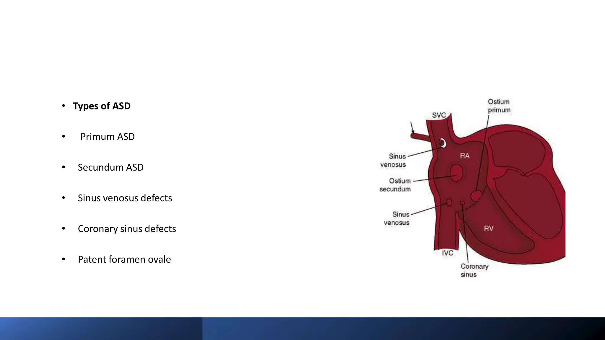
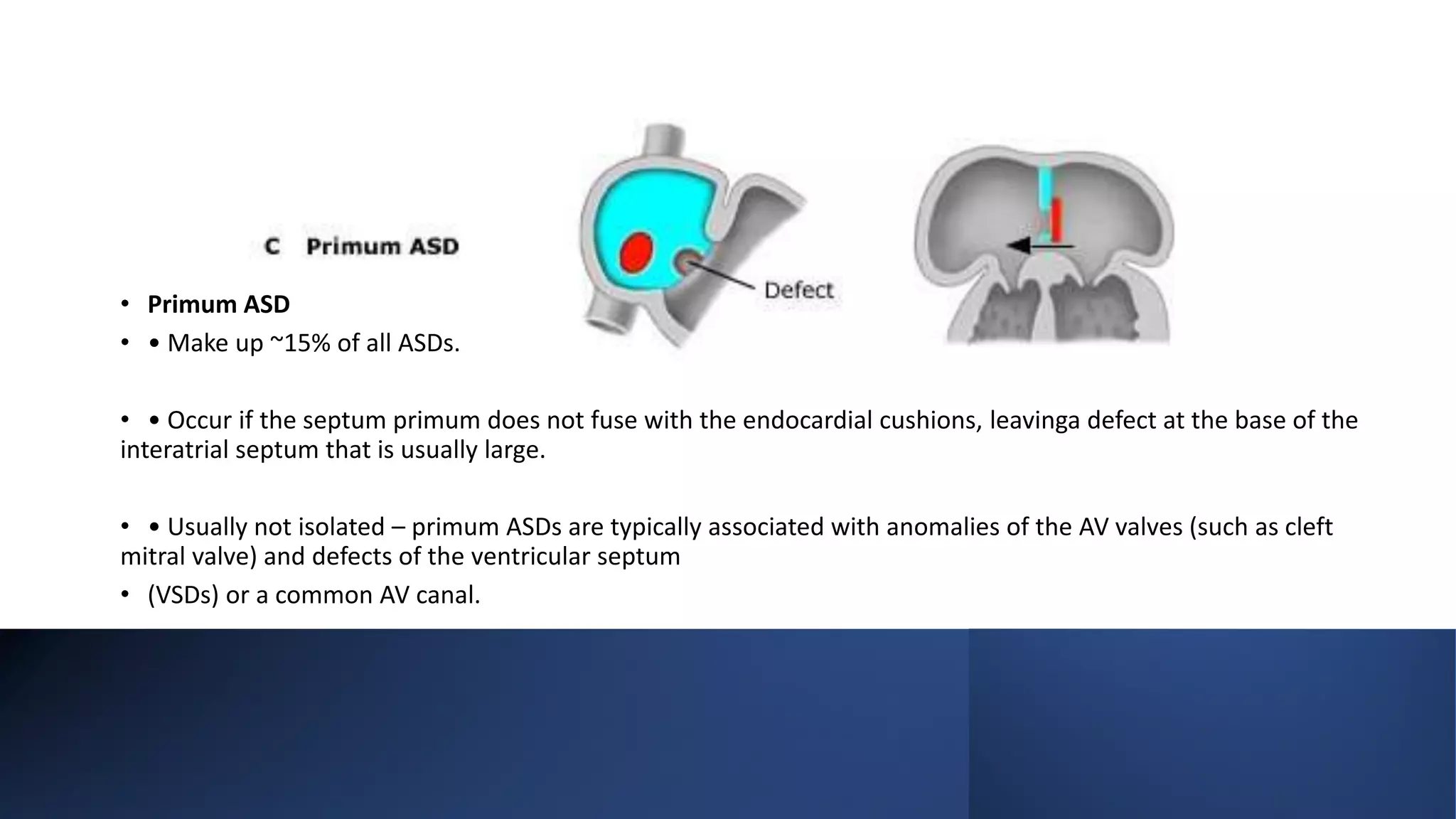
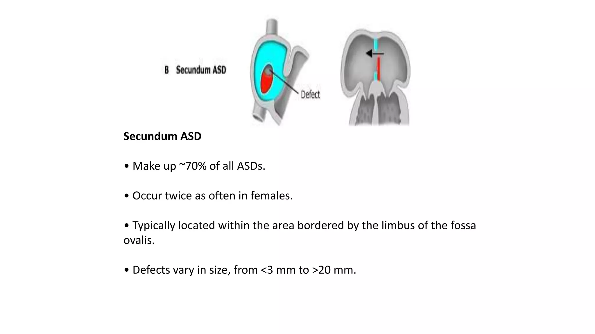
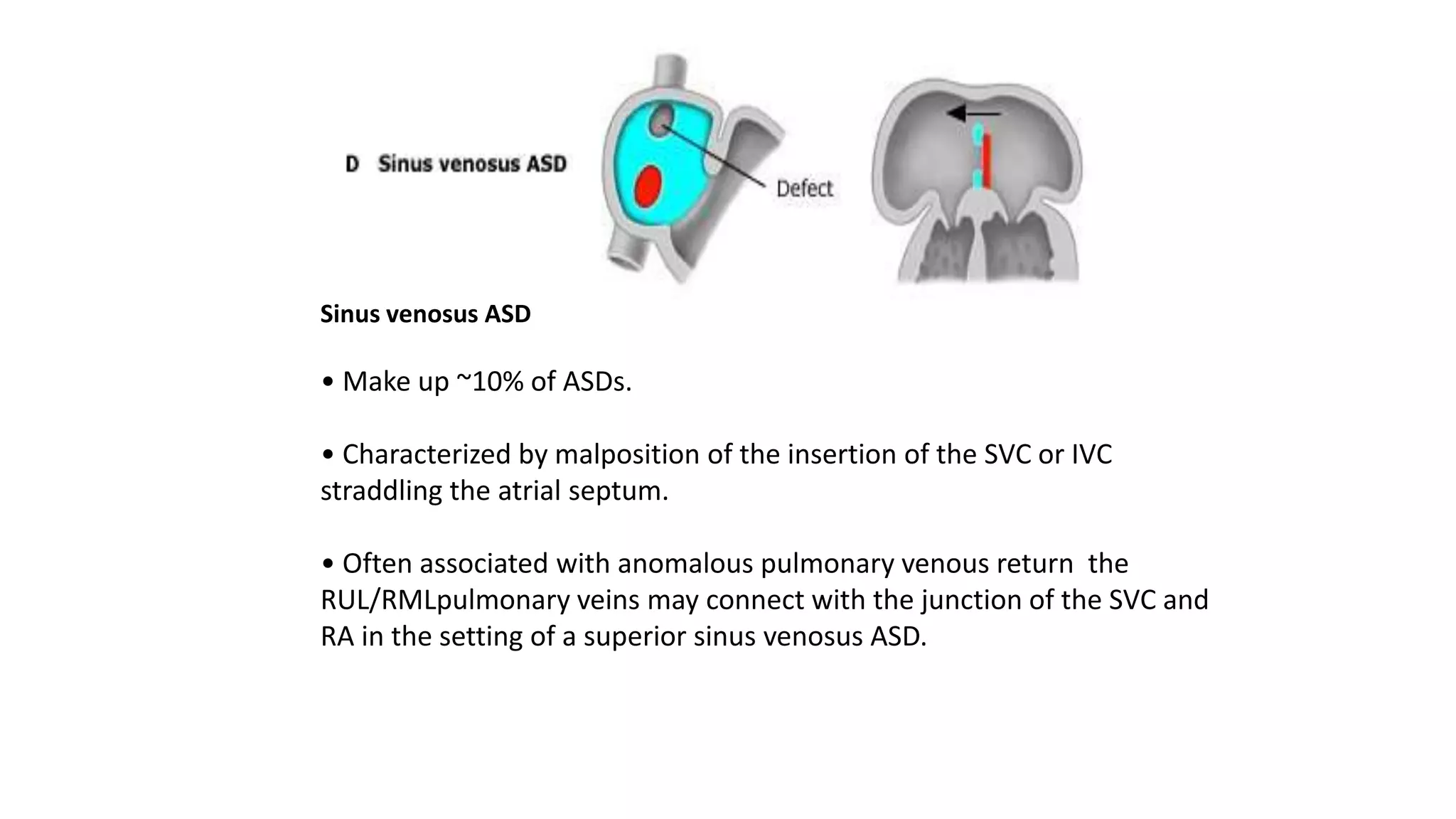
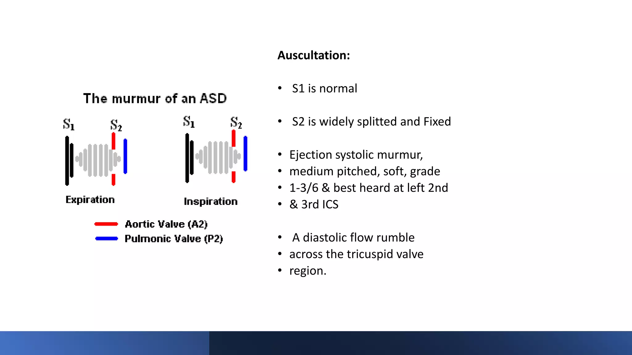
TAPVC defines the anomaly in which the pulmonary veins have no connection with the left atrium. Rather, the pulmonary veins connect directly to one of the systemic veins (TAPVC) or drain in to right atrium.
A PFO or ASD is present essentially in those who survive after birth
When pulmonary veins drain anomalously into the right atrium either because of complete absence of the interatrial septum or malattachment of the septum primum , then it is known as total anomalous pulmonary venous drainage.
When some or all of the pulmonary veins drain anomalously in to RA or its tributaries without being abnormally connected, the terms partially anomalous pulmonary venous drainage (PAPVD) or totally anomalous pulmonary venous drainage (TAPVD) with normal pulmonary venous connections are used.
The Norwood procedure is the first of three surgeries required to treat single-ventricle conditions such as hypoplastic left heart syndrome (HLHS). Because the left side of the heart can’t be fixed, the series of surgeries rebuilds other parts of the heart.
The Norwood procedure is performed in the baby’s first or second week of life.to redirect the blood flow.
Three goals for the Norwood procedure:
1, Build a new aorta.
2, Direct blood from the right ventricle through the new aorta and on to the rest of the body.
3, Direct the right ventricle to pump blood to the lungs until the next surgery.
A "hole" in the wall that separates the top two chambers of the heart.
This defect allows oxygen-rich blood to leak into the oxygen-poor blood chambers in the heart. ASD is a defect in the septum between the heart's two upper chambers (atria). The septum is a wall that separates the heart's left and right sides
Regarding the Post neonatal resuscitation. Always assess how the resuscitation went - mistakes made and scope for improvement - the Quality Improvement initiatives to improve the Quality of Neonatal Resuscitation - that in turn improves neonatal outcomes.
The neonate required resuscitation must be monitored for potential effects of difficult perinatal transition even though the Neonatal Resuscitation goes successfully. Based on the risk factors and the extent of resuscitation required - the newborn must be monitored. Not every post resuscitation baby needs an NICU monitoring
Post-neonatal resuscitation debriefing and monitoring are crucial aspects of newborn care, aimed at improving outcomes and enhancing team performance in future resuscitations. Debriefing following a resuscitation event allows healthcare teams to discuss the case, assess actions taken, and identify any areas for improvement. This reflective process not only helps in reinforcing correct practices but also in rectifying any gaps in the resuscitation process. It fosters a culture of learning and continuous improvement among neonatologists, pediatricians, and other team members involved.
Post-resuscitation neonatal monitoring is equally vital. After a resuscitation, newborns require close observation to ensure stable organ function and to detect any complications arising from the period of asphyxia or the resuscitation itself. Monitoring includes assessing vital signs, cardiovascular and respiratory function, and neurological status. This vigilant surveillance helps in early detection of issues such as hypoxic-ischemic encephalopathy or other organ dysfunctions, which can be critical in preventing long-term disabilities.
Together, debriefing and monitoring form an integrated approach to post-resuscitation care, ensuring that both the medical team and the newborn receive comprehensive support tailored to enhance recovery and outcomes following critical resuscitation efforts.
Neonatal Resuscitation Programme (NRP) - Preparation for Birth.pptxVannalaRaju2
The Neonatal Resuscitation Program (NRP) is an essential training protocol designed to equip healthcare professionals with the skills necessary to handle emergencies during childbirth, particularly those involving newborns who suffer from birth asphyxia. This guide is meticulously crafted for neonatologists, pediatricians, nurses, and other medical staff involved in the delivery room. The ultimate goal of the NRP is to reduce neonatal mortality rates and improve outcomes for newborns who do not breathe spontaneously or adequately at birth.
Understanding Birth Asphyxia
Birth asphyxia occurs when a newborn fails to establish regular breathing at birth, which can lead to insufficient oxygen reaching the brain and other organs, potentially causing lasting damage or even mortality. Prompt and effective resuscitation could mean the difference between life and long-term disability, or death. The conditions requiring neonatal resuscitation can include physiological challenges, congenital defects, or complications during delivery.
Role of Neonatologists and Pediatricians
Neonatologists and pediatricians are crucial in the NRP process. They are specifically trained to manage and mitigate risks associated with neonatal asphyxia. These specialists use their expertise to quickly assess the newborn's condition and administer life-saving interventions according to NRP guidelines. The guidelines recommend sequences of actions that include initial assessment, airway clearance, and effective ventilation strategies.
NRP Guidelines and Protocols
The NRP guidelines provide a systematic approach starting with the initial steps of warming the newborn to stimulate breathing, clearing the airway, and then providing gentle ventilation if necessary. These steps are critical to stabilize the newborns' condition. If the initial interventions are not successful, advanced resuscitation techniques such as chest compressions and administration of medications may be required.
Training and Certification
The NRP program offers comprehensive training that includes both theoretical knowledge and practical simulations to prepare healthcare providers for real-life scenarios. The training focuses on developing proficiency in decision-making and motor skills required for neonatal resuscitation. Certification in the NRP is often a requirement for healthcare professionals working in maternity wards and birthing centers, emphasizing its importance in clinical settings.
Impact on Neonatal Mortality
Neonatal mortality, particularly those cases related to birth asphyxia, remains a significant global health challenge. The implementation of NRP protocols has been shown to dramatically improve survival rates and health outcomes for affected newborns. Countries that have adopted widespread NRP training and guidelines report lower rates of neonatal mortality and better overall results in the management of complicated deliveries.
Scarlet Fever in Children and its complicationsVannalaRaju2
Scarlet fever is a bacterial illness that arises from a streptococcal infection, specifically the group A Streptococcus bacteria. This infection often follows strep throat, pharyngitis, or tonsillitis. Characterized by a distinct red rash, which feels like sandpaper, scarlet fever commonly affects children. The rash typically starts on the chest and spreads across the body. Accompanying symptoms include a high fever and a sore throat. If untreated, scarlet fever can lead to serious complications such as rheumatic fever. Prompt treatment with antibiotics is crucial to manage the infection and prevent further health issues.
Teething is a natural process: Teething is a normal part of a child's development when their teeth start to emerge through the gums.
Timing varies: The timing of teething can vary from child to child, but it typically starts around 6 months of age and continues until around 2 to 3 years old.
Common symptoms: Common signs of teething include drooling, irritability, gum swelling, chewing on objects, and disrupted sleep.
Soothing techniques: Provide teething rings, chilled (not frozen) toys, or gentle gum massages to help soothe your child's discomfort during teething.
Dental care starts early: As soon as your child's first tooth emerges, it's important to start brushing with a small amount of fluoride toothpaste and schedule their first dental visit by their first birthday.
- Video recording of this lecture in English language: https://youtu.be/lK81BzxMqdo
- Video recording of this lecture in Arabic language: https://youtu.be/Ve4P0COk9OI
- Link to download the book free: https://nephrotube.blogspot.com/p/nephrotube-nephrology-books.html
- Link to NephroTube website: www.NephroTube.com
- Link to NephroTube social media accounts: https://nephrotube.blogspot.com/p/join-nephrotube-on-social-media.html
Prix Galien International 2024 Forum ProgramLevi Shapiro
June 20, 2024, Prix Galien International and Jerusalem Ethics Forum in ROME. Detailed agenda including panels:
- ADVANCES IN CARDIOLOGY: A NEW PARADIGM IS COMING
- WOMEN’S HEALTH: FERTILITY PRESERVATION
- WHAT’S NEW IN THE TREATMENT OF INFECTIOUS,
ONCOLOGICAL AND INFLAMMATORY SKIN DISEASES?
- ARTIFICIAL INTELLIGENCE AND ETHICS
- GENE THERAPY
- BEYOND BORDERS: GLOBAL INITIATIVES FOR DEMOCRATIZING LIFE SCIENCE TECHNOLOGIES AND PROMOTING ACCESS TO HEALTHCARE
- ETHICAL CHALLENGES IN LIFE SCIENCES
- Prix Galien International Awards Ceremony
micro teaching on communication m.sc nursing.pdfAnurag Sharma
Microteaching is a unique model of practice teaching. It is a viable instrument for the. desired change in the teaching behavior or the behavior potential which, in specified types of real. classroom situations, tends to facilitate the achievement of specified types of objectives.
Flu Vaccine Alert in Bangalore Karnatakaaddon Scans
As flu season approaches, health officials in Bangalore, Karnataka, are urging residents to get their flu vaccinations. The seasonal flu, while common, can lead to severe health complications, particularly for vulnerable populations such as young children, the elderly, and those with underlying health conditions.
Dr. Vidisha Kumari, a leading epidemiologist in Bangalore, emphasizes the importance of getting vaccinated. "The flu vaccine is our best defense against the influenza virus. It not only protects individuals but also helps prevent the spread of the virus in our communities," he says.
This year, the flu season is expected to coincide with a potential increase in other respiratory illnesses. The Karnataka Health Department has launched an awareness campaign highlighting the significance of flu vaccinations. They have set up multiple vaccination centers across Bangalore, making it convenient for residents to receive their shots.
To encourage widespread vaccination, the government is also collaborating with local schools, workplaces, and community centers to facilitate vaccination drives. Special attention is being given to ensuring that the vaccine is accessible to all, including marginalized communities who may have limited access to healthcare.
Residents are reminded that the flu vaccine is safe and effective. Common side effects are mild and may include soreness at the injection site, mild fever, or muscle aches. These side effects are generally short-lived and far less severe than the flu itself.
Healthcare providers are also stressing the importance of continuing COVID-19 precautions. Wearing masks, practicing good hand hygiene, and maintaining social distancing are still crucial, especially in crowded places.
Protect yourself and your loved ones by getting vaccinated. Together, we can help keep Bangalore healthy and safe this flu season. For more information on vaccination centers and schedules, residents can visit the Karnataka Health Department’s official website or follow their social media pages.
Stay informed, stay safe, and get your flu shot today!
Couples presenting to the infertility clinic- Do they really have infertility...Sujoy Dasgupta
Dr Sujoy Dasgupta presented the study on "Couples presenting to the infertility clinic- Do they really have infertility? – The unexplored stories of non-consummation" in the 13th Congress of the Asia Pacific Initiative on Reproduction (ASPIRE 2024) at Manila on 24 May, 2024.
TEST BANK for Operations Management, 14th Edition by William J. Stevenson, Ve...kevinkariuki227
TEST BANK for Operations Management, 14th Edition by William J. Stevenson, Verified Chapters 1 - 19, Complete Newest Version.pdf
These simplified slides by Dr. Sidra Arshad present an overview of the non-respiratory functions of the respiratory tract.
Learning objectives:
1. Enlist the non-respiratory functions of the respiratory tract
2. Briefly explain how these functions are carried out
3. Discuss the significance of dead space
4. Differentiate between minute ventilation and alveolar ventilation
5. Describe the cough and sneeze reflexes
Study Resources:
1. Chapter 39, Guyton and Hall Textbook of Medical Physiology, 14th edition
2. Chapter 34, Ganong’s Review of Medical Physiology, 26th edition
3. Chapter 17, Human Physiology by Lauralee Sherwood, 9th edition
4. Non-respiratory functions of the lungs https://academic.oup.com/bjaed/article/13/3/98/278874
MANAGEMENT OF ATRIOVENTRICULAR CONDUCTION BLOCK.pdfJim Jacob Roy
Cardiac conduction defects can occur due to various causes.
Atrioventricular conduction blocks ( AV blocks ) are classified into 3 types.
This document describes the acute management of AV block.
Explore natural remedies for syphilis treatment in Singapore. Discover alternative therapies, herbal remedies, and lifestyle changes that may complement conventional treatments. Learn about holistic approaches to managing syphilis symptoms and supporting overall health.
Factory Supply Best Quality Pmk Oil CAS 28578–16–7 PMK Powder in Stockrebeccabio
Factory Supply Best Quality Pmk Oil CAS 28578–16–7 PMK Powder in Stock
Telegram: bmksupplier
signal: +85264872720
threema: TUD4A6YC
You can contact me on Telegram or Threema
Communicate promptly and reply
Free of customs clearance, Double Clearance 100% pass delivery to USA, Canada, Spain, Germany, Netherland, Poland, Italy, Sweden, UK, Czech Republic, Australia, Mexico, Russia, Ukraine, Kazakhstan.Door to door service
Hot Selling Organic intermediates
Pulmonary Thromboembolism - etilogy, types, medical- Surgical and nursing man...VarunMahajani
Disruption of blood supply to lung alveoli due to blockage of one or more pulmonary blood vessels is called as Pulmonary thromboembolism. In this presentation we will discuss its causes, types and its management in depth.
2. • Introduction
• An atrial septal defect (ASD) is a hole of variable size in the atrial septum.
• ASD is detected in 1 child per 1500 live births, and accounts for 5-10% of congenital heart
defects.
• ASDs make up 30-40% of all congenital heart disease detected in adults (second only to
bicuspid aortic valve).
• ASDs occur in women 2-3 times more common than men.
3. • ASDs can occur in different anatomic portions of the atrial septum.
• ASDs can be isolated or occur with other congenital cardiac anomalies.
• Functional consequences of ASDs are related to the anatomic location of the defect, its
size, and the presence or absence of other cardiac anomalies.
5. • Types of ASD
• Primum ASD
• Secundum ASD
• Sinus venosus defects
• Coronary sinus defects
• Patent foramen ovale
6. • Primum ASD
• • Make up ~15% of all ASDs.
• • Occur if the septum primum does not fuse with the endocardial cushions, leavinga defect at the base of the
interatrial septum that is usually large.
• • Usually not isolated – primum ASDs are typically associated with anomalies of the AV valves (such as cleft
mitral valve) and defects of the ventricular septum
• (VSDs) or a common AV canal.
7. Secundum ASD
• Make up ~70% of all ASDs.
• Occur twice as often in females.
• Typically located within the area bordered by the limbus of the fossa
ovalis.
• Defects vary in size, from <3 mm to >20 mm.
8. • Secundum ASD
• • May be associated with other ASDs.
• • Multiple defects can be seen if the floor of the fossa ovalis (AKA valve of the foramen
ovale) is fenestrated.
• • Ten to twenty percent have a functional mitral valve prolapse.
• • May be related to changing LV geometry associated with RV volume overload
9. Sinus venosus ASD
• Make up ~10% of ASDs.
• Characterized by malposition of the insertion of the SVC or IVC
straddling the atrial septum.
• Often associated with anomalous pulmonary venous return the
RUL/RMLpulmonary veins may connect with the junction of the SVC and
RA in the setting of a superior sinus venosus ASD.
10. • Coronary Sinus Septal Defects
• • Less than 1% of ASDs
• • Defects in the inferior/anterior atrial septum region that
• includes the coronary sinus orifice.
• • Defect of at least a portion of the common wall separating the coronary sinus and the left
atrium – AKA “unroofed coronary sinus”
• • Can be associated with a persistent left SVC draining into the coronary sinus.
12. • Patent Foramen Ovale
• •Not truly an “ASD” because no
• septal tissue is missing.
• •Oxygenated blood from the IVC
• crosses the foramen ovale in utero.
• •At birth, the flap normally closes
• due to Reduced right heart pressure and PVR
• • Elevated LA pressure.
• • Flap fusion is complete by age two
• in 70-75% of children; the
• remainder have a PFO.
13. LEFT-TO-RIGHT SHUNT
• In early infancy, when pulmonary resistance is high, left and right ventricular
compliances are similar, and net shunting through an ASD is typically slight.
• As the left ventricle matures, it becomes less compliant in diastole than the
right, and left atrial pressure rises. This drives a left-to-right shunt at the atrial
level in the presence of an ASD.
• With age, the disparity between systemic and pulmonary resistance, and in
turn between left and right ventricular compliance, results in increased left-to-
right shunting and advancing right ventricular volume loading.
HEMODYNAMICS
14. Desaturated blood enters
the right atrium from the
vena cava at a volume of
3L/min/m2 and mixes with
an additional 3 L of fully
saturated blood shunting
left to right across the ASD
HEMODYNAMICS
15. Results in :
increase in oxygen saturation
in the right atrium.
Six liters of blood flows
through the tricuspid valve
and causes a mid-diastolic
flow rumble.
Oxygen saturation may be
slightly higher in the right
ventricle because of
incomplete mixing at the
atrial level.
16. The full 6 L flows across
the right ventricular
outflow tract and causes a
systolic ejection flow
murmur.
Six liters returns to the left
atrium, with 3 L shunting left to
right across the defect and 3 L
crossing the mitral valve to be
ejected by the left ventricle into
the ascending aorta.
18. • Over time, right ventricular
volume load results in dilation
and hypertrophy, eventually
affecting the function of both
ventricles.
• Atrial enlargement may
contribute to the late incidence
of atrial fibrillation. Right
ventricular volume overload is
noted to occur as a rule when
ASDs are larger than 6 mm in
diameter
• Volume-induced
hypertrophy of the right
ventricle produces a loss
of coronary reserve and
eventual impairment of
right ventricular systolic
and diastolic function.
• Left ventricular
functional reserve is
diminished by adulthood
in most patients with ASD.
19. • • Although left ventricular systolic function may be normal at rest, the left ventricle
exhibits a subnormal diastolic dimension, and a loss of functional reserve at exercise.
•
• • In general, the functional loss in the left and right ventricles is normalized 6 months
following ASD closure in children and young adults.
20. • NATURAL HISTORY
• In patients with an ASD <3 mm in size
• diagnosed before 3 months of age,
• spontaneous closure occurs in 100% of
• patients at 1½ years of age.
• Spontaneous closure occurs more than 80%
• in patients with defects between 3-8 mm
• before 1½ years of age.
• An ASD with a diameter > 8 mm rarely closes spontaneously.
21. • Natural History of ASDs
• • Most ASDs <8mm close spontaneously in infants.
• • Spontaneous closure is unusual in children and adults; defects often become progressively larger.
• • Most patients with a significant shunt flow ratio (Qp:Qs > 2:1) will be symptomatic and require closure by age40.
• • Increasing size of the ASD may preclude percutaneous closure.
• • Weight is more affected then height
• Association
• • Holt oram syndrome
• • Patau’s syndrome
• • Edward’s syndrome
22. • • If untreated, pulmonary hypertension and subsequent CCF may develop during or after
third decade, and reversal of shunt may occur (rare), it may be progressive with pregnancy.
• • With or without surgery, atrial arrhythmias (flutter or fibrillation) may occur in adults.
• • Cerebrovascular accident, resulting from paradoxical embolization through an ASD, is a
rare complication.
Infective endocarditis does not occur in patients with isolated ASDs.
Mitral stenosis may occur as a result of rheumatic fever in a case of
ASD (Lutembacher syndrome).
23. • SYMPTOMS AND SIGNS
• Vary with the size of defect.
• Small defect:
• Asymptomatic and is usually diagnosed during a routine health check up.
• Large defect:
• Symptomatic and patients usually present with
• Failure to thrive.
• Easy fatigability.
• Increased perspiration
• Recurrent Pulmonary infections.
• Platypnea
24. On examination
• General examination
• Appearance: Usually normal
• Heart rate: Normal
• Respiratory rate: Normal
• Weight and height: may be less than 10th centile.
25. • Precordium
• Inspection:
• Slight prominence of precordium
• Palpation:
• Apex beat may be shifted to left
• P2 may be palpable
• Left parasternal heave may be
• present
26. Auscultation:
• S1 is normal
• S2 is widely splitted and Fixed
• Ejection systolic murmur,
• medium pitched, soft, grade
• 1-3/6 & best heard at left 2nd
• & 3rd ICS
• A diastolic flow rumble
• across the tricuspid valve
• region.
27. INVESTIGATIONS
Routine tests :
(CBC, septic screening, s.electrolyte, s.
creatinine, blood grouping, coagulation
profile, etc)
should be done before management.
Diagnostic Investigations includes-
-X-ray
-Ecg
-Echocardiography
-Sometimes cardiac catheterization
29. • Enlarged ‘p’ wave indicating Right atrial hypertrophy.
• rsR’ seen and tall R wave Indicating RBBB and RVH.
• Also note that the aVF is predominantly upwards as compared to
Lead I indicating Right Axis Deviation.
30. Echocardiogram
Primary diagnostic
imaging modality for ASD.
Provides:
- exact localization of ASD
- size of ASD
- measurement of septal
rims
- Confirmation of the shunt
- Abnormal motion of
ventricular septum.
- Associated lesions can be
identified
31. Cardiac catheterization
Patients with the classic features of a
hemodynamically significant ASD on physical
examination and chest radiography, in whom
echocardiographic identification of an isolated
secundum ASD is made, need not undergo diagnostic
catheterization before repair.
Exception:
an older patient, in whom pulmonary vascular
resistance may be a concern.
33. MANAGEMENT
• Patients with small shunts and normal RV size are generally
asymptomatic and require no therapy but need longtime follow up for
spontaneous closure
• Moderate to large shunt and/or symptomatic ASD should be
managed with following strategies:
• Medical therapy
• Interventional therapy
• Surgical therapy
34. Medical management
• Aim to reduce volume overload and to strengthen functions of heart
muscles.
Symptomatic children :
• Diuretics:
These agents relieve ventricular overload,
peripheral and pulmonary congestion
• Digoxin:
Helps to strengthen the heart muscle, enabling it to pump more
efficiently
35. Afterload reducers:
• - Enalapril
• - Captopril
• Exercise restriction is no necessary
• Prophylaxis for infective endocarditis is not
Indicated
• Atrial arrythmias : Appropriate Antiarrhythmic
drugs.
• Atrial fibrillation : Antiarrhythmic drugs and anticoagulants.
36. • Irreversible PAH :dobutamine, calcium channel
blockers (high dose), diuretics, prostacycline, sildenafil
or oxygen therapy.
• Treatment of Other complications, likepulmonary
infections, thrombo- embolic events or heart failure
should also be treated accordingly.
37. .
Closure of ASD :
• In patients with small secundum ASDs and
minimal left-to-right shunts without right ventricular enlargement, closure is not required
Indications of ASD closure-
• All symptomatic patients
• Asymptomatic patients with- Qp : Qs ratio of at least 2 : 1
• Right ventricular enlargement
• Time of closure- usually after the 1st yr and
before entry into school
38. Interventional therapy
Indication:
1. Echocardiographic evidence of ostium secundum ASD
2. Clinical evidence of RV volume load ( i.e. 1.5:1 degree of left to right
shunt or RV enlargement)
3. ASD diameter less than 36 mm
4. Presence of sufficient rim of tissue( at least 5 mm)
5. Patient with fenestrated Fontan lateral tunnel if temporary balloon
occlusion is tolerated
39. Contraindication:
• Sinus venosus, coronary sinus or primum ASD
• Extensive congenital cardiac anomaly
• Known sepsis within one month prior to implantation or any untreated
systemic infection prior to device placement.
• Bleeding disorder, untreated ulcer or any other contraindications to aspirin
therapy.
• Demonstrated intracardiac thrombi on echo.
• Any patient whose size or condition would cause to be a poor candidate for
cardiac catheterization.
41. Advantages of device
closure-
• It is safe and cost-effective
than surgery
• Successful implantation
rates more than 96%,
• Fewer complications:
Major<1%,
• Shortened hospitalization
• Avoidance of pain and
residual thoracotomy scars
• Reduced need for blood
products.
Disadvantages of device
closure-
• Higher rate of small
residual leak
42. Complications of Device Closure:
• Device misalignment/embolization
• Device erosion of atrial wall or aorta
• Device impingement on adjacent structures AV valve,
• Coronary sinus, SVC, Pulmonary veins, Aorta
• Infection including endocarditis
• Thromboembolic Complication
• Allergic reaction
• Valvular regurgitation
• Residual shunt
43. Surgical management
• Surgical closure has been the “gold standard” formof treatment of
ASD.
• Surgeons need proper training and expertise in performing
operations.
• The surgical approach can be by right thoracotomy or sternotomy,
and more limited incisions are feasible with either approach.
44. • Procedure- Simple
suture or patch closure
• Timing-
Surgery is usually
delayed until the patient
is 2 to 4years of age
because the
• possibility of
spontaneousclosure
exists.
• In infancy- If CCF not
respond to medical
management
45.
46. Indications:
• ASD with RA and RV enlargement with / without
symptoms.
• ASD minimum diameter > 10 mm on echocardiography
• A sinus venosus, coronary sinus or primum ASD.
• Chronic atrial arrythmia with ASD (concomitant Maze
procedure)
Contraindications:
• Patients with severe irreversible PAH & reverse shunt
SPO2 < 90%
47. Advantages of Surgery-
• Can be performed in any
type of ASD
• Associated anatomical
abnormality can be
corrected concurrently.
• Excellent late outcome.
Disadvantages of Surgery-
• Costly
• Needs expertise hands
• Prolong Hospital stay
• pain and residual
thoracotomy scars
49. Follow – Up After Surgical Closure:
• Early postoperative follow-up:
• -Symptoms of undue fever, fatigue, vomiting, chest pain, or
• abdominal pain
• ( may represent post pericardiotomy syndrome with
• tamponade and needs immediate evaluation with
• echocardiography.)
• Annual clinical F/U: (if following conditions persist or
• develop)
• - PAH.
• - Atrial arrhythmias.
• - RV or LV dysfunction.
• - Coexisting valvular or other cardiac lesion
50. TAKE –HOME MESSAGES
• Atrial septal defects are relatively common CHD
• Early symptoms are usually rare except very large
• deffect.
• Any kind of closure is safe and effective and
• associated with improved life expectancy
• A comprehensive treatment plan should include
• input from the primary care provider, the
• Paediatric Cardiologist and the Paediatric
• Cardiovascular surgeon.
51. PROGNOSIS:
• Patients generally survive up to adulthood
without
• surgical or percutaneous intervention mainly
with
• small to moderate size ASD and many patients
live to
• advanced age.
• The results after surgical or device closure in
• children with moderate to large shunts are
excellent.
• Mortality is less than 2% after surgical closure of
• uncomplicated ASD
• Mortality and morbidity increase with pulmonary
• vascular disease