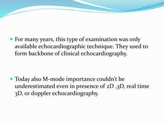
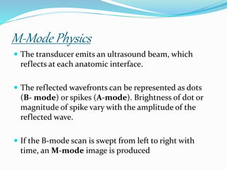
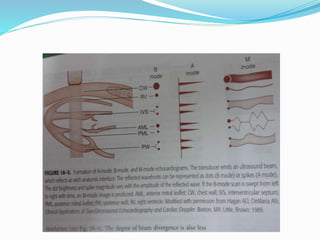
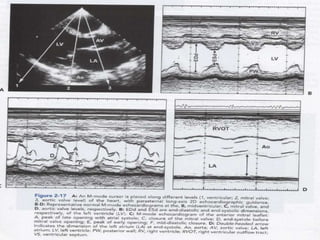
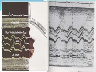
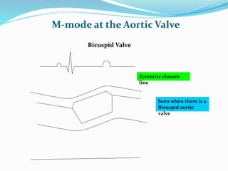
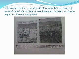
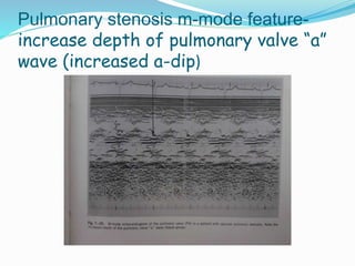
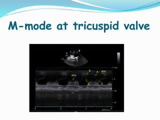
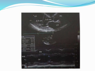
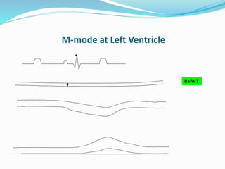
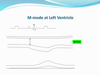
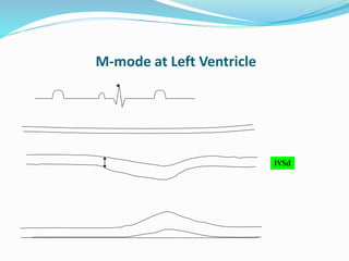
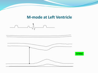
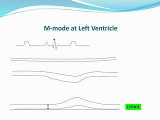
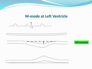
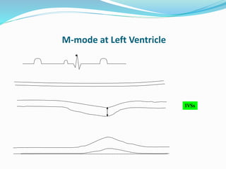
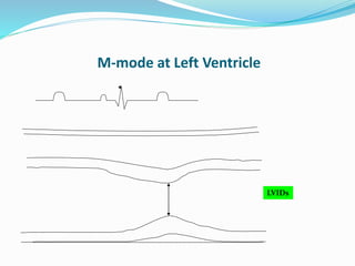
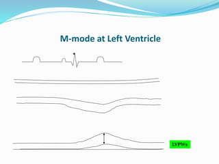
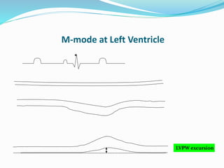
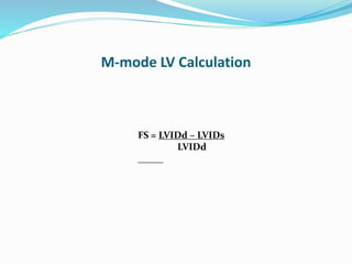
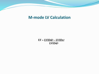
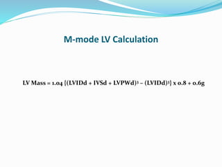
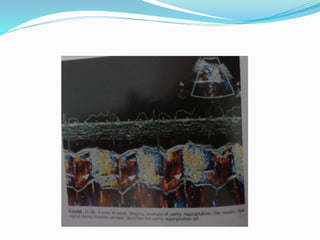
Dr. Amit Kumar
Senior Resident, Department of Cardiology
R.N.T Medical College
Udaipur , India
 For many years, this type of examination was only
available echocardiographic technique. They used to
form backbone of clinical echocardiography.
 Today also M-mode importance couldn’t be
underestimated even in presence of 2D ,3D, real time
3D, or doppler echocardiography.
M-Mode Physics
 The transducer emits an ultrasound beam, which
reflects at each anatomic interface.
 The reflected wavefronts can be represented as dots
(B- mode) or spikes (A-mode). Brightness of dot or
magnitude of spike vary with the amplitude of the
reflected wave.
 If the B-mode scan is swept from left to right with
time, an M-mode image is produced
M-Mode Physics….
 M-mode has got better temporal resolution and
thus subtle abnormalities in motion and timing is
better appreciated. For eg. systolic anterior motion of
mitral valve in HCM & RV diastolic collapse in
tamponade.
 Because of its high sampling frequency( upto 1000
pulses per second), M-mode has excellent axial
resolution and is useful in identifying the relative
location of structures and measuring range of motion.
 M-mode echocardiography is use to evaluate the
morphology of structures ; movement and velocity
of cardiac valves and walls; and timing of cardiac
events.
M-mode Evaluation
 Amplitude
 Velocity
 Time intervals
 Morphology
Amplitude = Y2 –Y1
Y1
Y2
Amplitude Measurement
Distance
Time
Time interval = T2 – T1
T2T1
Time Measurement
Distance
Time
dy = Y2 –Y1
Y1
Y2
Slope Measurement
T1 T2
dt = T2 – T1
Slope = dy/dt = velocity
M-mode at the Mitral Valve
 The mitral valve has 2 leaflets – anterior
and posterior.
Mitral stenosis: M-mode
features
 Decrease EF slope.
 Paradoxical anterior diastolic motion of PML.
 Seperation between leaflets is decreased.
 Thickening of leaflets.
 Early diastolic dip of IVS.
 Reduced mitral valve leaflet excursion( D-E excursion)
 Earlier pliability for BMV used to be decided on basis of D-
E amplitude. A MV with D-E amplitude of 20mm or more
is usually considered pliable.
Mitral Stenosis
Mitral regurgitation: m-mode
features
 Indirect evidences- LA enlargement, LV enlargement
 Exaggerated septal motion (1cm)
 LAE with systolic expansion of the posterior left atrial
wall.
Mitral valve prolapse : m-
mode features
 Thick redundant mitral valve leaflets.
 Mid to late systolic sagging back of the anterior,
posterior or both MV leaflet >2mm from C-D point of
MV.
 Holosystolic sagging back of the anterior, posterior, or
both MV leaflet >3mm from the C-D point of MV.
Flail mitral leaflet : m-mode
features
 Coarse diastolic fluttering of mitral leaflets.
 Flail mitral leaflet may appear within LA
Infective endocarditis: m-
mode features
 Valve leaflet appear thickened, “smudged”, “shaggy”.
 Vegetation on a valve leaflet usually doesn’t restrict
valve motion.
Mitral Valve Endocarditis
LA myxoma: m-mode
features
 Blunted E point of the mitral valve.
 Decrease E-F slope.
 Heavy band of echoes behind the anterior mitral
leaflet in diastole.
 Echo free space at anterior mitral leaflet at onset of
diastole prior to dense echoes from tumor
Premature closure of Mitral
valve: m-mode features
 When C-point of the mitral valve occurs before the
onset of the QRS complex.
Fluttering of AML in aortic
regurgitation
“B” bump due to elevated EDP
B-bump or notch
Hypertrophic Cardiomyopathy
Systolic anterior motion of
mitral valve s/o dynamic LVOT
obstruction
M-mode at the Aortic Valve
 The aortic valve has 3 cusps – right coronary,
left coronary and non-coronary cusps.
 The cusps imaged in the PLAX view are the
right coronary and the non-coronary cusps.
 Leaflet may show fine systolic fluttering in
healthy individuals.
M-mode at the Aortic Valve
Coronary
cusp
Non-coronary cusp
Anterior aortic root
Posterior aortic root
Left Atrium
M-mode at the Aortic Valve
LA dimension
Cusp Separation (1.5-2.5cms in adult)
Aortic root
M-mode at the Aortic Valve
LA dimension
Measurements are made
from leading edge to
leading edge.
Aortic stenosis: m-mode
features
 Thickening valve leaflets.
 Decreased excursion of valve leaflet.
 Absence of systolic flutter of aortic valve leaflet.
Critical Aortic Stenosis
M-mode at the Aortic Valve
Bicuspid Valve
Eccentric closure
line
Seen when there is a
Bicuspid aortic
valve
Aortic regurgitation: m-mode
features
 Diastolic fluttering of AML.
 Diastolic fluttering of aortic valve.
 Premature closure of mitral valve.
 Premature opening of aortic valve.
 Dilated LV.
Fluttering of AML in aortic
regurgitation
Premature opening of Aortic
valve in AR
Diastolic fluttering of aortic
valve in AR
coarse fluttering of aortic
valve cusp- SUBAORTIC
MEMBRANE
Early closure of AV due to Severe LV dysfun
M-mode in a pt with LV dysfunction-
showing rounded closure of
AV,indicating decrease forward flow
at end of systole
Systolic notching of AV in
HCM
a- downward motion, concides with A-wave of MV; b- represents
onset of ventricular systole; c- max downward position ; d- closure
begins; e- closure is completed
 In adults it is unusual to record more than posterior
leaflet of the pulmonary valve.
 In children or in pt with unusually large pulmonary
arteries, one may also record anterior leaflet.
 In reality one can rarely record the entire excursion of
the pulmonar valve throughout cardiac cycle in adults.
M-mode at Pulmonary valve
Pulmonary stenosis m-mode feature-
increase depth of pulmonary valve “a”
wave (increased a-dip)
Pulmonary HTN with “Flying W sign”
Pulmonary hypertension m-mode
feature- loss of A-dip of pulmonary
valve
M-mode at tricuspid valve
M-mode at Left Ventricle
RVWT
M-mode at Left Ventricle
RVIDd
M-mode at Left Ventricle
IVSd
M-mode at Left Ventricle
LVIDd
M-mode at Left Ventricle
LVPWd
M-mode at Left Ventricle
IVS excursion
M-mode at Left Ventricle
IVSs
M-mode at Left Ventricle
LVIDs
M-mode at Left Ventricle
LVPWs
M-mode at Left Ventricle
LVPW excursion
• FS
• EF
• LV mass
M-mode LV Calculation
FS = LVIDd – LVIDs
LVIDd
M-mode LV Calculation
EF = LVIDd3 – LVIDs3
LVIDd3
M-mode LV Calculation
LV Mass = 1.04 {(LVIDd + IVSd + LVPWd)3 – (LVIDd)3} x 0.8 + 0.6g
LV M-mode parameters
range
 Ventricular end-diastolic dimension- 37 to 56mm
 Ventricular end-systolic dimension- 26 to 36mm
 LV diastolic IVS thickness- 7to 11mm
 IVS excursion – 6 to 11mm
 IVS % thickening- 27 to 70%
 LV posterior diastolic wall thickness- 7 to 11mm
 LVPW excursion- 9 to 14mm
 LVPW % thickening- 25 to 80%
 %FS- 28-41%
 EF- 48-78%
LV parameters….
 Stroke volume- 75 to 100cc
 Cardiac output- 4 to 8L/min
 Cardiac index- 2.4 to 4.2L/min/m2
 LV mass- male< 294gm; female<198gm
 Mitral valve EF slope- 50 to 150mm/sec
 DE Excursion- 15 to 25 mm
 Mitral valve E-point septal seperation- <7mm
Increase LVW thickness
Ischemia
Ischemia
 Normal Wall thickness
 No systolic thickening
 Reduced Motion
Post Infarct
Post Infarct
 Thin echogenic wall
 No systolic thickening
 Reduced Motion
Dilated Cardiomyopathy
Paradoxic Septal Motion
Normal E point to septal separation is < 6 mm
With reduced lvef, EPSS may be increased.
help in differentiating pleural
effusion from pericardial
effusion
 If the ultrasonic beam is directed towards the left
atrium :
1)Gradual decrease in the echo free space–
pericardial effusion
2)Sudden cessation of echo free space- pleural
effusion
Quantitation of pericardial fluid can be done by m-mode echo,
but 2D-echocardiography gives a better idea esp. in c/o large or
loculated effusion
Cardiac tamponade- m
mode features
 Compressed RV (RVID<7mm)
 Increase in RV dimension with inspiration and
simultaneously decrease in LV dimension during
inspiration.
 Decrease mitral valve EF-slope with inspiration.
 Decrease mitral valve DE-amplitude with inspiration.
 RV diastolic collapse.(specific)
 RA diastolic collapse.(sensitive)
 Dilated IVC with blunted respiratory changes.
Cardiac tamponade- RV early
diastolic collapse
Constrictive pericarditis: m-
mode features
 Pericardial thickening
 Paradoxical septal motion
 Septal bounce( abrupt displacement of the IVS during early
diastole)
 Flattening of mid & late diastolic motion of the posterior
LV wall.
 Rapid early diastolic, or E-F, slope of the mitral valve.
 Rapid downward motion of the posterior aortic wall in
early diastole.
 Premature opening of pulmonary valve
 Dilated IVC with blunted respiratory changes.
Constrictive pericarditis-
septal bounce
Motion of the posterior aortic wall
reflects the filling and emptying patterns
of the left atrium.
 With impaired LA emptying, the aortic wall motion is
reduced during the rapid emptying phase, or the first third
of diastole.
 LA emptying index-
 If the first third of diastole does not represent at least 40%
of the total amplitude of the aortic wall motion during
diastole, then restriction to ventricular filling is suspected.
LA emptying index:
Decreased in c/o mitral stenosis
Increased in c/o mitral regurgitation
Color doppler m-mode
imaging
 Used to determine velocity of propagation (Vp) of LV
inflow.
 Determination of width of AR jet.
 Duration of MR.
Velocity of propagation
TAPSE (tricuspid annular plane
systolic excursion)
TAPSE – a measure for
assessing RV function
 TAPSE reference range 15 to 20mm
 Mildly abnormal- 13 to 15mm
 Moderately abnormal- 10 to 12mm
 Severely abnormal- <10mm
TAPSE
M mode echo

M mode echo

  • 1.
    Dr. Amit Kumar SeniorResident, Department of Cardiology R.N.T Medical College Udaipur , India
  • 2.
     For manyyears, this type of examination was only available echocardiographic technique. They used to form backbone of clinical echocardiography.  Today also M-mode importance couldn’t be underestimated even in presence of 2D ,3D, real time 3D, or doppler echocardiography.
  • 3.
    M-Mode Physics  Thetransducer emits an ultrasound beam, which reflects at each anatomic interface.  The reflected wavefronts can be represented as dots (B- mode) or spikes (A-mode). Brightness of dot or magnitude of spike vary with the amplitude of the reflected wave.  If the B-mode scan is swept from left to right with time, an M-mode image is produced
  • 5.
    M-Mode Physics….  M-modehas got better temporal resolution and thus subtle abnormalities in motion and timing is better appreciated. For eg. systolic anterior motion of mitral valve in HCM & RV diastolic collapse in tamponade.  Because of its high sampling frequency( upto 1000 pulses per second), M-mode has excellent axial resolution and is useful in identifying the relative location of structures and measuring range of motion.
  • 6.
     M-mode echocardiographyis use to evaluate the morphology of structures ; movement and velocity of cardiac valves and walls; and timing of cardiac events.
  • 7.
    M-mode Evaluation  Amplitude Velocity  Time intervals  Morphology
  • 8.
    Amplitude = Y2–Y1 Y1 Y2 Amplitude Measurement Distance Time
  • 9.
    Time interval =T2 – T1 T2T1 Time Measurement Distance Time
  • 10.
    dy = Y2–Y1 Y1 Y2 Slope Measurement T1 T2 dt = T2 – T1 Slope = dy/dt = velocity
  • 13.
    M-mode at theMitral Valve  The mitral valve has 2 leaflets – anterior and posterior.
  • 15.
    Mitral stenosis: M-mode features Decrease EF slope.  Paradoxical anterior diastolic motion of PML.  Seperation between leaflets is decreased.  Thickening of leaflets.  Early diastolic dip of IVS.  Reduced mitral valve leaflet excursion( D-E excursion)  Earlier pliability for BMV used to be decided on basis of D- E amplitude. A MV with D-E amplitude of 20mm or more is usually considered pliable.
  • 16.
  • 17.
    Mitral regurgitation: m-mode features Indirect evidences- LA enlargement, LV enlargement  Exaggerated septal motion (1cm)  LAE with systolic expansion of the posterior left atrial wall.
  • 20.
    Mitral valve prolapse: m- mode features  Thick redundant mitral valve leaflets.  Mid to late systolic sagging back of the anterior, posterior or both MV leaflet >2mm from C-D point of MV.  Holosystolic sagging back of the anterior, posterior, or both MV leaflet >3mm from the C-D point of MV.
  • 22.
    Flail mitral leaflet: m-mode features  Coarse diastolic fluttering of mitral leaflets.  Flail mitral leaflet may appear within LA
  • 25.
    Infective endocarditis: m- modefeatures  Valve leaflet appear thickened, “smudged”, “shaggy”.  Vegetation on a valve leaflet usually doesn’t restrict valve motion.
  • 27.
  • 28.
    LA myxoma: m-mode features Blunted E point of the mitral valve.  Decrease E-F slope.  Heavy band of echoes behind the anterior mitral leaflet in diastole.  Echo free space at anterior mitral leaflet at onset of diastole prior to dense echoes from tumor
  • 30.
    Premature closure ofMitral valve: m-mode features  When C-point of the mitral valve occurs before the onset of the QRS complex.
  • 32.
    Fluttering of AMLin aortic regurgitation
  • 33.
    “B” bump dueto elevated EDP B-bump or notch
  • 34.
    Hypertrophic Cardiomyopathy Systolic anteriormotion of mitral valve s/o dynamic LVOT obstruction
  • 37.
    M-mode at theAortic Valve  The aortic valve has 3 cusps – right coronary, left coronary and non-coronary cusps.  The cusps imaged in the PLAX view are the right coronary and the non-coronary cusps.  Leaflet may show fine systolic fluttering in healthy individuals.
  • 38.
    M-mode at theAortic Valve Coronary cusp Non-coronary cusp Anterior aortic root Posterior aortic root Left Atrium
  • 40.
    M-mode at theAortic Valve LA dimension Cusp Separation (1.5-2.5cms in adult) Aortic root
  • 41.
    M-mode at theAortic Valve LA dimension Measurements are made from leading edge to leading edge.
  • 42.
    Aortic stenosis: m-mode features Thickening valve leaflets.  Decreased excursion of valve leaflet.  Absence of systolic flutter of aortic valve leaflet.
  • 43.
  • 44.
    M-mode at theAortic Valve Bicuspid Valve Eccentric closure line Seen when there is a Bicuspid aortic valve
  • 45.
    Aortic regurgitation: m-mode features Diastolic fluttering of AML.  Diastolic fluttering of aortic valve.  Premature closure of mitral valve.  Premature opening of aortic valve.  Dilated LV.
  • 46.
    Fluttering of AMLin aortic regurgitation
  • 47.
    Premature opening ofAortic valve in AR
  • 48.
    Diastolic fluttering ofaortic valve in AR
  • 49.
    coarse fluttering ofaortic valve cusp- SUBAORTIC MEMBRANE
  • 50.
    Early closure ofAV due to Severe LV dysfun M-mode in a pt with LV dysfunction- showing rounded closure of AV,indicating decrease forward flow at end of systole
  • 51.
  • 53.
    a- downward motion,concides with A-wave of MV; b- represents onset of ventricular systole; c- max downward position ; d- closure begins; e- closure is completed
  • 54.
     In adultsit is unusual to record more than posterior leaflet of the pulmonary valve.  In children or in pt with unusually large pulmonary arteries, one may also record anterior leaflet.  In reality one can rarely record the entire excursion of the pulmonar valve throughout cardiac cycle in adults.
  • 55.
  • 56.
    Pulmonary stenosis m-modefeature- increase depth of pulmonary valve “a” wave (increased a-dip)
  • 57.
    Pulmonary HTN with“Flying W sign” Pulmonary hypertension m-mode feature- loss of A-dip of pulmonary valve
  • 59.
  • 62.
    M-mode at LeftVentricle RVWT
  • 63.
    M-mode at LeftVentricle RVIDd
  • 64.
    M-mode at LeftVentricle IVSd
  • 65.
    M-mode at LeftVentricle LVIDd
  • 66.
    M-mode at LeftVentricle LVPWd
  • 67.
    M-mode at LeftVentricle IVS excursion
  • 68.
    M-mode at LeftVentricle IVSs
  • 69.
    M-mode at LeftVentricle LVIDs
  • 70.
    M-mode at LeftVentricle LVPWs
  • 71.
    M-mode at LeftVentricle LVPW excursion
  • 72.
    • FS • EF •LV mass M-mode LV Calculation
  • 73.
    M-mode LV Calculation FS= LVIDd – LVIDs LVIDd
  • 74.
    M-mode LV Calculation EF= LVIDd3 – LVIDs3 LVIDd3
  • 75.
    M-mode LV Calculation LVMass = 1.04 {(LVIDd + IVSd + LVPWd)3 – (LVIDd)3} x 0.8 + 0.6g
  • 76.
    LV M-mode parameters range Ventricular end-diastolic dimension- 37 to 56mm  Ventricular end-systolic dimension- 26 to 36mm  LV diastolic IVS thickness- 7to 11mm  IVS excursion – 6 to 11mm  IVS % thickening- 27 to 70%  LV posterior diastolic wall thickness- 7 to 11mm  LVPW excursion- 9 to 14mm  LVPW % thickening- 25 to 80%  %FS- 28-41%  EF- 48-78%
  • 77.
    LV parameters….  Strokevolume- 75 to 100cc  Cardiac output- 4 to 8L/min  Cardiac index- 2.4 to 4.2L/min/m2  LV mass- male< 294gm; female<198gm  Mitral valve EF slope- 50 to 150mm/sec  DE Excursion- 15 to 25 mm  Mitral valve E-point septal seperation- <7mm
  • 78.
  • 79.
  • 80.
    Ischemia  Normal Wallthickness  No systolic thickening  Reduced Motion
  • 81.
  • 82.
    Post Infarct  Thinechogenic wall  No systolic thickening  Reduced Motion
  • 83.
  • 84.
  • 85.
    Normal E pointto septal separation is < 6 mm With reduced lvef, EPSS may be increased.
  • 89.
    help in differentiatingpleural effusion from pericardial effusion  If the ultrasonic beam is directed towards the left atrium : 1)Gradual decrease in the echo free space– pericardial effusion 2)Sudden cessation of echo free space- pleural effusion
  • 92.
    Quantitation of pericardialfluid can be done by m-mode echo, but 2D-echocardiography gives a better idea esp. in c/o large or loculated effusion
  • 93.
    Cardiac tamponade- m modefeatures  Compressed RV (RVID<7mm)  Increase in RV dimension with inspiration and simultaneously decrease in LV dimension during inspiration.  Decrease mitral valve EF-slope with inspiration.  Decrease mitral valve DE-amplitude with inspiration.  RV diastolic collapse.(specific)  RA diastolic collapse.(sensitive)  Dilated IVC with blunted respiratory changes.
  • 97.
    Cardiac tamponade- RVearly diastolic collapse
  • 100.
    Constrictive pericarditis: m- modefeatures  Pericardial thickening  Paradoxical septal motion  Septal bounce( abrupt displacement of the IVS during early diastole)  Flattening of mid & late diastolic motion of the posterior LV wall.  Rapid early diastolic, or E-F, slope of the mitral valve.  Rapid downward motion of the posterior aortic wall in early diastole.  Premature opening of pulmonary valve  Dilated IVC with blunted respiratory changes.
  • 103.
  • 106.
    Motion of theposterior aortic wall reflects the filling and emptying patterns of the left atrium.
  • 107.
     With impairedLA emptying, the aortic wall motion is reduced during the rapid emptying phase, or the first third of diastole.  LA emptying index-  If the first third of diastole does not represent at least 40% of the total amplitude of the aortic wall motion during diastole, then restriction to ventricular filling is suspected.
  • 109.
    LA emptying index: Decreasedin c/o mitral stenosis Increased in c/o mitral regurgitation
  • 111.
    Color doppler m-mode imaging Used to determine velocity of propagation (Vp) of LV inflow.  Determination of width of AR jet.  Duration of MR.
  • 112.
  • 115.
    TAPSE (tricuspid annularplane systolic excursion)
  • 116.
    TAPSE – ameasure for assessing RV function  TAPSE reference range 15 to 20mm  Mildly abnormal- 13 to 15mm  Moderately abnormal- 10 to 12mm  Severely abnormal- <10mm
  • 117.