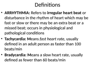
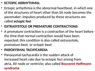
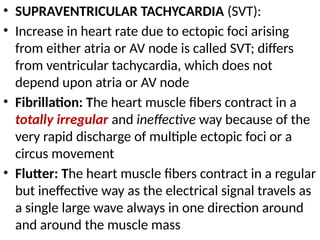
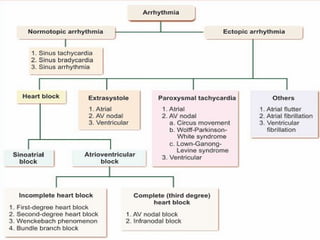
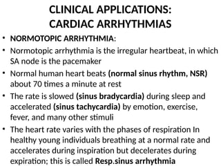
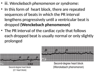
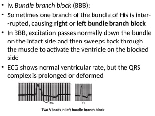
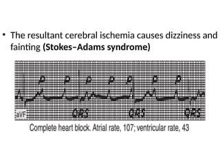
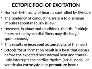
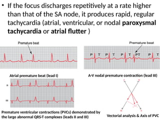
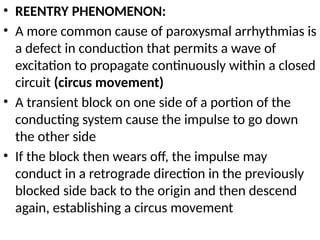
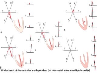
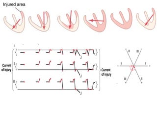
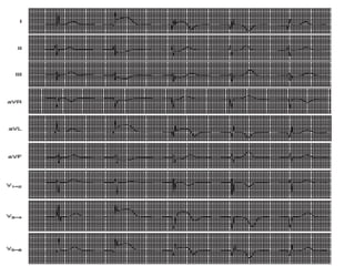
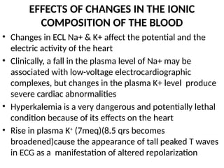
CARDIAC ARRYTHMIAS
KAMRAN
Definitions
• ARRHYTHMIA: Refers to irregular heart beat or
disturbance in the rhythm of heart which may be
fast or slow or there may be an extra beat or a
missed beat; occurs in physiological and
pathological conditions
• Tachycardia: Means fast heart rate, usually
defined in an adult person as faster than 100
beats/min
• Bradycardia: Means a slow heart rate, usually
defined as fewer than 60 beats/min
• ECTOPIC ARRHYTHMIA:
• Ectopic arrhythmia is the abnormal heartbeat, in which one
of the structures of heart other than SA node becomes the
pacemaker; impulses produced by these structures are
called ectopic foci
• EXTRASYSTOLE OR PREMATURE CONTRACTIONS:
• A premature contraction is a contraction of the heart before
the time that normal contraction would have been
expected; this condition is also called extrasystole,
premature beat, or ectopic beat
• PAROXYSMAL TACHYCARDIA
• Paroxysmal tachycardia is the sudden attack of
increased heart rate due to ectopic foci arising from
atria, AV node or ventricle; also called Bouveret Hoffmann
syndrome
• SUPRAVENTRICULAR TACHYCARDIA (SVT):
• Increase in heart rate due to ectopic foci arising
from either atria or AV node is called SVT; differs
from ventricular tachycardia, which does not
depend upon atria or AV node
• Fibrillation: The heart muscle fibers contract in a
totally irregular and ineffective way because of the
very rapid discharge of multiple ectopic foci or a
circus movement
• Flutter: The heart muscle fibers contract in a regular
but ineffective way as the electrical signal travels as
a single large wave always in one direction around
and around the muscle mass
CLINICAL APPLICATIONS:
CARDIAC ARRHYTHMIAS
• NORMOTOPIC ARRHYTHMIA:
• Normotopic arrhythmia is the irregular heartbeat, in which
SA node is the pacemaker
• Normal human heart beats (normal sinus rhythm, NSR)
about 70 times a minute at rest
• The rate is slowed (sinus bradycardia) during sleep and
accelerated (sinus tachycardia) by emotion, exercise,
fever, and many other stimuli
• The heart rate varies with the phases of respiration In
healthy young individuals breathing at a normal rate and
accelerates during inspiration but decelerates during
expiration; this is called Resp.sinus arrhythmia
Respiratory sinus arrhythmia (RSA)
Sinus tachycardia (lead I)
Sinus bradycardia (lead III)
• Sick Sinus Syndrome:
• Sick sinus syndrome (bradycardia-tachycardia
syndrome; sinus node dysfunction) is a collection of
heart rhythm disorders that include sinus
bradycardias, tachycardias and bradycardia-tachycardia
(alternating slow and fast heart rhythms)
• Uncommon and is usually found in people older
than 50, in whom the cause is often a nonspecific,
scar-like degeneration of the heart’s conduction
system
• When found in younger people, especially in
children, a common cause of sick sinus syndrome is
heart surgery, especially on the upper chambers
ABNORMAL PACEMAKERS
• Normal pacemaker of the heart is SA node and its
discharge rate is more rapid than that of the other
parts of the conduction system
• This is the reason that the SA node normally
controls the heart rate
• The AV node and other portions of the conduction
system can, in abnormal situations, become the
cardiac pacemaker
• In addition, diseased atrial and ventricular muscle
fibers can have their membrane potentials reduced
and discharge repetitively
• Heart Block:
• Heart block is the blockage of impulses generated
by SA node in the conductive system and this results
in improper or no impulse delivered to heart muscles,
resulting in ectopic arrhythmia
• Two types:
• 1. Sinoatrial block
• 2. Atrioventricular block
• 1. Sinoatrial Block – AV Nodal Rhythm:
• In rare instances, the impulse from the sinus node is
blocked before it enters the atrial muscle
• In ECG, it appears as sudden cessation of P waves,
with resultant standstill of the atria
• However, the ventricles pick up a
new rhythm from the impulse
usually originating from the A-V
node which takes over the
pacemaker function
• In ECG, the rate of the ventricular
QRS-T complex is slowed but not
otherwise altered
• SA block is due to the defect in
internodal fibers and it occurs
suddenly
• Any part of the AV node can starts
beating with decreased rate of 40 to
60/minute,ie., upper, middle or
lower part of AV node
• Upper nodal rhythm of AV node: the appearance of
inverted ‘P’ wave in ECG;
• The middle nodal rhythm: all chambers of the heart
contract simultaneously, thus, no appearance of ‘P’
wave or its merging with QRS complex;
• In lower nodal rhythm, QRS complex appears prior
to P wave and R-P interval is obtained instead of P-R
interval; called reversed heart block
• Atrioventricular Block:
• The only way by which impulses ordinarily can pass
from the atria into the ventricles is through the A-V
bundle, also known as the bundle of His
• Defect in the bundle or its branches either lead to
delay of impulses from atria to ventricles
(incomplete heart block) or no delivery of impulses
(complete heart block)
• In case of incomplete heart block, Impulses reach
ventricles late but in case of complete heart block,
the ventricles beat in their own rhythm,
independent of atrial beat
• Incomplete heart block is of four types:
i. First degree heart block
ii. Second degree heart block
iii. Wenckebach phenomenon
iv. Bundle branch block
• i. First degree heart block:
• When the conduction of impulses through AV node is
very slow, i.e. the AV nodal delay is longer, it is called 1st
degree heart block; common in young adults and
atheletes; rheumatic fever & some drugs
• In ECG, the P-R interval appears to be longer more than
0.2 second (normal 0.16 seconds); no other symptoms
• ii. Second degree heart block:
• Some of the impulses produced by SA node fail
to reach the ventricles; also called the partial heart
block
• The ventricular contraction occurs for every 2, 3 or 4
atrial contractions, i.e. 2 : 1, 3 : 1 or 4
• In ECG, the QRST complex is missing accordingly
Prolonged P-R interval caused by first degree
A-V heart block (lead II)
Second degree A-V block, showing occasional
failure of the ventricles to receive the
excitatory signals (lead V3)
• iii. Wenckebach phenomenon or syndrome:
• In this form of heart block, there are repeated
sequences of beats in which the PR interval
lengthens progressively until a ventricular beat is
dropped (Wenckebach phenomenon)
• The PR interval of the cardiac cycle that follows
each dropped beat is usually normal or only slightly
prolonged
• iv. Bundle branch block (BBB):
• Sometimes one branch of the bundle of His is inter-
-rupted, causing right or left bundle branch block
• In BBB, excitation passes normally down the bundle
on the intact side and then sweeps back through
the muscle to activate the ventricle on the blocked
side
• ECG shows normal ventricular rate, but the QRS
complex is prolonged or deformed
Two V leads in left bundle branch block
• Block can also occur in the anterior or posterior
fascicle of the left bundle branch, producing the
condition called hemiblock or fascicular block
• Left anterior hemiblock produces abnormal left axis
deviation in the ECG, whereas left posterior hemi-
-block produces abnormal right axis deviation
• 2. Complete Heart block 3rd
degree HB:
• Also called complete AV block or third degree heart
block
• In this case, conduction from the atria to the
ventricles is completely interrupted and the
ventricles beat at a low rate (idioventricular
rhythm) independently of the atria
• The block may be due to disease in the AV node (AV
nodal block) or in the conducting system below the
node (infranodal block)
• In AV nodal block, the unaffected tissue becomes
the pacemaker and the rate of the idioventricular
rhythm is approximately 45 beats/min
• In infranodal block due to disease in the bundle of
His, the ventricular pacemaker is located more
peripherally in the conduction system and the
ventricular rate is lower averaging 35 beats/min,
(may low as 15 beats/min in individual cases)
• The resultant cerebral ischemia causes dizziness and
fainting (Stokes–Adams syndrome)
ECTOPIC FOCI OF EXCITATION
• Normal rhythmicity of heart is controlled by SAnode
• The tendency of conducting system to discharge
impulses spontaneously is low
• However, in abnormal conditions, the His–Purkinje
fibers or the myocardial fibers may discharge
spontaneously
• This results in increased automaticity of the heart
• Ectopic focus formation result in a beat that occurs
before the expected next normal beat and transie-
-ntly interrupts the cardiac rhythm (atrial, nodal, or
ventricular extrasystole or premature beat )
• If the focus discharges repetitively at a rate higher
than that of the SA node, it produces rapid, regular
tachycardia (atrial, ventricular, or nodal paroxysmal
tachycardia or atrial flutter )
Atrial premature beat (lead I) A-V nodal premature contraction (lead III)
Premature ventricular contractions (PVCs) demonstrated by
the large abnormal QRS-T complexes (leads II and III) Vectorial analysis & Axis of PVC
• REENTRY PHENOMENON:
• A more common cause of paroxysmal arrhythmias is
a defect in conduction that permits a wave of
excitation to propagate continuously within a closed
circuit (circus movement)
• A transient block on one side of a portion of the
conducting system cause the impulse to go down
the other side
• If the block then wears off, the impulse may
conduct in a retrograde direction in the previously
blocked side back to the origin and then descend
again, establishing a circus movement
ATRIAL ARRHYTHMIAS
• Excitation spreading from ectopic focus in the atria
stimulates the AV node prematurely and is conducted to
the ventricles
• The P waves of atrial extrasystoles are abnormal, but the
QRST configurations are usually normal
• The excitation may depolarize the SA node, which must
repolarize and then depolarize to the firing level before it
can initiate the next normal beat
• This results a pause between the extrasystole and the
next normal beat
• The length of this pause is usually equal to the interval
between the normal beats preceding the extrasystole,
and the rhythm is “reset”
• Atrial tachycardia occurs when an atrial focus
discharges regularly or there is reentrant activity
producing atrial rates up to 220/min
• P wave in ECG is inverted, with normal QRST in
paroxysmal atrial tachycardia
• The atrial rate is 200–350/min in atrial flutter
• Max. number of impulses conducted by AV node is
about 230 to 240 /minute and there is large counter
clockwise circus movement in the right atrium
• So, during atrial flutter, the second degree of heart
block occurs
• This produces a characteristic sawtooth pattern of
flutter waves due to atrial contractions
• Prolonged atrial flutter may lead to AF or HF
• In atrial fibrillation (AF), the atria beat very rapidly (300–
500/min) in a completely irregular and disorganized fashion
• Because the AV node discharges at irregular intervals, the
ventricles also beat at a completely irregular rate, usually
80–160/min
• The condition can be paroxysmal or chronic, and in some
cases may be genetic predisposition
• Most of the cases are due to multiple concurrently
circulating reentrant excitation waves in both atria
• Some cases seem to be produced by discharge of one or
more ectopic foci
• Many of these foci appear to be located in the pulmonary
veins as much as 4 cm from the heart
VENTRICULAR ARRHYTHMIAS
• Premature beats originating from ventricular ectopic
focus slowly spread to the rest of ventricles and usually
have bizarrely shaped prolonged QRS
• They are usually incapable of exciting the bundle of
His, and retrograde conduction to the atria therefore
does not occur
• In the meantime, the next succeeding normal SA nodal
impulse depolarizes the atria
• The P wave is usually buried in the QRS of the
extrasystole
• If the normal impulse reaches the ventricles, they are still
in the refractory period following depolarization from the
ectopic focus
• However, the second succeeding impulse from the
SA node produces a normal beat
• Thus, ventricular premature beats are followed by a
compensatory pause that is often longer than the
pause after an atrial extrasystole
• Furthermore, ventricular premature beats do not
interrupt the regular discharge of the SA node,
whereas atrial premature beats often interrupt and
“reset” the normal rhythm
• Atrial and ventricular premature beats are not
strong enough to produce a pulse at the wrist if
they occur early in diastole; improper diastole filling
• Paroxysmal VT arise as a result of a series of rapid,
regular ventricular depolarizations usually due to a
circus movement involving the ventricles
• Torsade de pointes is a form of VT in which the QRS
morphology varies
• SV tachycardias (paroxysmal nodal tachycardia) can
be distinguished from paroxysmal VT by use of the
HBE; in SVT, a His bundle H deflection is present,
whereas in VT, there is none
• Ventricular premature beats, in the absence of
ischemic heart disease, are usually benign
• In VF, the ventricular muscle contract in a totally
irregular and ineffective way due to very rapid
discharge of multiple ventricular ectopic foci or a
circus movement
• VF can be produced by an electric shock or an extra-
systole during a critical interval, the vulnerable
period
• This period coincides in time with the midportion of
the T wave, i.e., the time when some of the ventri-
-cular muscles are depolarized, some in the state of
repolarizing, and some completely repolarized
• The fibrillating ventricles cannot pump blood
effectively, and circulation of the blood stops
ACCELERATED AV CONDUCTION
• Normally, the only conducting pathway between
the atria and the ventricles is the AV node
• However in some individuals, who are prone to
attacks of paroxysmal atrial arrhythmias, have an
additional aberrant muscular or nodal tissue
connection (bundle of Kent) between the atria and
ventricles
• This conducts impulses more rapidly than the slowly
conducting AV node, and the ventricles are excited
early resulting in VT
• VT is more serious because cardiac output is ↓, and
VF is an occasional complication of VT
• On ECG, it appears as a short PR interval and a
prolonged QRS deflection slurred on the upstroke,
with a normal interval between the start of the P
wave and the end of the QRS complex (“PJ interval”)
• This represents accelerated AV conduction and the
syndrome is called Wolf–Parkinson White syndrome
• The paroxysmal atrial tachycardias seen in this
syndrome often follow an atrial premature beat
• This beat conducts normally down the AV node but
spreads to the ventricular end of the aberrant
bundle, and the impulse is transmitted retrograde to
the atrium
Accelerated AV conduction
Normal sinus beat
Short PR interval; wide, slurred QRS complex normal
PJ interval (Wolf –Parkinson–White syndrome)
Short PR interval, short PJ interval, normal
QRS complex (Ganong–Levine syndrome)
• A circus movement is thus established
• Less commonly, an atrial premature beat finds the AV
node refractory but reaches the ventricles via the
bundle of Kent, setting up a circus movement in which
the impulse passes from the ventricles to the atria via
the AV node
• Attacks of paroxysml SVT, usually nodal tachycardia,
are seen in individuals with short PR intervals and
normal QRS complexes (Lown–Ganong–Levine
syndrome)
• In this condition, depolarization presumably passes
from the atria to the ventricles via bundle of Kent
that bypasses the AV node but enters the
intraventricular conducting system distal to the node
CURRENT OF INJURY
• Current of injury means flow of current from an
injured region of heart to the unaffected part
• When ischemia occurs in any part of the ventricular
musculature due to coronary occlusion, that part of
ventricle becomes depolarized either partially or
completely and the repolarization does not occur
• It causes flow of current from affected (depolarized)
part to unaffected part of the ventricular muscle
• Current of injury in myocardial infarction affects the
ECG pattern and cardiac vector
• In ECG, the J point and ST segments are displaced
• Cardiac Axis:
• In the infarction of anterior wall of the
ventricle, the cardiac axis (vector) is deviated
to right up to +150° due to current of injury
and in the posterior wall infarction, there is
left axis deviation up to –95°
Shaded areas of the ventricles are depolarized (−); nonshaded areas are still polarized (+)
EFFECTS OF CHANGES IN THE IONIC
COMPOSITION OF THE BLOOD
• Changes in ECL Na+ & K+ affect the potential and the
electric activity of the heart
• Clinically, a fall in the plasma level of Na+ may be
associated with low-voltage electrocardiographic
complexes, but changes in the plasma K+ level produce
severe cardiac abnormalities
• Hyperkalemia is a very dangerous and potentially lethal
condition because of its effects on the heart
• Rise in plasma K+
(7meq)(8.5 qrs becomes
broadened)cause the appearance of tall peaked T waves
in ECG as a manifestation of altered repolarization
• Paralysis of the atria and prolongation of the QRS
complexes occur at even higher conc.
• Ventricular arrhythmias may develop
• The fibers eventually become unexcitable, and the heart
stops in diastole
• Conversely, a ↓ in the plasma K+ level causes prolong-
-ation of the PR interval, prominent U wave, & occasio- -
nally, late T wave inversion in the precardial leads
• ↑ in extracellular Ca2+ conc. ↑ myocardial contractility
• When large amounts of Ca2+ are infused into
experimental animals, the heart relaxes less during
diastole and eventually stops in systole(calcium rigor)
• Hypocalcemia causes prolongation of the ST segment
and consequently of the QT interval
arrhthmias (1).pptx......dr kamran .......

arrhthmias (1).pptx......dr kamran .......

  • 1.
  • 2.
    Definitions • ARRHYTHMIA: Refersto irregular heart beat or disturbance in the rhythm of heart which may be fast or slow or there may be an extra beat or a missed beat; occurs in physiological and pathological conditions • Tachycardia: Means fast heart rate, usually defined in an adult person as faster than 100 beats/min • Bradycardia: Means a slow heart rate, usually defined as fewer than 60 beats/min
  • 3.
    • ECTOPIC ARRHYTHMIA: •Ectopic arrhythmia is the abnormal heartbeat, in which one of the structures of heart other than SA node becomes the pacemaker; impulses produced by these structures are called ectopic foci • EXTRASYSTOLE OR PREMATURE CONTRACTIONS: • A premature contraction is a contraction of the heart before the time that normal contraction would have been expected; this condition is also called extrasystole, premature beat, or ectopic beat • PAROXYSMAL TACHYCARDIA • Paroxysmal tachycardia is the sudden attack of increased heart rate due to ectopic foci arising from atria, AV node or ventricle; also called Bouveret Hoffmann syndrome
  • 4.
    • SUPRAVENTRICULAR TACHYCARDIA(SVT): • Increase in heart rate due to ectopic foci arising from either atria or AV node is called SVT; differs from ventricular tachycardia, which does not depend upon atria or AV node • Fibrillation: The heart muscle fibers contract in a totally irregular and ineffective way because of the very rapid discharge of multiple ectopic foci or a circus movement • Flutter: The heart muscle fibers contract in a regular but ineffective way as the electrical signal travels as a single large wave always in one direction around and around the muscle mass
  • 6.
    CLINICAL APPLICATIONS: CARDIAC ARRHYTHMIAS •NORMOTOPIC ARRHYTHMIA: • Normotopic arrhythmia is the irregular heartbeat, in which SA node is the pacemaker • Normal human heart beats (normal sinus rhythm, NSR) about 70 times a minute at rest • The rate is slowed (sinus bradycardia) during sleep and accelerated (sinus tachycardia) by emotion, exercise, fever, and many other stimuli • The heart rate varies with the phases of respiration In healthy young individuals breathing at a normal rate and accelerates during inspiration but decelerates during expiration; this is called Resp.sinus arrhythmia
  • 7.
    Respiratory sinus arrhythmia(RSA) Sinus tachycardia (lead I) Sinus bradycardia (lead III)
  • 8.
    • Sick SinusSyndrome: • Sick sinus syndrome (bradycardia-tachycardia syndrome; sinus node dysfunction) is a collection of heart rhythm disorders that include sinus bradycardias, tachycardias and bradycardia-tachycardia (alternating slow and fast heart rhythms) • Uncommon and is usually found in people older than 50, in whom the cause is often a nonspecific, scar-like degeneration of the heart’s conduction system • When found in younger people, especially in children, a common cause of sick sinus syndrome is heart surgery, especially on the upper chambers
  • 9.
    ABNORMAL PACEMAKERS • Normalpacemaker of the heart is SA node and its discharge rate is more rapid than that of the other parts of the conduction system • This is the reason that the SA node normally controls the heart rate • The AV node and other portions of the conduction system can, in abnormal situations, become the cardiac pacemaker • In addition, diseased atrial and ventricular muscle fibers can have their membrane potentials reduced and discharge repetitively
  • 10.
    • Heart Block: •Heart block is the blockage of impulses generated by SA node in the conductive system and this results in improper or no impulse delivered to heart muscles, resulting in ectopic arrhythmia • Two types: • 1. Sinoatrial block • 2. Atrioventricular block • 1. Sinoatrial Block – AV Nodal Rhythm: • In rare instances, the impulse from the sinus node is blocked before it enters the atrial muscle • In ECG, it appears as sudden cessation of P waves, with resultant standstill of the atria
  • 11.
    • However, theventricles pick up a new rhythm from the impulse usually originating from the A-V node which takes over the pacemaker function • In ECG, the rate of the ventricular QRS-T complex is slowed but not otherwise altered • SA block is due to the defect in internodal fibers and it occurs suddenly • Any part of the AV node can starts beating with decreased rate of 40 to 60/minute,ie., upper, middle or lower part of AV node
  • 12.
    • Upper nodalrhythm of AV node: the appearance of inverted ‘P’ wave in ECG; • The middle nodal rhythm: all chambers of the heart contract simultaneously, thus, no appearance of ‘P’ wave or its merging with QRS complex; • In lower nodal rhythm, QRS complex appears prior to P wave and R-P interval is obtained instead of P-R interval; called reversed heart block • Atrioventricular Block: • The only way by which impulses ordinarily can pass from the atria into the ventricles is through the A-V bundle, also known as the bundle of His
  • 13.
    • Defect inthe bundle or its branches either lead to delay of impulses from atria to ventricles (incomplete heart block) or no delivery of impulses (complete heart block) • In case of incomplete heart block, Impulses reach ventricles late but in case of complete heart block, the ventricles beat in their own rhythm, independent of atrial beat • Incomplete heart block is of four types: i. First degree heart block ii. Second degree heart block iii. Wenckebach phenomenon iv. Bundle branch block
  • 14.
    • i. Firstdegree heart block: • When the conduction of impulses through AV node is very slow, i.e. the AV nodal delay is longer, it is called 1st degree heart block; common in young adults and atheletes; rheumatic fever & some drugs • In ECG, the P-R interval appears to be longer more than 0.2 second (normal 0.16 seconds); no other symptoms • ii. Second degree heart block: • Some of the impulses produced by SA node fail to reach the ventricles; also called the partial heart block • The ventricular contraction occurs for every 2, 3 or 4 atrial contractions, i.e. 2 : 1, 3 : 1 or 4 • In ECG, the QRST complex is missing accordingly
  • 15.
    Prolonged P-R intervalcaused by first degree A-V heart block (lead II) Second degree A-V block, showing occasional failure of the ventricles to receive the excitatory signals (lead V3)
  • 16.
    • iii. Wenckebachphenomenon or syndrome: • In this form of heart block, there are repeated sequences of beats in which the PR interval lengthens progressively until a ventricular beat is dropped (Wenckebach phenomenon) • The PR interval of the cardiac cycle that follows each dropped beat is usually normal or only slightly prolonged
  • 17.
    • iv. Bundlebranch block (BBB): • Sometimes one branch of the bundle of His is inter- -rupted, causing right or left bundle branch block • In BBB, excitation passes normally down the bundle on the intact side and then sweeps back through the muscle to activate the ventricle on the blocked side • ECG shows normal ventricular rate, but the QRS complex is prolonged or deformed Two V leads in left bundle branch block
  • 18.
    • Block canalso occur in the anterior or posterior fascicle of the left bundle branch, producing the condition called hemiblock or fascicular block • Left anterior hemiblock produces abnormal left axis deviation in the ECG, whereas left posterior hemi- -block produces abnormal right axis deviation • 2. Complete Heart block 3rd degree HB: • Also called complete AV block or third degree heart block • In this case, conduction from the atria to the ventricles is completely interrupted and the ventricles beat at a low rate (idioventricular rhythm) independently of the atria
  • 19.
    • The blockmay be due to disease in the AV node (AV nodal block) or in the conducting system below the node (infranodal block) • In AV nodal block, the unaffected tissue becomes the pacemaker and the rate of the idioventricular rhythm is approximately 45 beats/min • In infranodal block due to disease in the bundle of His, the ventricular pacemaker is located more peripherally in the conduction system and the ventricular rate is lower averaging 35 beats/min, (may low as 15 beats/min in individual cases)
  • 20.
    • The resultantcerebral ischemia causes dizziness and fainting (Stokes–Adams syndrome)
  • 21.
    ECTOPIC FOCI OFEXCITATION • Normal rhythmicity of heart is controlled by SAnode • The tendency of conducting system to discharge impulses spontaneously is low • However, in abnormal conditions, the His–Purkinje fibers or the myocardial fibers may discharge spontaneously • This results in increased automaticity of the heart • Ectopic focus formation result in a beat that occurs before the expected next normal beat and transie- -ntly interrupts the cardiac rhythm (atrial, nodal, or ventricular extrasystole or premature beat )
  • 22.
    • If thefocus discharges repetitively at a rate higher than that of the SA node, it produces rapid, regular tachycardia (atrial, ventricular, or nodal paroxysmal tachycardia or atrial flutter ) Atrial premature beat (lead I) A-V nodal premature contraction (lead III) Premature ventricular contractions (PVCs) demonstrated by the large abnormal QRS-T complexes (leads II and III) Vectorial analysis & Axis of PVC
  • 23.
    • REENTRY PHENOMENON: •A more common cause of paroxysmal arrhythmias is a defect in conduction that permits a wave of excitation to propagate continuously within a closed circuit (circus movement) • A transient block on one side of a portion of the conducting system cause the impulse to go down the other side • If the block then wears off, the impulse may conduct in a retrograde direction in the previously blocked side back to the origin and then descend again, establishing a circus movement
  • 24.
    ATRIAL ARRHYTHMIAS • Excitationspreading from ectopic focus in the atria stimulates the AV node prematurely and is conducted to the ventricles • The P waves of atrial extrasystoles are abnormal, but the QRST configurations are usually normal • The excitation may depolarize the SA node, which must repolarize and then depolarize to the firing level before it can initiate the next normal beat • This results a pause between the extrasystole and the next normal beat • The length of this pause is usually equal to the interval between the normal beats preceding the extrasystole, and the rhythm is “reset”
  • 25.
    • Atrial tachycardiaoccurs when an atrial focus discharges regularly or there is reentrant activity producing atrial rates up to 220/min • P wave in ECG is inverted, with normal QRST in paroxysmal atrial tachycardia • The atrial rate is 200–350/min in atrial flutter • Max. number of impulses conducted by AV node is about 230 to 240 /minute and there is large counter clockwise circus movement in the right atrium • So, during atrial flutter, the second degree of heart block occurs • This produces a characteristic sawtooth pattern of flutter waves due to atrial contractions
  • 26.
    • Prolonged atrialflutter may lead to AF or HF • In atrial fibrillation (AF), the atria beat very rapidly (300– 500/min) in a completely irregular and disorganized fashion • Because the AV node discharges at irregular intervals, the ventricles also beat at a completely irregular rate, usually 80–160/min • The condition can be paroxysmal or chronic, and in some cases may be genetic predisposition • Most of the cases are due to multiple concurrently circulating reentrant excitation waves in both atria • Some cases seem to be produced by discharge of one or more ectopic foci • Many of these foci appear to be located in the pulmonary veins as much as 4 cm from the heart
  • 28.
    VENTRICULAR ARRHYTHMIAS • Prematurebeats originating from ventricular ectopic focus slowly spread to the rest of ventricles and usually have bizarrely shaped prolonged QRS • They are usually incapable of exciting the bundle of His, and retrograde conduction to the atria therefore does not occur • In the meantime, the next succeeding normal SA nodal impulse depolarizes the atria • The P wave is usually buried in the QRS of the extrasystole • If the normal impulse reaches the ventricles, they are still in the refractory period following depolarization from the ectopic focus
  • 29.
    • However, thesecond succeeding impulse from the SA node produces a normal beat • Thus, ventricular premature beats are followed by a compensatory pause that is often longer than the pause after an atrial extrasystole • Furthermore, ventricular premature beats do not interrupt the regular discharge of the SA node, whereas atrial premature beats often interrupt and “reset” the normal rhythm • Atrial and ventricular premature beats are not strong enough to produce a pulse at the wrist if they occur early in diastole; improper diastole filling
  • 30.
    • Paroxysmal VTarise as a result of a series of rapid, regular ventricular depolarizations usually due to a circus movement involving the ventricles • Torsade de pointes is a form of VT in which the QRS morphology varies • SV tachycardias (paroxysmal nodal tachycardia) can be distinguished from paroxysmal VT by use of the HBE; in SVT, a His bundle H deflection is present, whereas in VT, there is none • Ventricular premature beats, in the absence of ischemic heart disease, are usually benign
  • 31.
    • In VF,the ventricular muscle contract in a totally irregular and ineffective way due to very rapid discharge of multiple ventricular ectopic foci or a circus movement • VF can be produced by an electric shock or an extra- systole during a critical interval, the vulnerable period • This period coincides in time with the midportion of the T wave, i.e., the time when some of the ventri- -cular muscles are depolarized, some in the state of repolarizing, and some completely repolarized • The fibrillating ventricles cannot pump blood effectively, and circulation of the blood stops
  • 33.
    ACCELERATED AV CONDUCTION •Normally, the only conducting pathway between the atria and the ventricles is the AV node • However in some individuals, who are prone to attacks of paroxysmal atrial arrhythmias, have an additional aberrant muscular or nodal tissue connection (bundle of Kent) between the atria and ventricles • This conducts impulses more rapidly than the slowly conducting AV node, and the ventricles are excited early resulting in VT • VT is more serious because cardiac output is ↓, and VF is an occasional complication of VT
  • 34.
    • On ECG,it appears as a short PR interval and a prolonged QRS deflection slurred on the upstroke, with a normal interval between the start of the P wave and the end of the QRS complex (“PJ interval”) • This represents accelerated AV conduction and the syndrome is called Wolf–Parkinson White syndrome • The paroxysmal atrial tachycardias seen in this syndrome often follow an atrial premature beat • This beat conducts normally down the AV node but spreads to the ventricular end of the aberrant bundle, and the impulse is transmitted retrograde to the atrium
  • 35.
    Accelerated AV conduction Normalsinus beat Short PR interval; wide, slurred QRS complex normal PJ interval (Wolf –Parkinson–White syndrome) Short PR interval, short PJ interval, normal QRS complex (Ganong–Levine syndrome)
  • 36.
    • A circusmovement is thus established • Less commonly, an atrial premature beat finds the AV node refractory but reaches the ventricles via the bundle of Kent, setting up a circus movement in which the impulse passes from the ventricles to the atria via the AV node • Attacks of paroxysml SVT, usually nodal tachycardia, are seen in individuals with short PR intervals and normal QRS complexes (Lown–Ganong–Levine syndrome) • In this condition, depolarization presumably passes from the atria to the ventricles via bundle of Kent that bypasses the AV node but enters the intraventricular conducting system distal to the node
  • 37.
    CURRENT OF INJURY •Current of injury means flow of current from an injured region of heart to the unaffected part • When ischemia occurs in any part of the ventricular musculature due to coronary occlusion, that part of ventricle becomes depolarized either partially or completely and the repolarization does not occur • It causes flow of current from affected (depolarized) part to unaffected part of the ventricular muscle • Current of injury in myocardial infarction affects the ECG pattern and cardiac vector • In ECG, the J point and ST segments are displaced
  • 38.
    • Cardiac Axis: •In the infarction of anterior wall of the ventricle, the cardiac axis (vector) is deviated to right up to +150° due to current of injury and in the posterior wall infarction, there is left axis deviation up to –95°
  • 39.
    Shaded areas ofthe ventricles are depolarized (−); nonshaded areas are still polarized (+)
  • 42.
    EFFECTS OF CHANGESIN THE IONIC COMPOSITION OF THE BLOOD • Changes in ECL Na+ & K+ affect the potential and the electric activity of the heart • Clinically, a fall in the plasma level of Na+ may be associated with low-voltage electrocardiographic complexes, but changes in the plasma K+ level produce severe cardiac abnormalities • Hyperkalemia is a very dangerous and potentially lethal condition because of its effects on the heart • Rise in plasma K+ (7meq)(8.5 qrs becomes broadened)cause the appearance of tall peaked T waves in ECG as a manifestation of altered repolarization
  • 43.
    • Paralysis ofthe atria and prolongation of the QRS complexes occur at even higher conc. • Ventricular arrhythmias may develop • The fibers eventually become unexcitable, and the heart stops in diastole • Conversely, a ↓ in the plasma K+ level causes prolong- -ation of the PR interval, prominent U wave, & occasio- - nally, late T wave inversion in the precardial leads • ↑ in extracellular Ca2+ conc. ↑ myocardial contractility • When large amounts of Ca2+ are infused into experimental animals, the heart relaxes less during diastole and eventually stops in systole(calcium rigor) • Hypocalcemia causes prolongation of the ST segment and consequently of the QT interval