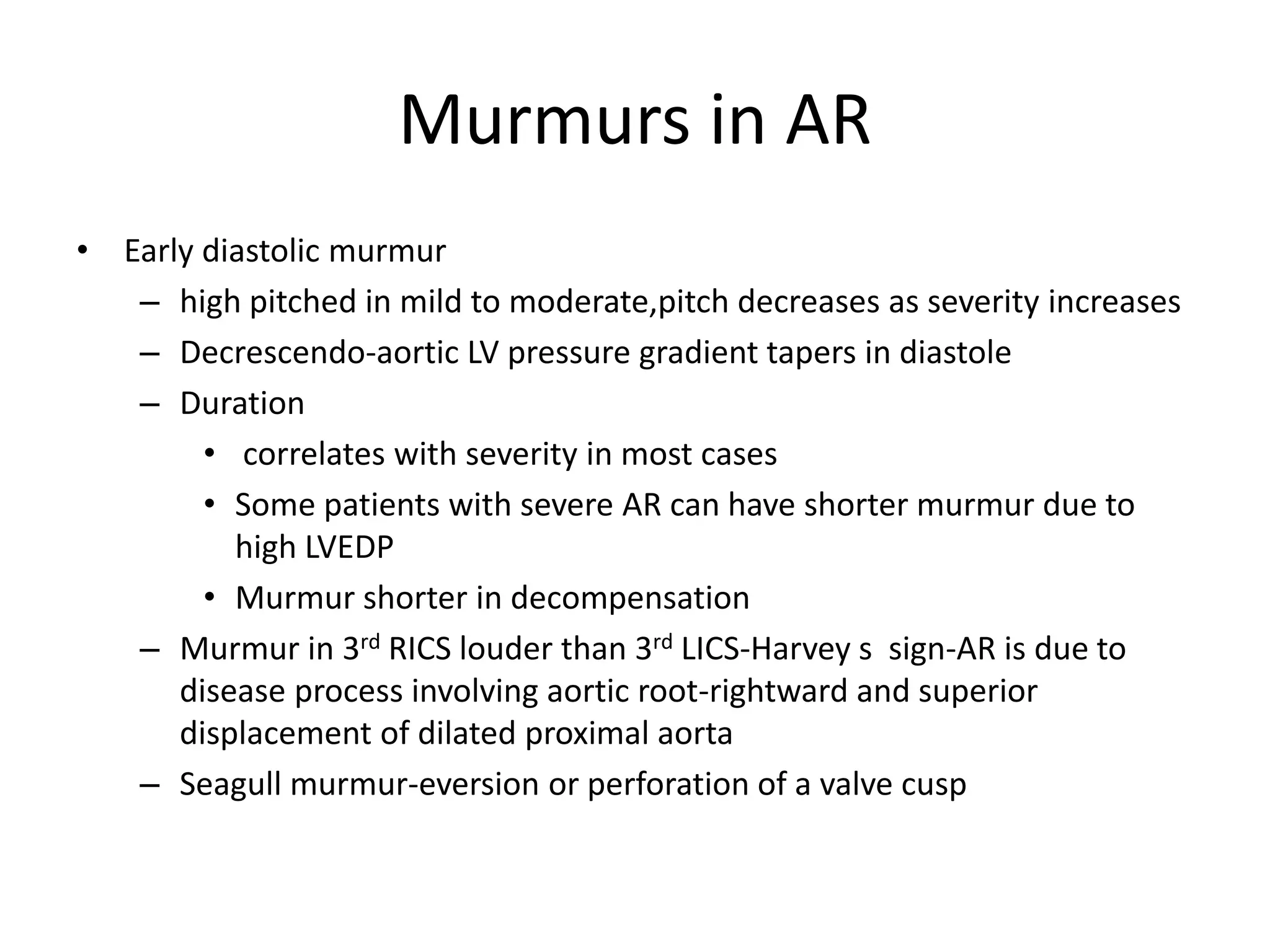
Clinical hemodynamic correlation
in aortic regurgitation
Etiology
• Aortic root disease
– Aortopathy
– Aortitis
– Age related aortic dilatation
• Valvular disease
– Calcific AS in older patients with AR
– Bicuspid aortic valve
– Cusp retraction or fibrosis
• c/c rheumatic
• Inflammatory
– Cusp perforation/tears
• Infective endocarditis
• Trauma
– Lack of cusp support
• Dissection of aorta
• VSD
Pathophysiology
• Volume overload –compensatory mechanisms
• LV EDV increases without increase in diastolic
pressure due to increased compliance
• LV preload reserve is maintained initially
– Eccentric hypertrophy
– Sarcomeres laid in series
– Preload at sarcomere level is near normal
– Normal contractile performance of each unit
contributes to enhanced stroke volume
Pathophysiology (continued)
• Increased afterload
– Increased chamber volume,increased systolic
pressure
– Increased systolic wall stress and afterload
– concentric LVH
• Continued increase in chamber volume and
afterload –matched by continued recruitment
of preload reserve and compensatory
hypertrophy
Pathophysiology(continued)
• Decompensation
– Afterload mismatch-reversible
– Impaired LV contractility-irreversible
A/c AR-pathophysiology
• Hemodynamically significant AR of sudden
onset,into a LV not previously subjected to
volume overload
• Volume overload is poorly tolerated
– Ventricular compliance is normal
– LV operating on steep portion of diastolic P/V
relation
– End diastolic LV pressure markedly increased
approaching aortic diastolic pressure
A/c AR-pathophysiology (ctd.)
• LV fails to increase stroke voume(not hypertrophied or
dilated)-Decrease in COP
• Increase in LVEDP causes rise in mean LA pressure and
PCWP-pulmonary edema
• Premature closure of MV –early crossover of pressures
• Diastolic MR
• Arterial BP-
• fall in systolic pressure
• Normal pulse pressure
• Diastolic pressure maintained by reflex increase in SVR in failure
c/c Vs a/c AR-hemodynamic
response
Variable A/c AR C/c AR c/c AR
decompensated
LVEDV Slight ↑ marked↑ Marked ↑
LV compliance normal increase Increased than
normal
LVEDP Marked ↑ modest↑ Marked increase
Forward stroke
volume
decreased normal decreased
Aortic systolic
presure
normal increased increased
Pulse pressure normal increased normal
Peripheral vascular
resistance
increased decreased increased
Hemodynamic assessment-c/c AR
• Elevated Ao.syst pressure
• Lowered Ao.diastolic pressure
• modest rise of LV pressures in diastole
• Premature closure of MV-when LV diastolic
pressure exceeds LA pressure-common in a/c AR
• Mean diastolic pressures rise with time and
severity of leak-rise in mean LA &PCWP
• Amplification of peak systolic pressure in
peripheral arteries
• Acute AR
– Regurgitation into non compliant LV -diastolic rise
of LV pressure&absence of A wave
– LV diastolic pressure exceeds LA pressure-
premature closure of MV
– Aortic and LV pressures equalise in diastole and
regurgitant flow &murmur ceases
Angiographic assessment
• Mild(1+)
– small amount of contrast
– never fills chamber
– cleared with each beat
• Moderate(2+)
– more contrast
– faint opacification of entire chamber
• Moderately severe(3+)
– LV well opacified
– equal in density with aorta
• Severe(4+)
– complete dense opacification of LV in one beat
– LV more densely opacified than aorta
Clinical features
• Asymptomatic phase longer
• Dyspnoea most common symptom
• Angina in 20% patients
– Decreased perfusion-low aortic diastolic pressure
– Increased myocardial oxygen demand
– Associated coronary atherosclerosis
– Osteal coronary invt.in syphilitic AR,takayasu arteritis
• Palpitations-
– awareness of forceful ventricular contraction
– Ventricular arrhythmias in decompensated stage
• Syncope-5 to 10%
Physical findings
• Elevated systolic pressure
• Low diastolic pressure
• Peripheral signs of AR-large stroke volume in
early systole with subsequent run off
• Hill s sign-exaggeration of peripheral
amplification
• Carotid thrill or shudder-more common in AS,but
also in AR
• Displacement of apical impulse
Heart sounds in AR
• S1-soft
– Increased LVEDP-earlier closure of MV
– Elevated diastolic pressure-less valve excursion
• S2-
– Soft A2 –valve structurally abnormal
– Delayed A2-prolonged LV ejection time
– P2 may be obscured by murmur
• S3 –
– in failure
• S4-
– Suggest decreased LV compliance&increased LVEDP
– Long PR interval
Murmurs in AR
• Early diastolic murmur
– high pitched in mild to moderate,pitch decreases as severity increases
– Decrescendo-aortic LV pressure gradient tapers in diastole
– Duration
• correlates with severity in most cases
• Some patients with severe AR can have shorter murmur due to
high LVEDP
• Murmur shorter in decompensation
– Murmur in 3rd RICS louder than 3rd LICS-Harvey s sign-AR is due to
disease process involving aortic root-rightward and superior
displacement of dilated proximal aorta
– Seagull murmur-eversion or perforation of a valve cusp
Murmurs in AR
• Systolic ejection murmur
– Increased LV stroke volume
– Abnormal Aortic valve
Murmurs in AR
• Austin Flint murmur
– low pitched,mid or late diastolic murmur
– Mechanism
• AR jet pushing AML
• Antegrade transmitral blood flow across a functionally narrowed
MV
• Diastolic MR
• Low pitched components of AR murmur heard best at apex
– Severe AR-reg. fraction>50%
– Severity of AR and AFM
• Mild-absent
• Moderate-may be present in late diastole
• Severe-earlier in timing,extend into presystole
• Very severe AR-premature closure of MV-absent presystolic
component
A/c AR
• Rapid onset of symptoms –
– rapid rise of LA pressure
– abrupt reduction of COP
• BP-
– Systolic pressure normal or slight fall
– elevated dia.pressure
– narrrow pulse pressure
• Acute rt heart failure can occur-elevated JVP
A/c AR
• Soft S1
• Soft A2,loud P2
• LV S3-rapid early diastolic filling
• Absent LVS4
• EDM
– Short -rapid diastolic equilibration of aortic and LV
pressures in diastole
– Low or medium pitch-
• Low gradient
• a/w CCF
• Austin Flint murmur presystolic component absent
Echocardiography
• Increased LV End Diastolic Dimensions ,near
normal end systolic dimensions and increased
contractility-compensated phase
• Increase in end systolic dimensions and
depressed contractility-decompensation
• M-Mode of MV
– Diastolic fluttering of AML in c/c AR
– Early closure of MV in a/c AR
• M-mode of AV
– Diastolic non coaptation,diastolic fluttering in c/c AR
– Premature opening of AV in a/c AR
THANK YOU

Aortic regurgitation

  • 1.
  • 2.
    Etiology • Aortic rootdisease – Aortopathy – Aortitis – Age related aortic dilatation • Valvular disease – Calcific AS in older patients with AR – Bicuspid aortic valve – Cusp retraction or fibrosis • c/c rheumatic • Inflammatory – Cusp perforation/tears • Infective endocarditis • Trauma – Lack of cusp support • Dissection of aorta • VSD
  • 5.
    Pathophysiology • Volume overload–compensatory mechanisms • LV EDV increases without increase in diastolic pressure due to increased compliance • LV preload reserve is maintained initially – Eccentric hypertrophy – Sarcomeres laid in series – Preload at sarcomere level is near normal – Normal contractile performance of each unit contributes to enhanced stroke volume
  • 6.
    Pathophysiology (continued) • Increasedafterload – Increased chamber volume,increased systolic pressure – Increased systolic wall stress and afterload – concentric LVH • Continued increase in chamber volume and afterload –matched by continued recruitment of preload reserve and compensatory hypertrophy
  • 7.
    Pathophysiology(continued) • Decompensation – Afterloadmismatch-reversible – Impaired LV contractility-irreversible
  • 8.
    A/c AR-pathophysiology • Hemodynamicallysignificant AR of sudden onset,into a LV not previously subjected to volume overload • Volume overload is poorly tolerated – Ventricular compliance is normal – LV operating on steep portion of diastolic P/V relation – End diastolic LV pressure markedly increased approaching aortic diastolic pressure
  • 9.
    A/c AR-pathophysiology (ctd.) •LV fails to increase stroke voume(not hypertrophied or dilated)-Decrease in COP • Increase in LVEDP causes rise in mean LA pressure and PCWP-pulmonary edema • Premature closure of MV –early crossover of pressures • Diastolic MR • Arterial BP- • fall in systolic pressure • Normal pulse pressure • Diastolic pressure maintained by reflex increase in SVR in failure
  • 10.
    c/c Vs a/cAR-hemodynamic response Variable A/c AR C/c AR c/c AR decompensated LVEDV Slight ↑ marked↑ Marked ↑ LV compliance normal increase Increased than normal LVEDP Marked ↑ modest↑ Marked increase Forward stroke volume decreased normal decreased Aortic systolic presure normal increased increased Pulse pressure normal increased normal Peripheral vascular resistance increased decreased increased
  • 11.
    Hemodynamic assessment-c/c AR •Elevated Ao.syst pressure • Lowered Ao.diastolic pressure • modest rise of LV pressures in diastole • Premature closure of MV-when LV diastolic pressure exceeds LA pressure-common in a/c AR • Mean diastolic pressures rise with time and severity of leak-rise in mean LA &PCWP • Amplification of peak systolic pressure in peripheral arteries
  • 13.
    • Acute AR –Regurgitation into non compliant LV -diastolic rise of LV pressure&absence of A wave – LV diastolic pressure exceeds LA pressure- premature closure of MV – Aortic and LV pressures equalise in diastole and regurgitant flow &murmur ceases
  • 15.
    Angiographic assessment • Mild(1+) –small amount of contrast – never fills chamber – cleared with each beat • Moderate(2+) – more contrast – faint opacification of entire chamber • Moderately severe(3+) – LV well opacified – equal in density with aorta • Severe(4+) – complete dense opacification of LV in one beat – LV more densely opacified than aorta
  • 16.
    Clinical features • Asymptomaticphase longer • Dyspnoea most common symptom • Angina in 20% patients – Decreased perfusion-low aortic diastolic pressure – Increased myocardial oxygen demand – Associated coronary atherosclerosis – Osteal coronary invt.in syphilitic AR,takayasu arteritis • Palpitations- – awareness of forceful ventricular contraction – Ventricular arrhythmias in decompensated stage • Syncope-5 to 10%
  • 17.
    Physical findings • Elevatedsystolic pressure • Low diastolic pressure • Peripheral signs of AR-large stroke volume in early systole with subsequent run off • Hill s sign-exaggeration of peripheral amplification • Carotid thrill or shudder-more common in AS,but also in AR • Displacement of apical impulse
  • 18.
    Heart sounds inAR • S1-soft – Increased LVEDP-earlier closure of MV – Elevated diastolic pressure-less valve excursion • S2- – Soft A2 –valve structurally abnormal – Delayed A2-prolonged LV ejection time – P2 may be obscured by murmur • S3 – – in failure • S4- – Suggest decreased LV compliance&increased LVEDP – Long PR interval
  • 19.
    Murmurs in AR •Early diastolic murmur – high pitched in mild to moderate,pitch decreases as severity increases – Decrescendo-aortic LV pressure gradient tapers in diastole – Duration • correlates with severity in most cases • Some patients with severe AR can have shorter murmur due to high LVEDP • Murmur shorter in decompensation – Murmur in 3rd RICS louder than 3rd LICS-Harvey s sign-AR is due to disease process involving aortic root-rightward and superior displacement of dilated proximal aorta – Seagull murmur-eversion or perforation of a valve cusp
  • 20.
    Murmurs in AR •Systolic ejection murmur – Increased LV stroke volume – Abnormal Aortic valve
  • 21.
    Murmurs in AR •Austin Flint murmur – low pitched,mid or late diastolic murmur – Mechanism • AR jet pushing AML • Antegrade transmitral blood flow across a functionally narrowed MV • Diastolic MR • Low pitched components of AR murmur heard best at apex – Severe AR-reg. fraction>50% – Severity of AR and AFM • Mild-absent • Moderate-may be present in late diastole • Severe-earlier in timing,extend into presystole • Very severe AR-premature closure of MV-absent presystolic component
  • 22.
    A/c AR • Rapidonset of symptoms – – rapid rise of LA pressure – abrupt reduction of COP • BP- – Systolic pressure normal or slight fall – elevated dia.pressure – narrrow pulse pressure • Acute rt heart failure can occur-elevated JVP
  • 23.
    A/c AR • SoftS1 • Soft A2,loud P2 • LV S3-rapid early diastolic filling • Absent LVS4 • EDM – Short -rapid diastolic equilibration of aortic and LV pressures in diastole – Low or medium pitch- • Low gradient • a/w CCF • Austin Flint murmur presystolic component absent
  • 24.
    Echocardiography • Increased LVEnd Diastolic Dimensions ,near normal end systolic dimensions and increased contractility-compensated phase • Increase in end systolic dimensions and depressed contractility-decompensation • M-Mode of MV – Diastolic fluttering of AML in c/c AR – Early closure of MV in a/c AR • M-mode of AV – Diastolic non coaptation,diastolic fluttering in c/c AR – Premature opening of AV in a/c AR
  • 28.

Editor's Notes