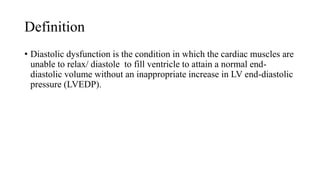
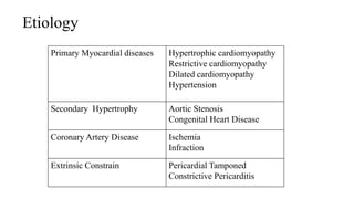
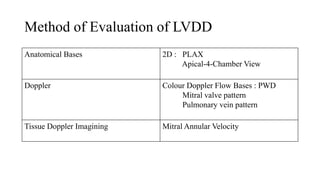
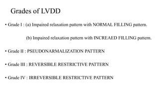
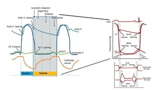
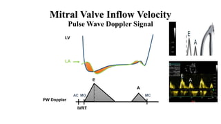
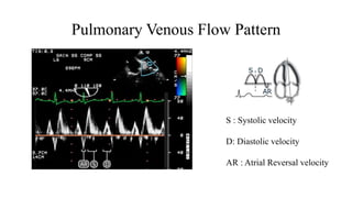
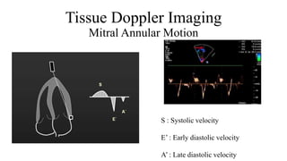
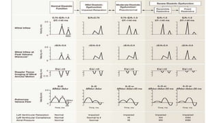
Left Ventricular Diastolic
Dysfunction
Name : Yukta Wankhede
PRN: 17040121108
Programme: B Sc. MT Cardiac Care Technology
Batch: 2017-2020
Definition
• Diastolic dysfunction is the condition in which the cardiac muscles are
unable to relax/ diastole to fill ventricle to attain a normal end-
diastolic volume without an inappropriate increase in LV end-diastolic
pressure (LVEDP).
Etiology
Primary Myocardial diseases Hypertrophic cardiomyopathy
Restrictive cardiomyopathy
Dilated cardiomyopathy
Hypertension
Secondary Hypertrophy Aortic Stenosis
Congenital Heart Disease
Coronary Artery Disease Ischemia
Infraction
Extrinsic Constrain Pericardial Tamponed
Constrictive Pericarditis
Anatomical Bases 2D : PLAX
Apical-4-Chamber View
Doppler Colour Doppler Flow Bases : PWD
Mitral valve pattern
Pulmonary vein pattern
Tissue Doppler Imagining Mitral Annular Velocity
Method of Evaluation of LVDD
Anatomical Bases
In 2D
• LV mass and dimensions
• LV hypertrophy is the commonest cause of diastolic dysfunction
Relative wall thickness
• LA volume and LA volume index
Grades of LVDD
• Grade I : (a) Impaired relaxation pattern with NORMAL FILLING pattern.
(b) Impaired relaxation pattern with INCREAED FILLING pattern.
• Grade II : PSEUDONARMALIZATION PATTERN
• Grade III : REVERSIBLE RESTRICTIVE PATTERN
• Grade IV : IRREVERSIBLE RESTRICTIVE PATTERN
E
A
IVRT
LV
LA
PW Doppler
AC MO MC
Mitral Valve Inflow Velocity
Pulse Wave Doppler Signal
Pulmonary Venous Flow Pattern
S : Systolic velocity
D: Diastolic velocity
AR : Atrial Reversal velocity
Tissue Doppler Imaging
Mitral Annular Motion
S : Systolic velocity
E’ : Early diastolic velocity
A’ : Late diastolic velocity
CASE STUDY
• An 88-year-old Japanese man presented to our emergency unit in a
comatose condition with a body temperature below the lower limit of
detection (<32°C when measured under the armpit; <34°C when
measured at the tympanic membrane) and no external injures.
• An electrocardiogram performed on admission showed sinus arrest
and junctional rhythm with a heart rate of 40 beats per minute. Osborn
waves were apparent in leads V4 to V6.
file:///C:/Users/USER/Downloads/Downloads/Cardiac%20Care/tudyyyyyyy/Left%20ventricular%20systolic%20dysfu
nction%20in%20a%20patient%20with%20accidental%20hypothermia_%20a%20case%20report%20_%20Journal%20
of%20Medical%20Case%20Reports%20_%20Full%20Text.html
Left ventricular diastolic dysfunction in echocardiography

Left ventricular diastolic dysfunction in echocardiography