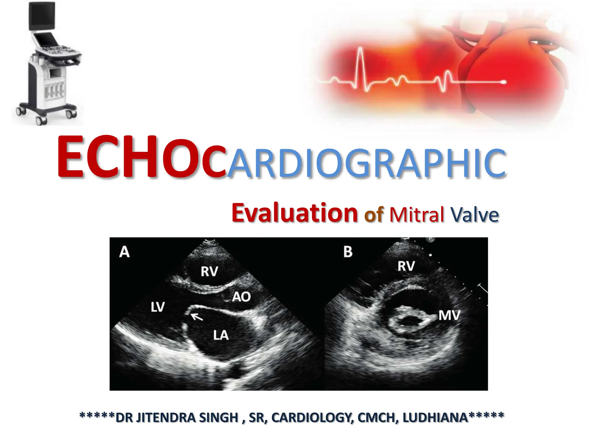
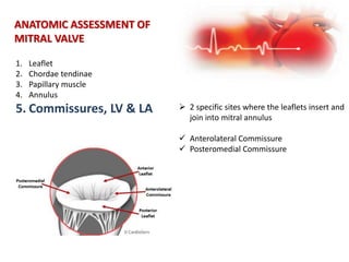
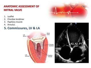
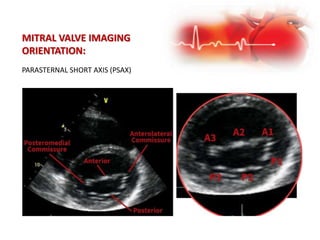
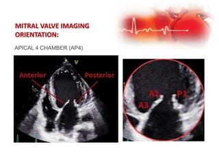
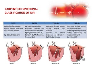
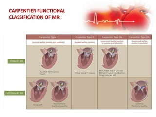
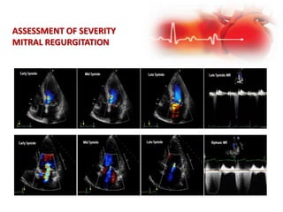
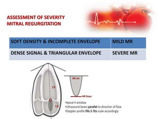
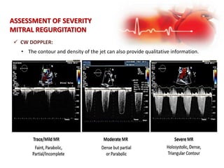
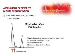
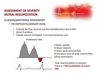
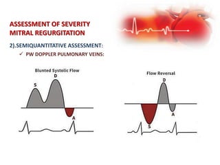
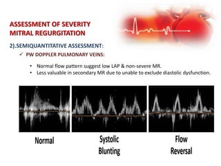
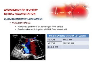
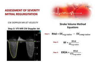
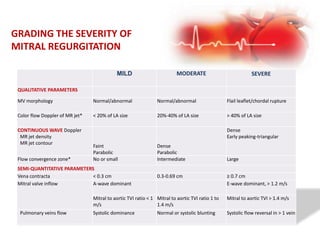
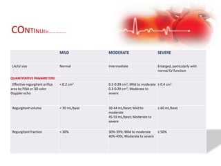
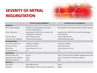
ECHOCARDIOGRAPHIC
Evaluation of Mitral Valve
*****DR JITENDRA SINGH , SR, CARDIOLOGY, CMCH, LUDHIANA*****
Q-Main questions to be answered by
cardiac imaging?
Q-MAIN QUESTIONS TO BE ANSWERED
BY CARDIOVASCULAR IMAGING?
WHAT IS THE MECHANISM OF MR?
 Leaflet morphology
 Leaflet motion (Carpentier type)
 Subvalvular involvement
 Annulus (dilation, calcium)
 LV size and function
 LA size and function
IS THE MR SEVERE?
 Quantitative (EROA, RV, RF)
 Qualitative (multiple)
WHAT IS THE EFFECT OF MR ON LV AND LA SIZE AND
LA/PULMONARY VENOUS PRESSURE?
 LV diameters, volumes, LVEF, GLS, fibrosis
 LA volume, pressure
 Estimated PA systolic pressure
ARE THERE ANATOMIC FEATURES THAT SUPPORT OR PRECLUDE A GIVEN
SURGICAL OR TRANSCATHETER APPROACH?
 Annulus size
 Leaflet length/thickening
 Subvalvular pathology
 Mitral annular calcium
 Potential for LVOT obstruction
 Others (device-specific)
Q-MAIN QUESTIONS TO BE ANSWERED
BY CARDIOVASCULAR IMAGING?
ANATOMIC ASSESSMENT OF
MITRAL VALVE
1. Leaflet
2. Chordae tendinae
3. Papillary muscle
4. Annulus
5. Commissures, LA & LV
 2 Leaflets with 3 Scallops
 Anterior Leaflet (AML): larger & thicker
 Dome-shaped
 Scallops: A1 (lateral), A2 (central), A3 (medial)
 Posterior Leaflet (PML): thinner & more flexible
 Crescent shaped
 Scallops: P1 (lateral), P2 (central), P3 (medial)
 Leaflets thin & pliable
 Scallops serve as segmental markers of leaflets
ANATOMIC ASSESSMENT OF
MITRAL VALVE
1. Leaflet
2. Chordae tendinae
3. Papillary muscle
4. Annulus
5. LV & LA
 Posterior leaflet is short in length, usually,
11–14 mm.
 Anterior leaflet is longer, normally 18–24
mm.
ANATOMIC ASSESSMENT OF
MITRAL VALVE
1.Leaflet
2. Chordae tendinae
3. Papillary muscle
4. Annulus
5. LV & LA
ANATOMIC ASSESSMENT OF
MITRAL VALVE
1. Leaflet
2. Chordae tendinae
3. Papillary muscle
4. Annulus
5. LV & LA
 Fibrous strings that attach specific portions of
mitral leaflets to papillary muscle tips.
 Normal average length is around 20mm
 Normal average thickness is 1-2mm
 Look for: thickening, fusion, calcification,
elongation, rupture.
ANATOMIC ASSESSMENT OF
MITRAL VALVE
1. Leaflet
2. Chordae tendinae
3. Papillary muscle
4. Annulus
5. LV & LA
 Primary chords insert into the FREE EDGE of the
leaflets, adjacent to the zone of coaptation.
Primary chords prevent leaflet prolapse by
ensuring leaflet tip coaptation throughout
systole.
 Secondary chords insert into the BODY OF THE
VENTRICULAR SURFACE OF THE LEAFLETS and
bear the significant systolic load by spreading it
evenly throughout the leaflets.
 Tertiary chords insert into both THE BASE OF THE
POSTERIOR LEAFLET AND BASAL LV WALL,
connecting the posterior leaflet and annulus to
the papillary muscle.
ANATOMIC ASSESSMENT OF
MITRAL VALVE
1. Leaflet
2. Chordae tendinae
3. Papillary muscle
4. Annulus
5. LV & LA
 ANTERO-LATERAL (A-L) PM is usually the
largest, comprised of two heads arising
from a single projection at the mid to apical
border between the lateral and infero-
lateral wall.
 POSTERO-MEDIAL (P-M) PM extends from
multiple myocardial projections from the
mid to apical inferior wall, comprising three
heads (anterior, intermediate and posterior)
ANATOMIC ASSESSMENT OF
MITRAL VALVE
1. Leaflet
2. Chordae tendinae
3. Papillary muscle
4. Mitral annulus
5. LV & LA
 Posterior mitral annulus is muscular.
 Anterior annulus consists of fibrous tissue
made up of the left and right trigones.
ANATOMIC ASSESSMENT OF
MITRAL VALVE
1. Leaflet
2. Chordae tendinae
3. Papillary muscle
4. Annulus
5. Commissures, LV & LA  2 specific sites where the leaflets insert and
join into mitral annulus
 Anterolateral Commissure
 Posteromedial Commissure
ANATOMIC ASSESSMENT OF
MITRAL VALVE
1. Leaflet
2. Chordae tendinae
3. Papillary muscle
4. Annulus
5. Commissures, LV & LA
MITRAL VALVE ZONES:
 Body (‘Smooth’) Zone: surface area on
leaflet body
 Coaptation (‘Rough’) Zone: represents
the coaptation area of leaflets.
 Crucial area to observe in assessment
of mitral valve function
MITRAL VALVE IMAGING
ORIENTATION:
4 standard views to visualize the mitral valve:
1. Parasternal long axis (PLAX)
2. Parasternal short axis (PSAX)
3. Apical 4 Chamber (AP4)
4. Apical 2 Chamber (AP2)
PARASTERNAL SHORT AXIS (PSAX)
MITRAL VALVE IMAGING
ORIENTATION:
MITRAL VALVE IMAGING
ORIENTATION:
APICAL 4 CHAMBER (AP4)
ETIOLOGICAL ASSESSMENT OF
MITRAL VALVE
Primary chronic MR
Primary MR is caused by a primary
abnormality of one or more
components of the valve
apparatus.
secondary chronic MR
Associated with LA dilation is
commonly associated with atrial
fibrillation or heart failure (HF) with
preserved ejection fraction
ETIOLOGY AND MECHANISM OF
MITRAL REGURGITATION:
ETIOLOGY OF MITRAL REGURGITATION MECHANISM OF MITRAL REGURGITATION
Atrial fibrillation Annular dilation, leaflet mal-coaptation
Acute ischemia Papillary muscle dysfunction or rupture
Congenital or genetic disorders; Marfan syndrome,
Ehlers-Danlos syndrome, Down syndrome
Leaflet prolapse, cleft or rudimentary leaflets
Endocarditis; infective and marantic Leaflet perforation, mal-coaptation, chordal rupture
Drugs; fenfluramine and dexfenfluramine Leaflets, chordae
Functional/secondary; dilated cardiomyopathy Left ventricular remolding, papillary muscle
displacement leading to leaflet tethering and
annulus dilation
Hypertrophic obstructive cardiomyopathy Systolic anterior motion of anterior mitral valve
leaflet
Myxomatous degeneration (primary)
(1) Barlow’s disease
(2) Fibroelastic deficiency
Leaflets prolapse
Rupture chordae
Mitral annular calcifications Annulus, leaflets
Rheumatic heart disease Leaflets, chordae
Radiation Leaflets, chordae
CARPENTIER FUNCTIONAL
CLASSIFICATION OF MR:
TYPE-1 TYPE-2 TYPE-3a TYPE-3b
Normal leaflet motion.
mitral annular dilatation
with normal leaflets.
Eg. DCM, Endocarditis
Excess leaflet motion.
Ruptured chordae and
the posterior leaflet flail
Eg.Degenrative valve ds,
Barlow's ds, Marfan synd.
Trauma, ICMP
Restricted leaflet motion
during systole and
diastole.
Leaflets are shown
thickened and retracted
Eg.RHD, Carcinoids
Restricted leaflet motion
during systole only.
Tethering of the posterior
leaflet secondary to
myocardial infarct
Eg. DCM, ICM
CARPENTIER FUNCTIONAL
CLASSIFICATION OF MR:
ASSESSMENT OF SEVERITY
MITRAL REGURGITATION
 Valve Morphology
 Color Flow Imaging
 CW Doppler
 PW Doppler
 Pulmonary Venous flow reversal
 Vena contracta
 Doppler volumetric method
 PISA
ASSESSMENT OF SEVERITY
MITRAL REGURGITATION
 COLOR FLOW IMAGING:-
• Size & extent of Jet into the LA increase with severity of MR.
• Regurgitant jet more than 40% of the LA area  Severe MR.
• Large eccentric jet adhering, swirling and reaching the posterior wall of
the left atrium  significant MR.
• Small jets appearing just beyond the mitral leaflets usually  Mild MR.
<4 cm2 or 20% of LA size Mild MR
4-10cm2 or 20-40% of LA size Moderate MR
>10cm2 or >40% of LA size Severe MR
 CW DOPPLER:
ASSESSMENT OF SEVERITY
MITRAL REGURGITATION
• Color flow Doppler is the primary method to evaluating the presence of
regurgitation. It provides us information on:
1. Location of regurgitation jet
2. Size of jet
3. Spatial orientation of jet area (width & length)
4. Flow convergence into regurgitant orifice
ASSESSMENT OF SEVERITY
MITRAL REGURGITATION
SOFT DENSITY & INCOMPLETE ENVELOPE MILD MR
DENSE SIGNAL & TRIANGULAR ENVELOPE SEVERE MR
ASSESSMENT OF SEVERITY
MITRAL REGURGITATION
 MR Vmax 4 m/s : indicate a low BP and high LAP
 MR Vmax 6 m/s : indicate a high BP and low LAP
 CW DOPPLER:
ASSESSMENT OF SEVERITY
MITRAL REGURGITATION
 CW DOPPLER:
• The contour and density of the jet can also provide qualitative information.
ASSESSMENT OF SEVERITY
MITRAL REGURGITATION
2).SEMIQUANTITATIVE ASSESSMENT:
ASSESSMENT OF SEVERITY
MITRAL REGURGITATION
 PW DOPPLER:
• The inflow pattern and velocity can help us identify supportive signs of severe
MR.
• Apical 4 window.
• Ultrasound beam parallel to direction of flow.
• Velocity informs us of the forward stroke volume (SV) across the mitral valve.
• E-Wave Dominant: supportive sign of severe MR
Increased E Wave (> 1.2 m/s)
Increased E/A Ratio
Shortened Deceleration Time
• Reliable to assess primary MR
Challenge for secondary
Unable to differentiate between LV filling pressures or regurgitation
• Dominant A-Wave inflow pattern excludes severe MR
2).SEMIQUANTITATIVE ASSESSMENT:
ASSESSMENT OF SEVERITY
MITRAL REGURGITATION
 PW DOPPLER:
2).SEMIQUANTITATIVE ASSESSMENT:
ASSESSMENT OF SEVERITY
MITRAL REGURGITATION
 PW DOPPLER PULMONARY VEINS:
• Evaluate for flow reversal and the hemodynamics due to MR..
• Apical 4 window.
• Sample volume of Doppler 1 cm into pulmonary vein.
S Wave: systolic
D Wave: diastolic
A Wave: peak reversal flow
Ar Duration: time of peak reversal flow
(atrial contraction)
Flow reversal pattern is present,
there is > 85% probability of severe
MR.
2).SEMIQUANTITATIVE ASSESSMENT:
ASSESSMENT OF SEVERITY
MITRAL REGURGITATION
 PW DOPPLER PULMONARY VEINS:
2).SEMIQUANTITATIVE ASSESSMENT:
ASSESSMENT OF SEVERITY
MITRAL REGURGITATION
 PW DOPPLER PULMONARY VEINS:
2).SEMIQUANTITATIVE ASSESSMENT:
ASSESSMENT OF SEVERITY
MITRAL REGURGITATION
 PW DOPPLER PULMONARY VEINS:
• Normal flow pattern suggest low LAP & non-severe MR.
• Less valuable in secondary MR due to unable to exclude diastolic dysfunction.
ASSESSMENT OF SEVERITY
MITRAL REGURGITATION
2).SEMIQUANTITATIVE ASSESSMENT:
 VENA CONTRACTA:
• Narrowest portion of jet as emerges from orifice
• Good marker to distinguish mild MR from severe MR
MR DIAGNOSTIC CRITERIA (JET WIDTH)
<0.3CM MILD MR
>0.7CM SEVERE MR
0.3-0.7 CM
ASSESSMENT OF SEVERITY
MITRAL REGURGITATION
2).SEMIQUANTITATIVE ASSESSMENT:
 MR VENA CONTRACTA WIDTH: STEP-BY-STEP
 Parasternal long-axis view
 Ultrasound beam perpendicular to flow
 Adjust the focus setting to the level of the MV
Zoom image
 Color Doppler sector NARROW around the valve
(maximize lateral & temporal resolution)
 Color scale 50-70 cm/sec
 Scroll frame by frame through the systolic cycle
for the largest jet area
 Measure the vena contracta- Narrowest portion
of the jet
 Toggle the color on and off to better visualize the
orifice
 MR VENA CONTRACTA WIDTH: PROS
 Semi-quantitative measure.
 A good marker to distinguish mild from severe.
 Can be used for both central and eccentric jets.
 Independent of driving pressure (blood pressure, etc.) for a fixed
MR orifice – Eg. Rheumatic heart, annular calcification,
endocarditis, etc
ASSESSMENT OF SEVERITY
MITRAL REGURGITATION
 MR VENA CONTRACTA WIDTH: CONS
 Small measurement errors lead to misclassification of MR
severity.
 MR NOT holosystolic (MVP) = overestimation
 Multiple jets = underestimation
 Elliptical-shape orifice (secondary MR) = underestimation
ASSESSMENT OF SEVERITY
MITRAL REGURGITATION
 Proximal Isovelocity Surface Area (PISA):
 Doppler volumetric method
 flow through the regurgitant orifice = flow through the isovelocity surface.
 PISA method is able to provide us with:
 Regurgitant Flow
 EROA
 Regurgitant Volume
 Zoom MV
 Color Doppler
 Shift Color Baseline DOWN (TTE) to 20-
40 cm/sec
 Freeze to obtain mid-systolic frame
 Measure radius (r): point of color
aliasing to vena contracta.
ASSESSMENT OF SEVERITY
MITRAL REGURGITATION
 Proximal Isovelocity Surface Area (PISA):
ASSESSMENT OF SEVERITY
MITRAL REGURGITATION
 Proximal Isovelocity Surface Area (PISA):
PISA = 2πr2
r
REGURGITANT FLOW RATE (RFR) = PISA x Valias (cm/sec)
Valias= aliasing velocity
ASSESSMENT OF SEVERITY
MITRAL REGURGITATION
EROAmax = RFR /VMRjet
RV = EROA x VTIMRjet
ASSESSMENT OF SEVERITY
MITRAL REGURGITATION
ASSESSMENT OF SEVERITY
MITRAL REGURGITATION
ASSESSMENT OF SEVERITY
MITRAL REGURGITATION
 Doppler volumetric method:
SV Method provides us with:
1. Regurgitant Volume (RVol)
2. Regurgitant Fraction (RF)
3. Effective Regurgitant Orifice Area (EROA)
calculate the SV of an orifice by obtaining two measures:
 Cross-sectional Area (CSA)
 Inflow VTI at the Annulus
ASSESSMENT OF SEVERITY
MITRAL REGURGITATION
Calculate SV Aortic Valve Outflow
and SV Mitral Valve Inflow
CW DOPPLER MR JET VELOCITY
ASSESSMENT OF SEVERITY
MITRAL REGURGITATION
GRADING THE SEVERITY OF
MITRAL REGURGITATION
MILD MODERATE SEVERE
QUALITATIVE PARAMETERS
MV morphology Normal/abnormal Normal/abnormal Flail leaflet/chordal rupture
Color flow Doppler of MR jet* < 20% of LA size 20%-40% of LA size > 40% of LA size
CONTINUOUS WAVE Doppler
MR jet density
MR jet contour
Faint
Parabolic
Dense
Parabolic
Dense
Early peaking-triangular
Flow convergence zone* No or small Intermediate Large
SEMI-QUANTITATIVE PARAMETERS
Vena contracta < 0.3 cm 0.3-0.69 cm ≥ 0.7 cm
Mitral valve inflow A-wave dominant E-wave dominant, > 1.2 m/s
Mitral to aortic TVI ratio < 1
m/s
Mitral to aortic TVI ratio 1 to
1.4 m/s
Mitral to aortic TVI > 1.4 m/s
Pulmonary veins flow Systolic dominance Normal or systolic blunting Systolic flow reversal in > 1 vein
MILD MODERATE SEVERE
LA/LV size Normal Intermediate Enlarged, particularly with
normal LV function
QUANTITATIVE PARAMETERS
Effective regurgitant orifice
area by PISA or 3D color
Doppler echo
< 0.2 cm2 0.2-0.29 cm2; Mild to moderate
0.3-0.39 cm2; Moderate to
severe
≥ 0.4 cm2
Regurgitant volume < 30 mL/beat 30-44 mL/beat; Mild to
moderate
45-59 mL/beat; Moderate to
severe
≥ 60 mL/beat
Regurgitant fraction < 30% 30%-39%; Mild to moderate
40%-49%; Moderate to severe
≥ 50%
CONTINUED…………..
SEVERITY OF MITRAL
REGURGITATION
 In patients with proportionate MR,
the magnitude of MR is equal to the
LVEDV.
 symmetrical displacement of the
papillary muscles, resulting in equal
tethering of the mitral valve leaflets.
 In patients with disproportionate
MR, the magnitude of MR exceeds
LVEDV. Unequal or discoordinated
activation or contraction of the
papillary muscles leading to
asymmetrical valve closure
SEVERITY OF MITRAL
REGURGITATION
MITRACLIP THERAPY IS A
SAFE, PROVEN,
MINIMALLY INVASIVE
TREATMENT OPTION
INDICATIONS FOR INTERVENTION
IN SEVERE PRIMARY MITRAL
REGURGITATION
INDICATIONS FOR MITRAL VALVE
INTERVENTION IN CHRONIC SEVERE
SECONDARY MITRAL REGURGITATION
MANAGEMENT OF PATIENTS WITH
SEVERE CHRONIC PRIMARY MR
MANAGEMENT OF PATIENTS WITH
CHRONIC SEVERE SECONDARY
MITRAL REGURGITATION
ECHOCARDIOGRAPHIC PARAMETERS
FOR MITRACLIP FEASIBILITY
FAVORABLE UNFAVORABLE CONTRAINDICATED
Etiology of MR Myxomatous valve
disease
Severe annular
dilation, > 50 mm or
EROA > 70.8 mm2
Rheumatic or endocarditis
valve disease
Location of MR Central, A2/P2
segments
Peripheral, A1/P1 or
A3/P3 segments
Perforated mitral leaflets or
clefts
Grasp zone
Calcification
Length
None
> 10 mm
Mild
7-10 mm
Moderate to severe
< 7 mm
Mitral valve
Area
Gradient
Length of posterior
leaflet
Leaflet mobility
> 4 cm2
< 4 mmHg
> 10 mm
Mobile
> 3.5 and < 4 cm2
> 4 and < 5 mmHg
7-10 mm
Restricted motion
< 3.5 cm2
> 5 mmHg
< 7 mm
Immobile
Primary MR Flail gap < 10 mm
Flail width < 15 mm
Flail gap > 10 mm
Flail width > 15 mm
Secondary MR Coaptation depth <
11 mm
Coaptation length > 2
mm
Coaptation depth >
11 mm
Coaptation length < 2
mm
UNFAVOURABLE
PARAMETERS FOR
PERCUTANEOUS MV REPAIR.
UNFAVOURABLE PARAMETERS FOR
MITRACLIP: PRIMARY MR
UNFAVOURABLE PARAMETERS FOR
MITRACLIP: SECONDARY MR
Large flail gap: separation of
prolapsing segment from
opposing leaflet >10 mm
LV end diastolic (LVED)
dimensions >70 mm
Flail width (size of prolapsing
segment) >15 mm
LVED volume >200 mL
Small MV orifice <4 cm2 LVED volume index >96 mL/m2
Posterior leaflet length:
Ideally >10 mm
Can be considered >6 mm
MITRAL VALVE ASSESSMENT ECHO 11.04.24.pptx

MITRAL VALVE ASSESSMENT ECHO 11.04.24.pptx

  • 1.
    ECHOCARDIOGRAPHIC Evaluation of MitralValve *****DR JITENDRA SINGH , SR, CARDIOLOGY, CMCH, LUDHIANA*****
  • 2.
    Q-Main questions tobe answered by cardiac imaging?
  • 3.
    Q-MAIN QUESTIONS TOBE ANSWERED BY CARDIOVASCULAR IMAGING? WHAT IS THE MECHANISM OF MR?  Leaflet morphology  Leaflet motion (Carpentier type)  Subvalvular involvement  Annulus (dilation, calcium)  LV size and function  LA size and function IS THE MR SEVERE?  Quantitative (EROA, RV, RF)  Qualitative (multiple) WHAT IS THE EFFECT OF MR ON LV AND LA SIZE AND LA/PULMONARY VENOUS PRESSURE?  LV diameters, volumes, LVEF, GLS, fibrosis  LA volume, pressure  Estimated PA systolic pressure
  • 4.
    ARE THERE ANATOMICFEATURES THAT SUPPORT OR PRECLUDE A GIVEN SURGICAL OR TRANSCATHETER APPROACH?  Annulus size  Leaflet length/thickening  Subvalvular pathology  Mitral annular calcium  Potential for LVOT obstruction  Others (device-specific) Q-MAIN QUESTIONS TO BE ANSWERED BY CARDIOVASCULAR IMAGING?
  • 5.
    ANATOMIC ASSESSMENT OF MITRALVALVE 1. Leaflet 2. Chordae tendinae 3. Papillary muscle 4. Annulus 5. Commissures, LA & LV  2 Leaflets with 3 Scallops  Anterior Leaflet (AML): larger & thicker  Dome-shaped  Scallops: A1 (lateral), A2 (central), A3 (medial)  Posterior Leaflet (PML): thinner & more flexible  Crescent shaped  Scallops: P1 (lateral), P2 (central), P3 (medial)  Leaflets thin & pliable  Scallops serve as segmental markers of leaflets
  • 6.
    ANATOMIC ASSESSMENT OF MITRALVALVE 1. Leaflet 2. Chordae tendinae 3. Papillary muscle 4. Annulus 5. LV & LA  Posterior leaflet is short in length, usually, 11–14 mm.  Anterior leaflet is longer, normally 18–24 mm.
  • 7.
    ANATOMIC ASSESSMENT OF MITRALVALVE 1.Leaflet 2. Chordae tendinae 3. Papillary muscle 4. Annulus 5. LV & LA
  • 8.
    ANATOMIC ASSESSMENT OF MITRALVALVE 1. Leaflet 2. Chordae tendinae 3. Papillary muscle 4. Annulus 5. LV & LA  Fibrous strings that attach specific portions of mitral leaflets to papillary muscle tips.  Normal average length is around 20mm  Normal average thickness is 1-2mm  Look for: thickening, fusion, calcification, elongation, rupture.
  • 9.
    ANATOMIC ASSESSMENT OF MITRALVALVE 1. Leaflet 2. Chordae tendinae 3. Papillary muscle 4. Annulus 5. LV & LA  Primary chords insert into the FREE EDGE of the leaflets, adjacent to the zone of coaptation. Primary chords prevent leaflet prolapse by ensuring leaflet tip coaptation throughout systole.  Secondary chords insert into the BODY OF THE VENTRICULAR SURFACE OF THE LEAFLETS and bear the significant systolic load by spreading it evenly throughout the leaflets.  Tertiary chords insert into both THE BASE OF THE POSTERIOR LEAFLET AND BASAL LV WALL, connecting the posterior leaflet and annulus to the papillary muscle.
  • 10.
    ANATOMIC ASSESSMENT OF MITRALVALVE 1. Leaflet 2. Chordae tendinae 3. Papillary muscle 4. Annulus 5. LV & LA  ANTERO-LATERAL (A-L) PM is usually the largest, comprised of two heads arising from a single projection at the mid to apical border between the lateral and infero- lateral wall.  POSTERO-MEDIAL (P-M) PM extends from multiple myocardial projections from the mid to apical inferior wall, comprising three heads (anterior, intermediate and posterior)
  • 11.
    ANATOMIC ASSESSMENT OF MITRALVALVE 1. Leaflet 2. Chordae tendinae 3. Papillary muscle 4. Mitral annulus 5. LV & LA  Posterior mitral annulus is muscular.  Anterior annulus consists of fibrous tissue made up of the left and right trigones.
  • 12.
    ANATOMIC ASSESSMENT OF MITRALVALVE 1. Leaflet 2. Chordae tendinae 3. Papillary muscle 4. Annulus 5. Commissures, LV & LA  2 specific sites where the leaflets insert and join into mitral annulus  Anterolateral Commissure  Posteromedial Commissure
  • 13.
    ANATOMIC ASSESSMENT OF MITRALVALVE 1. Leaflet 2. Chordae tendinae 3. Papillary muscle 4. Annulus 5. Commissures, LV & LA
  • 14.
    MITRAL VALVE ZONES: Body (‘Smooth’) Zone: surface area on leaflet body  Coaptation (‘Rough’) Zone: represents the coaptation area of leaflets.  Crucial area to observe in assessment of mitral valve function
  • 15.
    MITRAL VALVE IMAGING ORIENTATION: 4standard views to visualize the mitral valve: 1. Parasternal long axis (PLAX) 2. Parasternal short axis (PSAX) 3. Apical 4 Chamber (AP4) 4. Apical 2 Chamber (AP2)
  • 16.
    PARASTERNAL SHORT AXIS(PSAX) MITRAL VALVE IMAGING ORIENTATION:
  • 17.
  • 18.
    ETIOLOGICAL ASSESSMENT OF MITRALVALVE Primary chronic MR Primary MR is caused by a primary abnormality of one or more components of the valve apparatus. secondary chronic MR Associated with LA dilation is commonly associated with atrial fibrillation or heart failure (HF) with preserved ejection fraction
  • 19.
    ETIOLOGY AND MECHANISMOF MITRAL REGURGITATION: ETIOLOGY OF MITRAL REGURGITATION MECHANISM OF MITRAL REGURGITATION Atrial fibrillation Annular dilation, leaflet mal-coaptation Acute ischemia Papillary muscle dysfunction or rupture Congenital or genetic disorders; Marfan syndrome, Ehlers-Danlos syndrome, Down syndrome Leaflet prolapse, cleft or rudimentary leaflets Endocarditis; infective and marantic Leaflet perforation, mal-coaptation, chordal rupture Drugs; fenfluramine and dexfenfluramine Leaflets, chordae Functional/secondary; dilated cardiomyopathy Left ventricular remolding, papillary muscle displacement leading to leaflet tethering and annulus dilation Hypertrophic obstructive cardiomyopathy Systolic anterior motion of anterior mitral valve leaflet Myxomatous degeneration (primary) (1) Barlow’s disease (2) Fibroelastic deficiency Leaflets prolapse Rupture chordae Mitral annular calcifications Annulus, leaflets Rheumatic heart disease Leaflets, chordae Radiation Leaflets, chordae
  • 20.
    CARPENTIER FUNCTIONAL CLASSIFICATION OFMR: TYPE-1 TYPE-2 TYPE-3a TYPE-3b Normal leaflet motion. mitral annular dilatation with normal leaflets. Eg. DCM, Endocarditis Excess leaflet motion. Ruptured chordae and the posterior leaflet flail Eg.Degenrative valve ds, Barlow's ds, Marfan synd. Trauma, ICMP Restricted leaflet motion during systole and diastole. Leaflets are shown thickened and retracted Eg.RHD, Carcinoids Restricted leaflet motion during systole only. Tethering of the posterior leaflet secondary to myocardial infarct Eg. DCM, ICM
  • 21.
  • 22.
    ASSESSMENT OF SEVERITY MITRALREGURGITATION  Valve Morphology  Color Flow Imaging  CW Doppler  PW Doppler  Pulmonary Venous flow reversal  Vena contracta  Doppler volumetric method  PISA
  • 23.
    ASSESSMENT OF SEVERITY MITRALREGURGITATION  COLOR FLOW IMAGING:- • Size & extent of Jet into the LA increase with severity of MR. • Regurgitant jet more than 40% of the LA area  Severe MR. • Large eccentric jet adhering, swirling and reaching the posterior wall of the left atrium  significant MR. • Small jets appearing just beyond the mitral leaflets usually  Mild MR. <4 cm2 or 20% of LA size Mild MR 4-10cm2 or 20-40% of LA size Moderate MR >10cm2 or >40% of LA size Severe MR
  • 24.
     CW DOPPLER: ASSESSMENTOF SEVERITY MITRAL REGURGITATION • Color flow Doppler is the primary method to evaluating the presence of regurgitation. It provides us information on: 1. Location of regurgitation jet 2. Size of jet 3. Spatial orientation of jet area (width & length) 4. Flow convergence into regurgitant orifice
  • 25.
  • 26.
    SOFT DENSITY &INCOMPLETE ENVELOPE MILD MR DENSE SIGNAL & TRIANGULAR ENVELOPE SEVERE MR ASSESSMENT OF SEVERITY MITRAL REGURGITATION
  • 27.
     MR Vmax4 m/s : indicate a low BP and high LAP  MR Vmax 6 m/s : indicate a high BP and low LAP  CW DOPPLER: ASSESSMENT OF SEVERITY MITRAL REGURGITATION
  • 28.
     CW DOPPLER: •The contour and density of the jet can also provide qualitative information. ASSESSMENT OF SEVERITY MITRAL REGURGITATION
  • 29.
    2).SEMIQUANTITATIVE ASSESSMENT: ASSESSMENT OFSEVERITY MITRAL REGURGITATION  PW DOPPLER: • The inflow pattern and velocity can help us identify supportive signs of severe MR. • Apical 4 window. • Ultrasound beam parallel to direction of flow. • Velocity informs us of the forward stroke volume (SV) across the mitral valve. • E-Wave Dominant: supportive sign of severe MR Increased E Wave (> 1.2 m/s) Increased E/A Ratio Shortened Deceleration Time • Reliable to assess primary MR Challenge for secondary Unable to differentiate between LV filling pressures or regurgitation • Dominant A-Wave inflow pattern excludes severe MR
  • 30.
    2).SEMIQUANTITATIVE ASSESSMENT: ASSESSMENT OFSEVERITY MITRAL REGURGITATION  PW DOPPLER:
  • 31.
    2).SEMIQUANTITATIVE ASSESSMENT: ASSESSMENT OFSEVERITY MITRAL REGURGITATION  PW DOPPLER PULMONARY VEINS: • Evaluate for flow reversal and the hemodynamics due to MR.. • Apical 4 window. • Sample volume of Doppler 1 cm into pulmonary vein. S Wave: systolic D Wave: diastolic A Wave: peak reversal flow Ar Duration: time of peak reversal flow (atrial contraction) Flow reversal pattern is present, there is > 85% probability of severe MR.
  • 32.
    2).SEMIQUANTITATIVE ASSESSMENT: ASSESSMENT OFSEVERITY MITRAL REGURGITATION  PW DOPPLER PULMONARY VEINS:
  • 33.
    2).SEMIQUANTITATIVE ASSESSMENT: ASSESSMENT OFSEVERITY MITRAL REGURGITATION  PW DOPPLER PULMONARY VEINS:
  • 34.
    2).SEMIQUANTITATIVE ASSESSMENT: ASSESSMENT OFSEVERITY MITRAL REGURGITATION  PW DOPPLER PULMONARY VEINS: • Normal flow pattern suggest low LAP & non-severe MR. • Less valuable in secondary MR due to unable to exclude diastolic dysfunction.
  • 35.
    ASSESSMENT OF SEVERITY MITRALREGURGITATION 2).SEMIQUANTITATIVE ASSESSMENT:  VENA CONTRACTA: • Narrowest portion of jet as emerges from orifice • Good marker to distinguish mild MR from severe MR MR DIAGNOSTIC CRITERIA (JET WIDTH) <0.3CM MILD MR >0.7CM SEVERE MR 0.3-0.7 CM
  • 36.
    ASSESSMENT OF SEVERITY MITRALREGURGITATION 2).SEMIQUANTITATIVE ASSESSMENT:  MR VENA CONTRACTA WIDTH: STEP-BY-STEP  Parasternal long-axis view  Ultrasound beam perpendicular to flow  Adjust the focus setting to the level of the MV Zoom image  Color Doppler sector NARROW around the valve (maximize lateral & temporal resolution)  Color scale 50-70 cm/sec  Scroll frame by frame through the systolic cycle for the largest jet area  Measure the vena contracta- Narrowest portion of the jet  Toggle the color on and off to better visualize the orifice
  • 37.
     MR VENACONTRACTA WIDTH: PROS  Semi-quantitative measure.  A good marker to distinguish mild from severe.  Can be used for both central and eccentric jets.  Independent of driving pressure (blood pressure, etc.) for a fixed MR orifice – Eg. Rheumatic heart, annular calcification, endocarditis, etc ASSESSMENT OF SEVERITY MITRAL REGURGITATION  MR VENA CONTRACTA WIDTH: CONS  Small measurement errors lead to misclassification of MR severity.  MR NOT holosystolic (MVP) = overestimation  Multiple jets = underestimation  Elliptical-shape orifice (secondary MR) = underestimation
  • 38.
    ASSESSMENT OF SEVERITY MITRALREGURGITATION  Proximal Isovelocity Surface Area (PISA):  Doppler volumetric method  flow through the regurgitant orifice = flow through the isovelocity surface.  PISA method is able to provide us with:  Regurgitant Flow  EROA  Regurgitant Volume
  • 39.
     Zoom MV Color Doppler  Shift Color Baseline DOWN (TTE) to 20- 40 cm/sec  Freeze to obtain mid-systolic frame  Measure radius (r): point of color aliasing to vena contracta. ASSESSMENT OF SEVERITY MITRAL REGURGITATION  Proximal Isovelocity Surface Area (PISA):
  • 40.
    ASSESSMENT OF SEVERITY MITRALREGURGITATION  Proximal Isovelocity Surface Area (PISA): PISA = 2πr2 r REGURGITANT FLOW RATE (RFR) = PISA x Valias (cm/sec) Valias= aliasing velocity
  • 41.
    ASSESSMENT OF SEVERITY MITRALREGURGITATION EROAmax = RFR /VMRjet RV = EROA x VTIMRjet
  • 42.
  • 43.
  • 44.
    ASSESSMENT OF SEVERITY MITRALREGURGITATION  Doppler volumetric method: SV Method provides us with: 1. Regurgitant Volume (RVol) 2. Regurgitant Fraction (RF) 3. Effective Regurgitant Orifice Area (EROA) calculate the SV of an orifice by obtaining two measures:  Cross-sectional Area (CSA)  Inflow VTI at the Annulus
  • 45.
    ASSESSMENT OF SEVERITY MITRALREGURGITATION Calculate SV Aortic Valve Outflow and SV Mitral Valve Inflow
  • 46.
    CW DOPPLER MRJET VELOCITY ASSESSMENT OF SEVERITY MITRAL REGURGITATION
  • 47.
    GRADING THE SEVERITYOF MITRAL REGURGITATION MILD MODERATE SEVERE QUALITATIVE PARAMETERS MV morphology Normal/abnormal Normal/abnormal Flail leaflet/chordal rupture Color flow Doppler of MR jet* < 20% of LA size 20%-40% of LA size > 40% of LA size CONTINUOUS WAVE Doppler MR jet density MR jet contour Faint Parabolic Dense Parabolic Dense Early peaking-triangular Flow convergence zone* No or small Intermediate Large SEMI-QUANTITATIVE PARAMETERS Vena contracta < 0.3 cm 0.3-0.69 cm ≥ 0.7 cm Mitral valve inflow A-wave dominant E-wave dominant, > 1.2 m/s Mitral to aortic TVI ratio < 1 m/s Mitral to aortic TVI ratio 1 to 1.4 m/s Mitral to aortic TVI > 1.4 m/s Pulmonary veins flow Systolic dominance Normal or systolic blunting Systolic flow reversal in > 1 vein
  • 48.
    MILD MODERATE SEVERE LA/LVsize Normal Intermediate Enlarged, particularly with normal LV function QUANTITATIVE PARAMETERS Effective regurgitant orifice area by PISA or 3D color Doppler echo < 0.2 cm2 0.2-0.29 cm2; Mild to moderate 0.3-0.39 cm2; Moderate to severe ≥ 0.4 cm2 Regurgitant volume < 30 mL/beat 30-44 mL/beat; Mild to moderate 45-59 mL/beat; Moderate to severe ≥ 60 mL/beat Regurgitant fraction < 30% 30%-39%; Mild to moderate 40%-49%; Moderate to severe ≥ 50% CONTINUED…………..
  • 49.
    SEVERITY OF MITRAL REGURGITATION In patients with proportionate MR, the magnitude of MR is equal to the LVEDV.  symmetrical displacement of the papillary muscles, resulting in equal tethering of the mitral valve leaflets.  In patients with disproportionate MR, the magnitude of MR exceeds LVEDV. Unequal or discoordinated activation or contraction of the papillary muscles leading to asymmetrical valve closure
  • 50.
  • 51.
    MITRACLIP THERAPY ISA SAFE, PROVEN, MINIMALLY INVASIVE TREATMENT OPTION
  • 52.
    INDICATIONS FOR INTERVENTION INSEVERE PRIMARY MITRAL REGURGITATION
  • 53.
    INDICATIONS FOR MITRALVALVE INTERVENTION IN CHRONIC SEVERE SECONDARY MITRAL REGURGITATION
  • 54.
    MANAGEMENT OF PATIENTSWITH SEVERE CHRONIC PRIMARY MR
  • 55.
    MANAGEMENT OF PATIENTSWITH CHRONIC SEVERE SECONDARY MITRAL REGURGITATION
  • 56.
    ECHOCARDIOGRAPHIC PARAMETERS FOR MITRACLIPFEASIBILITY FAVORABLE UNFAVORABLE CONTRAINDICATED Etiology of MR Myxomatous valve disease Severe annular dilation, > 50 mm or EROA > 70.8 mm2 Rheumatic or endocarditis valve disease Location of MR Central, A2/P2 segments Peripheral, A1/P1 or A3/P3 segments Perforated mitral leaflets or clefts Grasp zone Calcification Length None > 10 mm Mild 7-10 mm Moderate to severe < 7 mm Mitral valve Area Gradient Length of posterior leaflet Leaflet mobility > 4 cm2 < 4 mmHg > 10 mm Mobile > 3.5 and < 4 cm2 > 4 and < 5 mmHg 7-10 mm Restricted motion < 3.5 cm2 > 5 mmHg < 7 mm Immobile Primary MR Flail gap < 10 mm Flail width < 15 mm Flail gap > 10 mm Flail width > 15 mm Secondary MR Coaptation depth < 11 mm Coaptation length > 2 mm Coaptation depth > 11 mm Coaptation length < 2 mm
  • 57.
    UNFAVOURABLE PARAMETERS FOR PERCUTANEOUS MVREPAIR. UNFAVOURABLE PARAMETERS FOR MITRACLIP: PRIMARY MR UNFAVOURABLE PARAMETERS FOR MITRACLIP: SECONDARY MR Large flail gap: separation of prolapsing segment from opposing leaflet >10 mm LV end diastolic (LVED) dimensions >70 mm Flail width (size of prolapsing segment) >15 mm LVED volume >200 mL Small MV orifice <4 cm2 LVED volume index >96 mL/m2 Posterior leaflet length: Ideally >10 mm Can be considered >6 mm