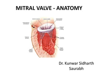
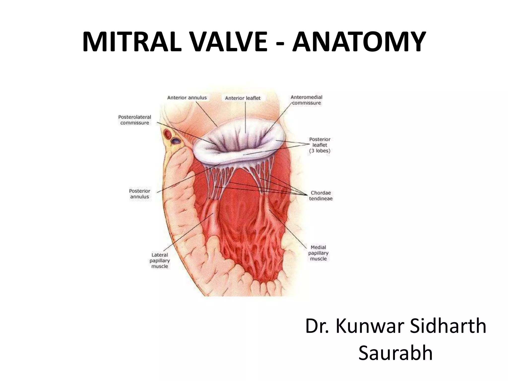
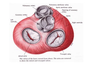
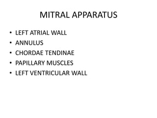
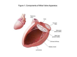
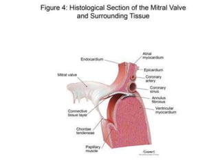
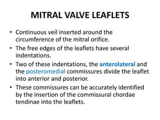
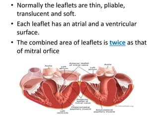
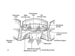
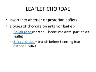
MITRAL VALVE - ANATOMY
Dr. Kunwar Sidharth
Saurabh
MITRAL VALVE
• ALSO KNOWN AS BICUSPID VALVE OR LEFT AV
VALVE.
• DUAL FLAP VALVE
• LIES BETWEEN LA AND LV
Cardiac Skeleton
• It is a high density single structure
of connective tissue (collagen) that forms and
anchors the valves and influences the forces
exerted through them.
• The cardiac skeleton separates and partitions
the atria from the ventricles
• The right and left fibrous rings of heart (anuli
fibrosi cordis) surround
the atrioventricular and arterial orifices.
• The right fibrous ring is known as the anulus
fibrosus dexter cordis, and the left is known as
the anulus fibrosus sinister cordis.
• The right fibrous trigone is continuous with the
central fibrous body.
• This is the strongest part of the fibrous cardiac
skeleton
• The valve rings, central body and skeleton of
the heart consisting of collagen are
impermeable to electrical propagation.
• The only channel allowed through this
collagen barrier is represented by a sinus that
opens up to the atrioventricular node and
exits to the bundle of His.
• The cardiac skeleton ensures that the
electrical and autonomic energy generated
above is ushered below and cannot return.
• The muscle origins/insertions of many of
the cardiomyocytes are anchored to opposite
sides of the valve rings.
MITRAL APPARATUS
• LEFT ATRIAL WALL
• ANNULUS
• CHORDAE TENDINAE
• PAPILLARY MUSCLES
• LEFT VENTRICULAR WALL
LEFT ATRIAL WALL
• Left atrial myocardium extends over the
proximal portion of the posterior leaflet.
• Left atrial enlargement can result in mitral
regurgitation by affecting posterior leaflet
• The anterior leaflet is not affected because of
its attachment to root of aorta
MITRAL ANNULUS
• Fibrous rings that connect with the leaflets.
• Not a continuous ring around the mitral
orifice.
• D- shaped
• The aortic valve is located between ventricular
septum and the mitral valve.
• The annulus functions as sphincter that
contracts and reduces the surface area of the
valve during the systole to ensure complete
closure of the leaflets.
• Annular dilatation of the mitral valve causes
poor leaflet apposition – resulting in MR.
MITRAL VALVE LEAFLETS
• Continuous veil inserted around the
circumference of the mitral orifice.
• The free edges of the leaflets have several
indentations.
• Two of these indentations, the anterolateral and
the posteromedial commissures divide the leaflet
into anterior and posterior.
• These commissures can be accurately identified
by the insertion of the commissural chordae
tendinae into the leaflets.
• Normally the leaflets are thin, pliable,
translucent and soft.
• Each leaflet has an atrial and a ventricular
surface.
• The combined area of leaflets is twice as that
of mitral orfice
Anterior Leaflet
• It is also anchored to the aortic root, unlike
the posterior leaflet.
• AKA Aortic, Septal, Greater or anteromedial
leaflet.
• The anterior leaflet is large and triangular in
shape, inserted on about 1/3rd of annulus.
• There are 2 zones – Rough and Clear , as per
the insertion of chordae tendinae.
• The clear zone is devoid of direct chordal
insertions.
• In continuation with aortic valve through aortic
mitral annulus and forms a boundary of LVOT.
• This region of continuity is 1/4th of the annulus,
corresponds to the region between half of left
coronary cusp, and half the non coronary cusp of
aortic valve.
• Limits of this attachment are demarcated by left
and right fibrous trigones.
• AV node and bundle of HIS are at risk of damage
near right trigone.
Posterior Leaflet
• Ventricular , Mural , Smaller or Posterolateral
leaflet. Scallop shaped.
• It has a wider attachment to the annulus than
the anterior leaflet. (2/3rd )
• 3 zones – Rough , Clear and Basal (receives
chordae directly from left ventricular
trabeculae)
SEGMENTAL CLASSIFICATION
A1 AND P1 –
ANTEROLATERAL
A2 AND P2 – MIDDLE
A3 AND P3 -
POSTEROMEDIAL
CHORDAE TENDINAE
• Small fibrous strings that originate either from the apical portion of
the papillary muscles or directly from the ventricular wall and insert
into the valve leaflets or muscle.
• Misalignment of leaflets, may put undue stress on chordae and may
cause rupture.
• Order of chordae –
– First order – inserted into free margin
– Second order – few mm back from free margin
– Third order – inserted at the base.
COMMISSURAL CHORDAE
• Chordae that insert into the interleaflet or
commissural areas located at the junction of
anterior and posterior leaflet.
• Two types –
– Posteromedial
– Anterolateral
• Shorter than the others and originate from
highest tip of papillary muscle.
LEAFLET CHORDAE
• Insert into anterior or posterior leaflets.
• 2 types of chordae on anterior leaflet-
– Rough zone chordae – insert into distal portion on
leaflet
– Strut chordae – branch before inserting into
anterior leaflet
PAPPILARY MUSCLES AND THE LEFT
VENTRICULAR WALL
• Muscular components of the apparatus.
• Normally arise from the apex and middle third
of left ventricular wall.
• Crescent shaped, conforms to the curvature of
the free wall of left ventricle.
• Anterolateral is larger than posteromedial
• LCx/LAD – SUPPLIES ANTEROMEDIAL
• RCA – SUPPLIES POSTEROMEDIAL
• Anterolateral – Attached to left half of anterior
and posterior leaflet by chordae tendinae
• Posterolateral – attached to right half.
• 4-12 chordae originating from each.
• Types of papillary muscle –
– Type 1 to 4.
• Design of mitral valve provides largest possible
orifice during diastolic phase.
• The valve opens as the anterior leaflet opens
and swings freely away from posterior leaflet
• Dimensions are enhanced by flexion of
anterior leaflet.
• During systole, anterior leaflet straightens and
extends towards posterior leaflet.
• Posterior leaflet acts as shelf to stop the
movement of anterior leaflet as they appose.
Mitral valve    anatomy - ppt by kunwar sidharth

Mitral valve anatomy - ppt by kunwar sidharth

  • 1.
    MITRAL VALVE -ANATOMY Dr. Kunwar Sidharth Saurabh
  • 2.
    MITRAL VALVE • ALSOKNOWN AS BICUSPID VALVE OR LEFT AV VALVE. • DUAL FLAP VALVE • LIES BETWEEN LA AND LV
  • 4.
    Cardiac Skeleton • Itis a high density single structure of connective tissue (collagen) that forms and anchors the valves and influences the forces exerted through them. • The cardiac skeleton separates and partitions the atria from the ventricles
  • 6.
    • The rightand left fibrous rings of heart (anuli fibrosi cordis) surround the atrioventricular and arterial orifices. • The right fibrous ring is known as the anulus fibrosus dexter cordis, and the left is known as the anulus fibrosus sinister cordis. • The right fibrous trigone is continuous with the central fibrous body. • This is the strongest part of the fibrous cardiac skeleton
  • 7.
    • The valverings, central body and skeleton of the heart consisting of collagen are impermeable to electrical propagation. • The only channel allowed through this collagen barrier is represented by a sinus that opens up to the atrioventricular node and exits to the bundle of His. • The cardiac skeleton ensures that the electrical and autonomic energy generated above is ushered below and cannot return. • The muscle origins/insertions of many of the cardiomyocytes are anchored to opposite sides of the valve rings.
  • 8.
    MITRAL APPARATUS • LEFTATRIAL WALL • ANNULUS • CHORDAE TENDINAE • PAPILLARY MUSCLES • LEFT VENTRICULAR WALL
  • 11.
    LEFT ATRIAL WALL •Left atrial myocardium extends over the proximal portion of the posterior leaflet. • Left atrial enlargement can result in mitral regurgitation by affecting posterior leaflet • The anterior leaflet is not affected because of its attachment to root of aorta
  • 12.
    MITRAL ANNULUS • Fibrousrings that connect with the leaflets. • Not a continuous ring around the mitral orifice. • D- shaped • The aortic valve is located between ventricular septum and the mitral valve.
  • 13.
    • The annulusfunctions as sphincter that contracts and reduces the surface area of the valve during the systole to ensure complete closure of the leaflets. • Annular dilatation of the mitral valve causes poor leaflet apposition – resulting in MR.
  • 14.
    MITRAL VALVE LEAFLETS •Continuous veil inserted around the circumference of the mitral orifice. • The free edges of the leaflets have several indentations. • Two of these indentations, the anterolateral and the posteromedial commissures divide the leaflet into anterior and posterior. • These commissures can be accurately identified by the insertion of the commissural chordae tendinae into the leaflets.
  • 15.
    • Normally theleaflets are thin, pliable, translucent and soft. • Each leaflet has an atrial and a ventricular surface. • The combined area of leaflets is twice as that of mitral orfice
  • 17.
    Anterior Leaflet • Itis also anchored to the aortic root, unlike the posterior leaflet. • AKA Aortic, Septal, Greater or anteromedial leaflet. • The anterior leaflet is large and triangular in shape, inserted on about 1/3rd of annulus. • There are 2 zones – Rough and Clear , as per the insertion of chordae tendinae.
  • 18.
    • The clearzone is devoid of direct chordal insertions. • In continuation with aortic valve through aortic mitral annulus and forms a boundary of LVOT. • This region of continuity is 1/4th of the annulus, corresponds to the region between half of left coronary cusp, and half the non coronary cusp of aortic valve. • Limits of this attachment are demarcated by left and right fibrous trigones. • AV node and bundle of HIS are at risk of damage near right trigone.
  • 20.
    Posterior Leaflet • Ventricular, Mural , Smaller or Posterolateral leaflet. Scallop shaped. • It has a wider attachment to the annulus than the anterior leaflet. (2/3rd ) • 3 zones – Rough , Clear and Basal (receives chordae directly from left ventricular trabeculae)
  • 21.
    SEGMENTAL CLASSIFICATION A1 ANDP1 – ANTEROLATERAL A2 AND P2 – MIDDLE A3 AND P3 - POSTEROMEDIAL
  • 22.
    CHORDAE TENDINAE • Smallfibrous strings that originate either from the apical portion of the papillary muscles or directly from the ventricular wall and insert into the valve leaflets or muscle. • Misalignment of leaflets, may put undue stress on chordae and may cause rupture. • Order of chordae – – First order – inserted into free margin – Second order – few mm back from free margin – Third order – inserted at the base.
  • 23.
    COMMISSURAL CHORDAE • Chordaethat insert into the interleaflet or commissural areas located at the junction of anterior and posterior leaflet. • Two types – – Posteromedial – Anterolateral • Shorter than the others and originate from highest tip of papillary muscle.
  • 24.
    LEAFLET CHORDAE • Insertinto anterior or posterior leaflets. • 2 types of chordae on anterior leaflet- – Rough zone chordae – insert into distal portion on leaflet – Strut chordae – branch before inserting into anterior leaflet
  • 25.
    PAPPILARY MUSCLES ANDTHE LEFT VENTRICULAR WALL • Muscular components of the apparatus. • Normally arise from the apex and middle third of left ventricular wall. • Crescent shaped, conforms to the curvature of the free wall of left ventricle. • Anterolateral is larger than posteromedial • LCx/LAD – SUPPLIES ANTEROMEDIAL • RCA – SUPPLIES POSTEROMEDIAL
  • 26.
    • Anterolateral –Attached to left half of anterior and posterior leaflet by chordae tendinae • Posterolateral – attached to right half. • 4-12 chordae originating from each. • Types of papillary muscle – – Type 1 to 4.
  • 27.
    • Design ofmitral valve provides largest possible orifice during diastolic phase. • The valve opens as the anterior leaflet opens and swings freely away from posterior leaflet • Dimensions are enhanced by flexion of anterior leaflet. • During systole, anterior leaflet straightens and extends towards posterior leaflet. • Posterior leaflet acts as shelf to stop the movement of anterior leaflet as they appose.