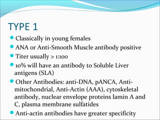
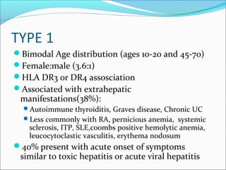
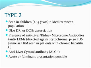
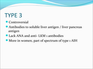
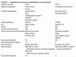
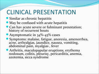
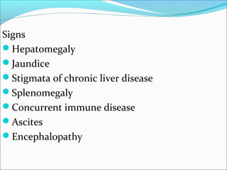
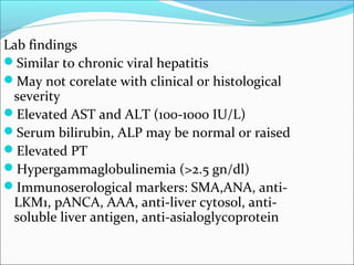
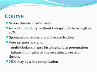
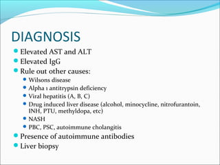
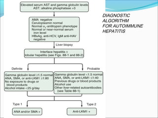
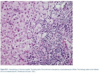
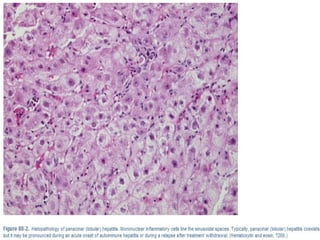
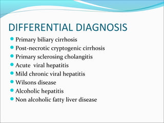
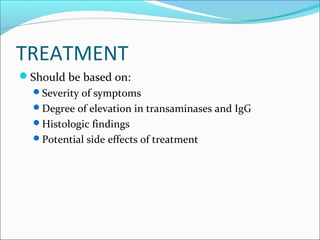
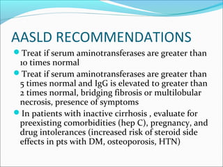
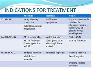
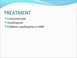
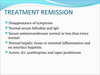
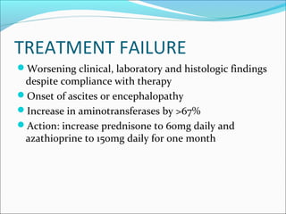
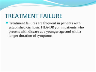
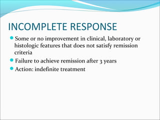
CHAIRED BY : Dr. Raminderpal Singh Sibia
PRESENTED BY : Dr. Rintu Sharma
Immunity
AUTOIMMUNE HEPATITIS
Chronic hepatitis of unknown etiology
Can progress to cirrhosis
Characteristics include:
presence of autoimmune antibody
evidence of hepatitis (interface being characteristic)
elevation of serum globulins
Continuing/unresolving hepatocellular inflammation
and necrosis
OTHER NAMES
Active chronic hepatitis or chronic active hepatitis
Chronic aggressive hepatitis
Lupoid hepatitis
Plasma cell hepatitis
Autoimmune chronic active hepatitis
Cases in which hepatotropic viruses, metabolic /
genetic derrangements and hepatotoxic drugs have
been excluded represent a spectrum of heterogenous
liver disorders of unknown cause, a proportion of
which are most likely autoimmune hepatitis.
BACKGROUND
First described in 1950’s
Accounts for 5.6% of liver transplants in the US
Affects women more than men (3.6:1)
If untreated approximately 40% die within 6 months
40% develop cirrhosis
EPIDEMIOLOGY
Frequency of AIH among patients with chronic liver
disease in North America is between 11%- 22%
Accounts for 5.6% of liver transplants in the US
Prevalence greatest among northern European white
persons
 Japenese have a lower frequency
PATHOGENESIS
Unknown mechanism but several proposed mechanisms
Genetically predisposed individual with exposure to an
environmental agent triggers the autoimmune pathogenic
process
Genetic predisposing factors:
HLA-DR3: early onset, severe form
HLA-DR4: caucasian, late onset, better response to steroids, higher
incidence of extrahepatic manifestations
IgG: part of the IgG molecule (mainly the heavy chain)
T-Cell receptors
PATHOGENESIS
EVIDENCE SUPPORTING AUTOIMMUNE
PATHOGENESIS
Histopathological lesions composed of cytotoxic Tcells
and plama cells
Circulating autoantibodies
Hyperglobulinemia
Other autoimmune disorders: thyroiditis, RA ,
autoimmune hemolytic anemia, ulcerative colitis,
membranoproliferative glomerulonephritis, diabetes
mellitus, celiac disease, sjogren’s syndrome
Hitocompatibility haplotypes assosciations
Response to steroids and immunosuppression.
PATHOGENESIS
Environmental Triggers: presumed to be certain
viruses, toxins, drugs
Drugs:
Oxyphenisatin
Methyldopa
Nitrofurantoin
Diclofenac
Minocycline
statins
CLASSIFICATION
TYPE 1
TYPE 2
OVERLAP SYNDROMES
TYPE 1
Classically in young females
ANA or Anti-Smooth Muscle antibody positive
Titer usually > 1:100
10% will have an antibody to Soluble Liver
antigens (SLA)
Other Antibodies: anti-DNA, pANCA, Anti-
mitochondrial, Anti-Actin (AAA), cytoskeletal
antibody, nuclear envelope proteins lamin A and
C, plasma membrane sulfatides
Anti-actin antibodies have greater specificity
TYPE 1
Bimodal Age distribution (ages 10-20 and 45-70)
Female:male (3.6:1)
HLA DR3 or DR4 assosciation
Associated with extrahepatic
manifestations(38%):
Autoimmune thyroiditis, Graves disease, Chronic UC
Less commonly with RA, pernicious anemia, systemic
sclerosis, ITP, SLE,coombs positive hemolytic anemia,
leucocytoclastic vasculitis, erythema nodosum
40% present with acute onset of symptoms
similar to toxic hepatitis or acute viral hepatitis
TYPE 2
Seen in children (2-14 years)in Meditteranean
population
DLA DR1 or DQB1 assosciation
Presence of anti-Liver/Kidney Microsome Antibodies
(anti- LKM1 )directed against cytochrome p450 2D6
{same as LKM seen in patients with chronic hepatitis
C}
Anti-Liver Cytosol antibody (ALC-1)
Acute or fulminant presentation possible
TYPE 3
Controversial
Antibodies to soluble liver antigen / liver pancreas
antigen
Lack ANA and anti- LKM 1 antibodies
More in women, part of spectrum of type 1 AIH
OVERLAP SYNDROMES
Primary Biliary Cirrhosis
Primary Sclerosing Cholangitis
5% of patients with chronic hepatitis C will have an
ANA titer of >1:100
A homogeneous pattern of staining is more common
in ANA positive autoimmune hepatitis compared to
that of ANA positive chronic hepatitis C
CLINICAL PRESENTATION
Similar as chronic hepatitis
May be confused with acute hepatitis
Can hav acute severe or fulminant presntation;
history of recurrent bouts
Asymptomatic in 34%-45% cases
Symptoms: malaise, fatigue, anorexia, amenorrhea,
acne, arthralgias, jaundice, nausea, vomiting,
abdominal pain, myalgias , fever
Arthritis, maculopapular eruptions, erythema
nodosum, colitis, pleurisy, pericarditis, anemia,
azotemia, sicca syndrome
Signs
Hepatomegaly
Jaundice
Stigmata of chronic liver disease
Splenomegaly
Concurrent immune disease
Ascites
Encephalopathy
Lab findings
Similar to chronic viral hepatitis
May not corelate with clinical or histological
severity
Elevated AST and ALT (100-1000 IU/L)
Serum bilirubin, ALP may be normal or raised
Elevated PT
Hypergammaglobulinemia (>2.5 gn/dl)
Immunoserological markers: SMA,ANA, anti-
LKM1, pANCA, AAA, anti-liver cytosol, anti-
soluble liver antigen, anti-asialoglycoprotein
Course
Severe disease in 20% cases
6-month mortality without therapy may be as high as
40%
Spontaneous remissions and exacerbations
Poor prognostic signs:
multilobular collapse histologically at presentation
failure of bilirubin to improve after 2 weeks of
therapy
HCC may be a late complication
DIAGNOSIS
Elevated AST and ALT
Elevated IgG
Rule out other causes:
Wilsons disease
Alpha 1 antitrypsin deficiency
Viral hepatitis (A, B, C)
Drug induced liver disease (alcohol, minocycline, nitrofurantoin,
INH, PTU, methyldopa, etc)
NASH
PBC, PSC, autoimmune cholangitis
Presence of autoimmune antibodies
Liver biopsy
Simplified scoring system
Greater specificity vs original scoring system ( 90% vs
73%)
Greater predictability ( 92% vs 82% )
Useful for excluding AIH in patients with other
conditions and concurrent immune features
Less sensitivity (95% vs 100 %)
DIAGNOSTIC
ALGORITHM
FOR AUTOIMMUNE
HEPATITIS
HISTOLOGY
Chronic hepatitis with marked piecemeal necrosis
and lobular involvement
Numerous plasma cells
Interface hepatitis: hallmark finding
Necroinflammatory activity
Evidence of hepatocellular regeneration (“rosette
formation” , regenerative “pseudolobules”)
Bile duct injuries and granulomas are uncommon
DIFFERENTIAL DIAGNOSIS
Primary biliary cirrhosis
Post-necrotic cryptogenic cirrhosis
Primary sclerosing cholangitis
Acute viral hepatitis
Mild chronic viral hepatitis
Wilsons disease
Alcoholic hepatitis
Non alcoholic fatty liver disease
TREATMENT
Should be based on:
Severity of symptoms
Degree of elevation in transaminases and IgG
Histologic findings
Potential side effects of treatment
AASLD RECOMMENDATIONS
Treat if serum aminotransferases are greater than
10 times normal
Treat if serum aminotransferases are greater than
5 times normal and IgG is elevated to greater than
2 times normal, bridging fibrosis or multilobular
necrosis, presence of symptoms
In patients with inactive cirrhosis , evaluate for
preexisting comorbidities (hep C), pregnancy, and
drug intolerances (increased risk of steroid side
effects in pts with DM, osteoporosis, HTN)
INDICATIONS FOR TREATMENT
Absolute Relative None
CLINICAL Incapacitating
symptoms
Mild or no
symptoms
Asymptomatic with
minimal lab
changes; previous
intolerance of
prednisolone/
azathioprine
Relentless clinical
progression
LABORATORY AST >10-foldULN AST 3-9.9 ULN AST<3 ULN
AST>5 fold ULN
Gammaglobulin
>2fold
AST>5 fold ULN
Gammaglobulin
<2fold
AST <3 fold ULN
HISTOLOGIC Bridging necrosis Interface hepatitis Inactive cirrhosis
Multilobular
necrosis
Portal hepatitis
Decompensated
cirrhosis
TREATMENT
Corticosteroids
Azathioprine
Children: azathioprine or 6MP
PREDNISONE ONLY
Prednisone 60mg PO daily with a taper down to
30mg at the 4th
week into treatment and then
maintenance of 20mg daily until reach endpoint
Reasons for Prednisone only:
Cytopenia
TPMT deficiency
Malignancy
Pregnancy
Therapy response expected in upto 80% of cases
Preferred treatment regimens
Combination therapy Single drug
therapy
Prednisolone
(mg/day)
Azathioprine (mg/d) Prednisolone
(mg/day)
30mg ? 1 week 50 mg until the end
point
60mg ? 1 week
20mg ? 1 week 40mg ? 1 week
15mg ? 2 weeks 30mg ? 2 weeks
10mg until the end
point
20mg until the end
point
COMBINATION THERAPY
Prednisone + Azathioprine
Prednisone: start at 30mg daily and taper down to
15mg at week 4, then maintain on 10mg daily until
therapy endpoint
Azathioprine 50mg daily
Side effects : Prednisone
Side effects : Azathioprine
TREATMENT REMISSION
Disappearance of symptoms
Normal serum bilirubin and IgG
Serum aminotransferases normal or less than twice
normal
Normal hepatic tissue or minimal inflammation and
no interface hepatitis.
Action: d/c azathioprine and taper prednisone
TREATMENT FAILURE
Worsening clinical, laboratory and histologic findings
despite compliance with therapy
Onset of ascites or encephalopathy
Increase in aminotransferases by >67%
Action: increase prednisone to 60mg daily and
azathioprine to 150mg daily for one month
TREATMENT FAILURE
Treatment failures are frequent in patients with
established cirrhosis, HLA-DR3 or in patients who
present with disease at a younger age and with a
longer duration of symptoms
INCOMPLETE RESPONSE
Some or no improvement in clinical, laboratory or
histologic features that does not satisfy remission
criteria
Failure to achieve remission after 3 years
Action: indefinite treatment
RELAPSE
An exacerbation after drug withdrawal in patients
who enter remission
Reappearance of histological disease
AST >3 folds ULN
Cirrhosis develops commonly
Reinstitute original treatment: azathioprine
continued indefinitely
Liver transplantation
LIVER TRANSPLANT
Patients with ascites and hepatic encephalopathy
(generally will have a poor prognosis, but consider
liver transplant if they have failed glucocorticoid
therapy.
Considered in patients with multilobar necrosis
and have at least one laboratory parameter which
does not normalize within 2 weeks of treatment
(theses patients have a high immediate mortality
rate)
LIVER TRANSPLANTATION
Considered in pts who worsen while on
glucocorticoid therapy.
Recurrence of disease after transplant is common in
those with AIH but has only been described in
patients who are not adequately immunosuppressed.
PROGNOSIS
PROGNOSIS
40% of all pts with AIH develop cirrhosis
54% develop esophageal varices within 2 years
Poor prognosis if has presence of ascites or hepatic
encephalopathy
13-20% of patients can have spontaneous resolution
Of patients who survive the most early and active stage of
disease, approximately 41% of them develop inactive
cirrhosis.
Of patients who have severe initial disease and survive the
first 2 years, typically survive long term.
Ai hepatitis dr rintu

Ai hepatitis dr rintu

  • 1.
    CHAIRED BY :Dr. Raminderpal Singh Sibia PRESENTED BY : Dr. Rintu Sharma Immunity
  • 2.
    AUTOIMMUNE HEPATITIS Chronic hepatitisof unknown etiology Can progress to cirrhosis Characteristics include: presence of autoimmune antibody evidence of hepatitis (interface being characteristic) elevation of serum globulins Continuing/unresolving hepatocellular inflammation and necrosis
  • 3.
    OTHER NAMES Active chronichepatitis or chronic active hepatitis Chronic aggressive hepatitis Lupoid hepatitis Plasma cell hepatitis Autoimmune chronic active hepatitis
  • 4.
    Cases in whichhepatotropic viruses, metabolic / genetic derrangements and hepatotoxic drugs have been excluded represent a spectrum of heterogenous liver disorders of unknown cause, a proportion of which are most likely autoimmune hepatitis.
  • 5.
    BACKGROUND First described in1950’s Accounts for 5.6% of liver transplants in the US Affects women more than men (3.6:1) If untreated approximately 40% die within 6 months 40% develop cirrhosis
  • 6.
    EPIDEMIOLOGY Frequency of AIHamong patients with chronic liver disease in North America is between 11%- 22% Accounts for 5.6% of liver transplants in the US Prevalence greatest among northern European white persons  Japenese have a lower frequency
  • 7.
    PATHOGENESIS Unknown mechanism butseveral proposed mechanisms Genetically predisposed individual with exposure to an environmental agent triggers the autoimmune pathogenic process Genetic predisposing factors: HLA-DR3: early onset, severe form HLA-DR4: caucasian, late onset, better response to steroids, higher incidence of extrahepatic manifestations IgG: part of the IgG molecule (mainly the heavy chain) T-Cell receptors
  • 8.
    PATHOGENESIS EVIDENCE SUPPORTING AUTOIMMUNE PATHOGENESIS Histopathologicallesions composed of cytotoxic Tcells and plama cells Circulating autoantibodies Hyperglobulinemia Other autoimmune disorders: thyroiditis, RA , autoimmune hemolytic anemia, ulcerative colitis, membranoproliferative glomerulonephritis, diabetes mellitus, celiac disease, sjogren’s syndrome Hitocompatibility haplotypes assosciations Response to steroids and immunosuppression.
  • 9.
    PATHOGENESIS Environmental Triggers: presumedto be certain viruses, toxins, drugs Drugs: Oxyphenisatin Methyldopa Nitrofurantoin Diclofenac Minocycline statins
  • 10.
  • 11.
    TYPE 1 Classically inyoung females ANA or Anti-Smooth Muscle antibody positive Titer usually > 1:100 10% will have an antibody to Soluble Liver antigens (SLA) Other Antibodies: anti-DNA, pANCA, Anti- mitochondrial, Anti-Actin (AAA), cytoskeletal antibody, nuclear envelope proteins lamin A and C, plasma membrane sulfatides Anti-actin antibodies have greater specificity
  • 12.
    TYPE 1 Bimodal Agedistribution (ages 10-20 and 45-70) Female:male (3.6:1) HLA DR3 or DR4 assosciation Associated with extrahepatic manifestations(38%): Autoimmune thyroiditis, Graves disease, Chronic UC Less commonly with RA, pernicious anemia, systemic sclerosis, ITP, SLE,coombs positive hemolytic anemia, leucocytoclastic vasculitis, erythema nodosum 40% present with acute onset of symptoms similar to toxic hepatitis or acute viral hepatitis
  • 13.
    TYPE 2 Seen inchildren (2-14 years)in Meditteranean population DLA DR1 or DQB1 assosciation Presence of anti-Liver/Kidney Microsome Antibodies (anti- LKM1 )directed against cytochrome p450 2D6 {same as LKM seen in patients with chronic hepatitis C} Anti-Liver Cytosol antibody (ALC-1) Acute or fulminant presentation possible
  • 14.
    TYPE 3 Controversial Antibodies tosoluble liver antigen / liver pancreas antigen Lack ANA and anti- LKM 1 antibodies More in women, part of spectrum of type 1 AIH
  • 16.
    OVERLAP SYNDROMES Primary BiliaryCirrhosis Primary Sclerosing Cholangitis 5% of patients with chronic hepatitis C will have an ANA titer of >1:100 A homogeneous pattern of staining is more common in ANA positive autoimmune hepatitis compared to that of ANA positive chronic hepatitis C
  • 18.
    CLINICAL PRESENTATION Similar aschronic hepatitis May be confused with acute hepatitis Can hav acute severe or fulminant presntation; history of recurrent bouts Asymptomatic in 34%-45% cases Symptoms: malaise, fatigue, anorexia, amenorrhea, acne, arthralgias, jaundice, nausea, vomiting, abdominal pain, myalgias , fever Arthritis, maculopapular eruptions, erythema nodosum, colitis, pleurisy, pericarditis, anemia, azotemia, sicca syndrome
  • 19.
    Signs Hepatomegaly Jaundice Stigmata of chronicliver disease Splenomegaly Concurrent immune disease Ascites Encephalopathy
  • 20.
    Lab findings Similar tochronic viral hepatitis May not corelate with clinical or histological severity Elevated AST and ALT (100-1000 IU/L) Serum bilirubin, ALP may be normal or raised Elevated PT Hypergammaglobulinemia (>2.5 gn/dl) Immunoserological markers: SMA,ANA, anti- LKM1, pANCA, AAA, anti-liver cytosol, anti- soluble liver antigen, anti-asialoglycoprotein
  • 21.
    Course Severe disease in20% cases 6-month mortality without therapy may be as high as 40% Spontaneous remissions and exacerbations Poor prognostic signs: multilobular collapse histologically at presentation failure of bilirubin to improve after 2 weeks of therapy HCC may be a late complication
  • 22.
    DIAGNOSIS Elevated AST andALT Elevated IgG Rule out other causes: Wilsons disease Alpha 1 antitrypsin deficiency Viral hepatitis (A, B, C) Drug induced liver disease (alcohol, minocycline, nitrofurantoin, INH, PTU, methyldopa, etc) NASH PBC, PSC, autoimmune cholangitis Presence of autoimmune antibodies Liver biopsy
  • 26.
    Simplified scoring system Greaterspecificity vs original scoring system ( 90% vs 73%) Greater predictability ( 92% vs 82% ) Useful for excluding AIH in patients with other conditions and concurrent immune features Less sensitivity (95% vs 100 %)
  • 27.
  • 28.
    HISTOLOGY Chronic hepatitis withmarked piecemeal necrosis and lobular involvement Numerous plasma cells Interface hepatitis: hallmark finding Necroinflammatory activity Evidence of hepatocellular regeneration (“rosette formation” , regenerative “pseudolobules”) Bile duct injuries and granulomas are uncommon
  • 31.
    DIFFERENTIAL DIAGNOSIS Primary biliarycirrhosis Post-necrotic cryptogenic cirrhosis Primary sclerosing cholangitis Acute viral hepatitis Mild chronic viral hepatitis Wilsons disease Alcoholic hepatitis Non alcoholic fatty liver disease
  • 32.
    TREATMENT Should be basedon: Severity of symptoms Degree of elevation in transaminases and IgG Histologic findings Potential side effects of treatment
  • 33.
    AASLD RECOMMENDATIONS Treat ifserum aminotransferases are greater than 10 times normal Treat if serum aminotransferases are greater than 5 times normal and IgG is elevated to greater than 2 times normal, bridging fibrosis or multilobular necrosis, presence of symptoms In patients with inactive cirrhosis , evaluate for preexisting comorbidities (hep C), pregnancy, and drug intolerances (increased risk of steroid side effects in pts with DM, osteoporosis, HTN)
  • 34.
    INDICATIONS FOR TREATMENT AbsoluteRelative None CLINICAL Incapacitating symptoms Mild or no symptoms Asymptomatic with minimal lab changes; previous intolerance of prednisolone/ azathioprine Relentless clinical progression LABORATORY AST >10-foldULN AST 3-9.9 ULN AST<3 ULN AST>5 fold ULN Gammaglobulin >2fold AST>5 fold ULN Gammaglobulin <2fold AST <3 fold ULN HISTOLOGIC Bridging necrosis Interface hepatitis Inactive cirrhosis Multilobular necrosis Portal hepatitis Decompensated cirrhosis
  • 35.
  • 36.
    PREDNISONE ONLY Prednisone 60mgPO daily with a taper down to 30mg at the 4th week into treatment and then maintenance of 20mg daily until reach endpoint Reasons for Prednisone only: Cytopenia TPMT deficiency Malignancy Pregnancy Therapy response expected in upto 80% of cases
  • 37.
    Preferred treatment regimens Combinationtherapy Single drug therapy Prednisolone (mg/day) Azathioprine (mg/d) Prednisolone (mg/day) 30mg ? 1 week 50 mg until the end point 60mg ? 1 week 20mg ? 1 week 40mg ? 1 week 15mg ? 2 weeks 30mg ? 2 weeks 10mg until the end point 20mg until the end point
  • 38.
    COMBINATION THERAPY Prednisone +Azathioprine Prednisone: start at 30mg daily and taper down to 15mg at week 4, then maintain on 10mg daily until therapy endpoint Azathioprine 50mg daily
  • 39.
    Side effects :Prednisone
  • 40.
    Side effects :Azathioprine
  • 41.
    TREATMENT REMISSION Disappearance ofsymptoms Normal serum bilirubin and IgG Serum aminotransferases normal or less than twice normal Normal hepatic tissue or minimal inflammation and no interface hepatitis. Action: d/c azathioprine and taper prednisone
  • 42.
    TREATMENT FAILURE Worsening clinical,laboratory and histologic findings despite compliance with therapy Onset of ascites or encephalopathy Increase in aminotransferases by >67% Action: increase prednisone to 60mg daily and azathioprine to 150mg daily for one month
  • 43.
    TREATMENT FAILURE Treatment failuresare frequent in patients with established cirrhosis, HLA-DR3 or in patients who present with disease at a younger age and with a longer duration of symptoms
  • 44.
    INCOMPLETE RESPONSE Some orno improvement in clinical, laboratory or histologic features that does not satisfy remission criteria Failure to achieve remission after 3 years Action: indefinite treatment
  • 45.
    RELAPSE An exacerbation afterdrug withdrawal in patients who enter remission Reappearance of histological disease AST >3 folds ULN Cirrhosis develops commonly Reinstitute original treatment: azathioprine continued indefinitely Liver transplantation
  • 46.
    LIVER TRANSPLANT Patients withascites and hepatic encephalopathy (generally will have a poor prognosis, but consider liver transplant if they have failed glucocorticoid therapy. Considered in patients with multilobar necrosis and have at least one laboratory parameter which does not normalize within 2 weeks of treatment (theses patients have a high immediate mortality rate)
  • 47.
    LIVER TRANSPLANTATION Considered inpts who worsen while on glucocorticoid therapy. Recurrence of disease after transplant is common in those with AIH but has only been described in patients who are not adequately immunosuppressed.
  • 49.
  • 50.
    PROGNOSIS 40% of allpts with AIH develop cirrhosis 54% develop esophageal varices within 2 years Poor prognosis if has presence of ascites or hepatic encephalopathy 13-20% of patients can have spontaneous resolution Of patients who survive the most early and active stage of disease, approximately 41% of them develop inactive cirrhosis. Of patients who have severe initial disease and survive the first 2 years, typically survive long term.