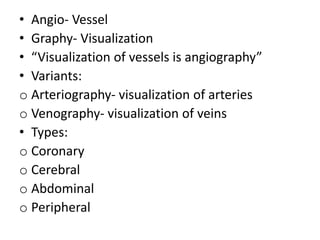
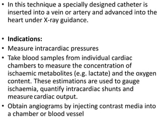
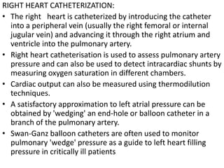
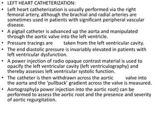
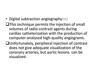
2. • Angio- Vessel
• Graphy- Visualization
• “Visualization of vessels is angiography”
• Variants:
o Arteriography- visualization of arteries
o Venography- visualization of veins
• Types:
o Coronary
o Cerebral
o Abdominal
o Peripheral
3.
4.
5.
6. • In this technique a specially designed catheter is
inserted into a vein or artery and advanced into the
heart under X-ray guidance.
• Indications:
• Measure intracardiac pressures
• Take blood samples from individual cardiac
chambers to measure the concentration of
ischaemic metabolites (e.g. lactate) and the oxygen
content. These estimations are used to gauge
ischaemia, quantify intracardiac shunts and
measure cardiac output.
• Obtain angiograms by injecting contrast media into
a chamber or blood vessel
7. RIGHT HEART CATHETERIZATION:
• The right heart is catheterized by introducing the catheter
into a peripheral vein (usually the right femoral or internal
jugular vein) and advancing it through the right atrium and
ventricle into the pulmonary artery.
• Right heart catheterisation is used to assess pulmonary artery
pressure and can also be used to detect intracardiac shunts by
measuring oxygen saturation in different chambers.
• Cardiac output can also be measured using thermodilution
techniques.
• A satisfactory approximation to left atrial pressure can be
obtained by 'wedging' an end-hole or balloon catheter in a
branch of the pulmonary artery.
• Swan-Ganz balloon catheters are often used to monitor
pulmonary 'wedge' pressure as a guide to left heart filling
pressure in critically ill patients
8. • LEFT HEART CATHETERIZATION:
• Left heart catheterization is usually performed via the right
femoral artery, although the brachial and radial arteries are
sometimes used in patients with significant peripheral vascular
disease.
• A pigtail catheter is advanced up the aorta and manipulated
through the aortic valve into the left ventricle.
• Pressure tracings are taken from the left ventricular cavity.
• The end diastolic pressure is invariably elevated in patients with
left ventricular dysfunction.
• A power injection of radio opaque contrast material is used to
opacify the left ventricular cavity (left ventriculography) and
thereby assesses left ventricular systolic function.
• The catheter is then withdrawn across the aortic valve into
the aorta and the ‘pullback’ gradient across the valve is measured.
• Aortography(a power injection into the aortic root) can be
performed to assess the aortic root and the presence and severity
of aortic regurgitation.
9. • Digital subtraction angiography:::::
This technique permits the injection of small
volumes of radio-contrast agents during
cardiac catheterization with the production of
computer-analysed high-quality angiograms.
Unfortunately, peripheral injection of contrast
does not give adequate visualization of the
coronary arteries, but aortic lesions can be
visualized.