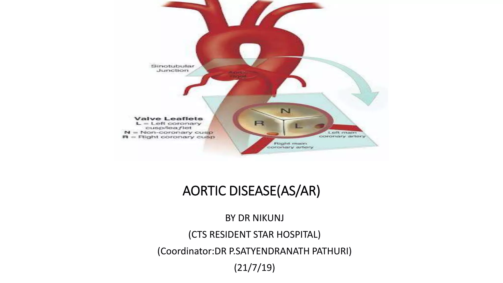
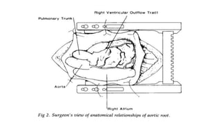
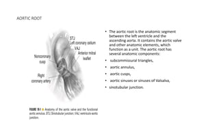
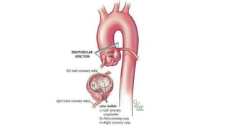
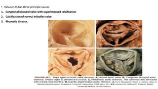
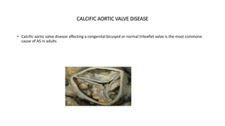
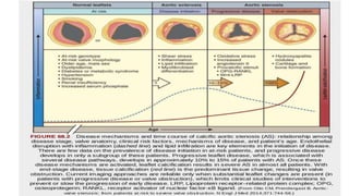
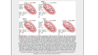
The document provides information on aortic valve disease including anatomy, etiology, and pathophysiology. It describes the key components of the aortic root including the aortic annulus, cusps, sinuses, and sinotubular junction. The three main causes of aortic stenosis are discussed as congenital bicuspid valve with calcification, calcification of a normal trileaflet valve, and rheumatic disease. The pathophysiology of aortic stenosis involves left ventricular pressure overload leading to hypertrophy and eventually decreased ejection fraction if severe stenosis is not corrected.
Electrical mapping of the heart is a medical procedure that is use to diagnose Arrhythmias in patients. This is done by using sensitive catheter to map the electrical activity in the chambers of the heart.
To begin an electrical mapping procedure, a thin tube called a catheter sheath is inserted into a small incision in the arm or upper thigh. This process is usually visualized using x-rays and a special dye that helps reveal the arteries (called angiography). This catheter is carefully guided through the blood vessels until it is inside the heart.
Evaluation of prosthetic valve function and clinical utility.Ramachandra Barik
Many of the prosthesis-related complications can be prevented or their impact minimized through optimal prosthesis selection in the individual patient and careful medical management and follow-up after implantation.
The heart has four valves; the other two are the mitral and the tricuspid valves. The aortic valve normally has three cusps or leaflets, although in 1–2% of the population it is found to congenitally have two leaflets
INTRODUCTION
EMBRYOLOGY AND DEVELOPMENT
HISTOLOGY OF ARTERIES
ARCH OF AORTA
SUBCLAVIAN ARTERY
Origin
Course and termination
Parts and clinical significance
Branches
Subclavian steal syndrome
CAROTID SYSTEM OF ARTERIES
Origin and termination
Branches
Surface marking
Ligation
Carotid sinus, carotid body and carotid pulse
EXTERNAL CAROTID ARTERY
Course
Branches
Relations
Surface marking
Ligation
BRANCHES OF ECA IN DETAIL
INTERNAL CAROTID ARTERY
Course and termination
Parts and branches
Clinical significance
CHAPTER 1 SEMESTER V - ROLE OF PEADIATRIC NURSE.pdfSachin Sharma
Pediatric nurses play a vital role in the health and well-being of children. Their responsibilities are wide-ranging, and their objectives can be categorized into several key areas:
1. Direct Patient Care:
Objective: Provide comprehensive and compassionate care to infants, children, and adolescents in various healthcare settings (hospitals, clinics, etc.).
This includes tasks like:
Monitoring vital signs and physical condition.
Administering medications and treatments.
Performing procedures as directed by doctors.
Assisting with daily living activities (bathing, feeding).
Providing emotional support and pain management.
2. Health Promotion and Education:
Objective: Promote healthy behaviors and educate children, families, and communities about preventive healthcare.
This includes tasks like:
Administering vaccinations.
Providing education on nutrition, hygiene, and development.
Offering breastfeeding and childbirth support.
Counseling families on safety and injury prevention.
3. Collaboration and Advocacy:
Objective: Collaborate effectively with doctors, social workers, therapists, and other healthcare professionals to ensure coordinated care for children.
Objective: Advocate for the rights and best interests of their patients, especially when children cannot speak for themselves.
This includes tasks like:
Communicating effectively with healthcare teams.
Identifying and addressing potential risks to child welfare.
Educating families about their child's condition and treatment options.
4. Professional Development and Research:
Objective: Stay up-to-date on the latest advancements in pediatric healthcare through continuing education and research.
Objective: Contribute to improving the quality of care for children by participating in research initiatives.
This includes tasks like:
Attending workshops and conferences on pediatric nursing.
Participating in clinical trials related to child health.
Implementing evidence-based practices into their daily routines.
By fulfilling these objectives, pediatric nurses play a crucial role in ensuring the optimal health and well-being of children throughout all stages of their development.
The dimensions of healthcare quality refer to various attributes or aspects that define the standard of healthcare services. These dimensions are used to evaluate, measure, and improve the quality of care provided to patients. A comprehensive understanding of these dimensions ensures that healthcare systems can address various aspects of patient care effectively and holistically. Dimensions of Healthcare Quality and Performance of care include the following; Appropriateness, Availability, Competence, Continuity, Effectiveness, Efficiency, Efficacy, Prevention, Respect and Care, Safety as well as Timeliness.
Antibiotic Stewardship by Anushri Srivastava.pptxAnushriSrivastav
Stewardship is the act of taking good care of something.
Antimicrobial stewardship is a coordinated program that promotes the appropriate use of antimicrobials (including antibiotics), improves patient outcomes, reduces microbial resistance, and decreases the spread of infections caused by multidrug-resistant organisms.
WHO launched the Global Antimicrobial Resistance and Use Surveillance System (GLASS) in 2015 to fill knowledge gaps and inform strategies at all levels.
ACCORDING TO apic.org,
Antimicrobial stewardship is a coordinated program that promotes the appropriate use of antimicrobials (including antibiotics), improves patient outcomes, reduces microbial resistance, and decreases the spread of infections caused by multidrug-resistant organisms.
ACCORDING TO pewtrusts.org,
Antibiotic stewardship refers to efforts in doctors’ offices, hospitals, long term care facilities, and other health care settings to ensure that antibiotics are used only when necessary and appropriate
According to WHO,
Antimicrobial stewardship is a systematic approach to educate and support health care professionals to follow evidence-based guidelines for prescribing and administering antimicrobials
In 1996, John McGowan and Dale Gerding first applied the term antimicrobial stewardship, where they suggested a causal association between antimicrobial agent use and resistance. They also focused on the urgency of large-scale controlled trials of antimicrobial-use regulation employing sophisticated epidemiologic methods, molecular typing, and precise resistance mechanism analysis.
Antimicrobial Stewardship(AMS) refers to the optimal selection, dosing, and duration of antimicrobial treatment resulting in the best clinical outcome with minimal side effects to the patients and minimal impact on subsequent resistance.
According to the 2019 report, in the US, more than 2.8 million antibiotic-resistant infections occur each year, and more than 35000 people die. In addition to this, it also mentioned that 223,900 cases of Clostridoides difficile occurred in 2017, of which 12800 people died. The report did not include viruses or parasites
VISION
Being proactive
Supporting optimal animal and human health
Exploring ways to reduce overall use of antimicrobials
Using the drugs that prevent and treat disease by killing microscopic organisms in a responsible way
GOAL
to prevent the generation and spread of antimicrobial resistance (AMR). Doing so will preserve the effectiveness of these drugs in animals and humans for years to come.
being to preserve human and animal health and the effectiveness of antimicrobial medications.
to implement a multidisciplinary approach in assembling a stewardship team to include an infectious disease physician, a clinical pharmacist with infectious diseases training, infection preventionist, and a close collaboration with the staff in the clinical microbiology laboratory
to prevent antimicrobial overuse, misuse and abuse.
to minimize the developme
Global launch of the Healthy Ageing and Prevention Index 2nd wave – alongside...ILC- UK
The Healthy Ageing and Prevention Index is an online tool created by ILC that ranks countries on six metrics including, life span, health span, work span, income, environmental performance, and happiness. The Index helps us understand how well countries have adapted to longevity and inform decision makers on what must be done to maximise the economic benefits that comes with living well for longer.
Alongside the 77th World Health Assembly in Geneva on 28 May 2024, we launched the second version of our Index, allowing us to track progress and give new insights into what needs to be done to keep populations healthier for longer.
The speakers included:
Professor Orazio Schillaci, Minister of Health, Italy
Dr Hans Groth, Chairman of the Board, World Demographic & Ageing Forum
Professor Ilona Kickbusch, Founder and Chair, Global Health Centre, Geneva Graduate Institute and co-chair, World Health Summit Council
Dr Natasha Azzopardi Muscat, Director, Country Health Policies and Systems Division, World Health Organisation EURO
Dr Marta Lomazzi, Executive Manager, World Federation of Public Health Associations
Dr Shyam Bishen, Head, Centre for Health and Healthcare and Member of the Executive Committee, World Economic Forum
Dr Karin Tegmark Wisell, Director General, Public Health Agency of Sweden
India Clinical Trials Market: Industry Size and Growth Trends [2030] Analyzed...Kumar Satyam
According to TechSci Research report, "India Clinical Trials Market- By Region, Competition, Forecast & Opportunities, 2030F," the India Clinical Trials Market was valued at USD 2.05 billion in 2024 and is projected to grow at a compound annual growth rate (CAGR) of 8.64% through 2030. The market is driven by a variety of factors, making India an attractive destination for pharmaceutical companies and researchers. India's vast and diverse patient population, cost-effective operational environment, and a large pool of skilled medical professionals contribute significantly to the market's growth. Additionally, increasing government support in streamlining regulations and the growing prevalence of lifestyle diseases further propel the clinical trials market.
Growing Prevalence of Lifestyle Diseases
The rising incidence of lifestyle diseases such as diabetes, cardiovascular diseases, and cancer is a major trend driving the clinical trials market in India. These conditions necessitate the development and testing of new treatment methods, creating a robust demand for clinical trials. The increasing burden of these diseases highlights the need for innovative therapies and underscores the importance of India as a key player in global clinical research.
R3 Stem Cells and Kidney Repair A New Horizon in Nephrology.pptxR3 Stem Cell
R3 Stem Cells and Kidney Repair: A New Horizon in Nephrology" explores groundbreaking advancements in the use of R3 stem cells for kidney disease treatment. This insightful piece delves into the potential of these cells to regenerate damaged kidney tissue, offering new hope for patients and reshaping the future of nephrology.
Defecation
Normal defecation begins with movement in the left colon, moving stool toward the anus. When stool reaches the rectum, the distention causes relaxation of the internal sphincter and an awareness of the need to defecate. At the time of defecation, the external sphincter relaxes, and abdominal muscles contract, increasing intrarectal pressure and forcing the stool out
The Valsalva maneuver exerts pressure to expel faeces through a voluntary contraction of the abdominal muscles while maintaining forced expiration against a closed airway. Patients with cardiovascular disease, glaucoma, increased intracranial pressure, or a new surgical wound are at greater risk for cardiac dysrhythmias and elevated blood pressure with the Valsalva maneuver and need to avoid straining to pass the stool.
Normal defecation is painless, resulting in passage of soft, formed stool
CONSTIPATION
Constipation is a symptom, not a disease. Improper diet, reduced fluid intake, lack of exercise, and certain medications can cause constipation. For example, patients receiving opiates for pain after surgery often require a stool softener or laxative to prevent constipation. The signs of constipation include infrequent bowel movements (less than every 3 days), difficulty passing stools, excessive straining, inability to defecate at will, and hard feaces
IMPACTION
Fecal impaction results from unrelieved constipation. It is a collection of hardened feces wedged in the rectum that a person cannot expel. In cases of severe impaction the mass extends up into the sigmoid colon.
DIARRHEA
Diarrhea is an increase in the number of stools and the passage of liquid, unformed feces. It is associated with disorders affecting digestion, absorption, and secretion in the GI tract. Intestinal contents pass through the small and large intestine too quickly to allow for the usual absorption of fluid and nutrients. Irritation within the colon results in increased mucus secretion. As a result, feces become watery, and the patient is unable to control the urge to defecate. Normally an anal bag is safe and effective in long-term treatment of patients with fecal incontinence at home, in hospice, or in the hospital. Fecal incontinence is expensive and a potentially dangerous condition in terms of contamination and risk of skin ulceration
HEMORRHOIDS
Hemorrhoids are dilated, engorged veins in the lining of the rectum. They are either external or internal.
FLATULENCE
As gas accumulates in the lumen of the intestines, the bowel wall stretches and distends (flatulence). It is a common cause of abdominal fullness, pain, and cramping. Normally intestinal gas escapes through the mouth (belching) or the anus (passing of flatus)
FECAL INCONTINENCE
Fecal incontinence is the inability to control passage of feces and gas from the anus. Incontinence harms a patient’s body image
PREPARATION AND GIVING OF LAXATIVESACCORDING TO POTTER AND PERRY,
An enema is the instillation of a solution into the rectum and sig
QA Paediatric dentistry department, Hospital Melaka 2020Azreen Aj
QA study - To improve the 6th monthly recall rate post-comprehensive dental treatment under general anaesthesia in paediatric dentistry department, Hospital Melaka
CHAPTER 1 SEMESTER V PREVENTIVE-PEDIATRICS.pdfSachin Sharma
This content provides an overview of preventive pediatrics. It defines preventive pediatrics as preventing disease and promoting children's physical, mental, and social well-being to achieve positive health. It discusses antenatal, postnatal, and social preventive pediatrics. It also covers various child health programs like immunization, breastfeeding, ICDS, and the roles of organizations like WHO, UNICEF, and nurses in preventive pediatrics.
1. AORTIC DISEASE(AS/AR)
BY DR NIKUNJ
(CTS RESIDENT STAR HOSPITAL)
(Coordinator:DR P.SATYENDRANATH PATHURI)
(21/7/19)
2. ANATOMY
• The aortic valve is the last of four cardiac valves through which the blood is pumped before it goes to the
rest of the body.
• It separates the left ventricular outflow tract from the aorta.
• Its main function is to prevent backward blood flow from the aorta to the left ventricle, while allowing the
blood to flow forward during systole with minimal resistance.
3.
4. AORTIC ROOT
• The aortic root is the anatomic segment
between the left ventricle and the
ascending aorta. It contains the aortic valve
and other anatomic elements, which
function as a unit. The aortic root has
several anatomic components:
• subcommissural triangles,
• aortic annulus,
• aortic cusps,
• aortic sinuses or sinuses of Valsalva,
• sinotubular junction.
5.
6. THE SUBCOMMISSURAL TRIANGLES
• The subcommissural triangles are
part of the left ventricular out fow
tract,
• The subcommissural triangles of
the noncoronary aortic cusp are
fibrous extension of the
intervalvular fibrous body and
membranous septum, whereas the
subcommissural triangle beneath
the left and the right aortic cusps is
an extension of the muscular
interventricular septum.
7. THE AORTIC ANNULUS
• The aortic annulus, a fibrous structure with a scalloped shape, attaches the aortic
valve to the left ventricle.
• It is attached directly to the myocardium in approximately 45% of its circumference,
and to fibrous structures in the remaining 55%
• The diameter of the aortic annulus is 10% to 20% larger than the diameter of the
sinotubular junction of the aortic root in young patients . As the number of elastic
fibers in the arterial wall decreases with age, the sinotubular junction dilates, and
its diameter tends to become equal to that of the aortic annulus in older patients.
8. • With dilation of the aortic annulus, the subcommissural triangles of the noncoronary cusp tend to become
more obtuse as the crescent shape of the aortic annulus along its fibrous insertion flattens.
9.
10.
11. CUSPS
• The normal aortic valve has three cusps. Each cusp has a semilunar shape and has a
base and a free margin. The base is attached to the aortic annulus in a crescent
fashion. The point at which the free margin of a cusp joins its base is the
commissure, and
• the ridge in the aortic wall that lies immediately above the commissures is the
sinotubular junction.
• At the mid point of each free edge is fibrou nodulus arantii on either side of
nodulus is extremely thin.
• The free margin of an aortic cusp extends from one of its commissures to the other.
The length of the free margin of an aortic cusp is approximately 1.5 times the length
of its base.
12. CUSPS
• The three aortic cusps often have different sizes in
a person, and the right and noncoronary cusps are
usually larger than the left cusp.
• The same cusp may have different sizes in
individuals with the same body surface area
• During diastole, the free margins and part of the
body of the three cusps touch each other
approximately in the center of the aortic root to
seal the aortic orifice.
• Thus, the average length of the free margins of
three aortic cusps must exceed the diameter of
the sinotubular junction to allow the cusps to
coapt centrally and render the aortic valve
competent .
13. • The aortic leaflets display three layers of connective tissue: the ventriculosa on the ventricular side,
the fibrosa on the aortic side, and the spongiosa between them
• The fibrosa is mainly composed of collagen fibers while the ventriculosa mostly consists of elastic
fibers.
• Between these two layers, the spongiosa is composed of a mucopolysaccharide gel-like substance
that facilitates the motion of the ventriculosa and fibrosa.
14. • If a pathologic process causes shortening of the length of the free
margin of a cusp, or if the sinotubular junction dilates, the cusps cannot
coapt centrally, resulting in aortic insufciency .
• If the length of a free margin is elongated, the cusp prolapses, and
depending on the degree of prolapse, aortic insufficiency
15.
16. AORTIC SINUSES, OR SINUSES OF VALSALVA
• The spaces contained between the aortic annulus
and the sinotubular junction are the aortic
sinuses. There are three cusps and three sinuses:
• left cusp and sinus,
• right cusp and sinus,
• noncoronary cusp and sinus.
• The left main coronary artery arises from the left
aortic sinus, and the right coronary artery arises
from the right aortic sinus.
• There are three sinuses of the aortic valve, each
related to the valve’s corresponding cusps. Each
sinus is divided into three areas a central part and
two adjacent parts, which are named according
to the valve cusps they adjoin.
• The noncoronary sinus is also refferred to as the
posterior aortic sinus.
17. AORTIC SINUSES, OR SINUSES OF VALSALVA
• The aortic sinuses facilitate closure of the aortic valve by creating eddies and currents between the cusps
and arterial wall .
• They also prevent the cusps from occluding the coronary artery orifices during systole, thus guaranteeing
myocardial perfusion during the entire cardiac cycle.
18. RIGHT CORONARY SINUS
• entire right coronary sinus lies adjacent to
the RVOT.
• central part lies adjacent to the crista
supraventricularis,
• left part is adjacent to the area of the
RVOT in the angle between the crista
supraventricularis and the pulmonary
valve.
• posterior (noncoronary) part of the right
coronary sinus is related to the area of the
right ventricle posteroinferior to the crista
supraventricularis.
• Inferiorly, the entire right coronary sinus
is related to the interventricular septum;
the muscular septum lies under the
central and left parts, while either
membranous or muscular septum may lie
under the posterior part of the right
coronary sinus.
19. NONCORONARY SINUS
• The atrialchambers with the intervening
atrial septum lie adjacent to the
noncoronary sinus.
• right and central parts of the
noncoronary sinus are related to the
right atrium and the interatrial septum,
• left part is related to the left atrium.
• Inferiorly, the right part, like the
posterior part of the right coronary
sinus, may be related either to the
membranous or the muscular septum
depending on the size of the
membranous septum. However,
beneath the central part of the
noncoronary sinus, the membranous
septum is a constant structure. The left
part of the noncoronary sinus inserts
into the anterior mitral leaflet
20. LEFT CORONARY SINUS
• posterior part of the left coronary
• it is related to the left atrium
posteriorly and to the anterior mitral
leaflet inferiorly.
• central part of the left aortic sinus is
the only part of the aortic root that is
not related to a cardiac chamber; it is
adjacent to the epicardium only.
• right part of the left coronary sinus lies
adjacent to the pulmonary trunk at the
level of the left pulmonary sinus.
inferior to it lies the muscular
interventricular septum.
21. • The aortic root of young individuals is elastic and very compliant. It expands and contracts during the
cardiac cycle.
• The normal aortic root has a fairly consistent shape, and the sizes of the cusps, the aortic annulus, the aortic
sinuses, and the sinotubular junction are somewhat interdependent.
• Thus, large cusps have a proportionally large annulus, sinus, and sinotubular junction.
22.
23. AORTIC STENOSIS Epidemiology and Etiology
• Isolated aortic stenosis (AS) is more common in men than in women and is found in 2% of people 65 years of
age and older.
• The most common causes of AS include agerelated calcific degeneration, bicuspid aortic valve, and
rheumatic aortic valve.
• The distribution of these causes varies across age groups and geographic regions.
• Age related degeneration is the most common cause of AS in patients older than 70 years.
• In contrast, bicuspid aortic valve calcification accounts for most surgical cases in patients younger than 70
years.
24. • Valvular AS has three principle causes
1. Congenital bicuspid valve with superimposed calcification
2. Calcification of normal trileaflet valve
3. Rhumatic disease.
25. • Fixed obstruction to LVOT may occur above the valve(supravalvular stenosis)
• Below the valve (subvalvular stenosis)
26. CONGENITAL BICUSPID VALVE WITH SUPERIMPOSED CALCIFICATION
• Congenital malformation can be
• Unicuspid: infancy
• Bicuspid :
• Ttricuspid :
• Domeshaped diaphragm:
27. CALCIFIC AORTIC VALVE DISEASE
• Calcific aortic valve disease affecting a congenital bicuspid or normal trileaflet valve is the most commone
cause of AS in adults
28.
29. RHEUMATIC AORTIC STENOSIS
• It Results from adhesions and fusion of the comissures and cusps and vasclarazation of the leaflets of the
valve ring
• Leading to retraction and stiffening of the free borders of the cusps
• Calcific nodules deveope on bothe surface and orifice is reduced to small round or triangular opening.
• It can be regurgitant as well as stenotic.
30.
31. PATHOPHYSIOLOGY
• No appreciable gradient exists across the normal aortic valve during systole.
• In AS, gradual obstruction of the left ventricular outflow leads to an increased left ventricular afterload
and left ventricular wall stress, elevated left ventricular systolic and diastolic pressures, decreased
aortic pressure, and prolonged left ventricular ejection time.
• Over time, this results in compensatory concentric left ventricular hypertrophy (LVH) to maintain
ejection fraction.
• In patients with chronic severe AS, this compensatory mechanism may become insufficient, leading to
gradual dilation and thinning of the left ventricle, and result in a decrease in ejection fraction and in
congestive heart failure.
32. PATHOPHYSIOLOGY
• Myocardial oxygen supply and demand is also altered in AS.
• LVH, increased systolic pressure, and prolonged ejection time result in increased myocardial oxygen
demand.
• Increased diastolic pressure and prolonged systolic ejection time result in decreased diastolic and myocardial
perfusion time and hence myocardial oxygen supply.
• The alteration in myocardial oxygen supply and demand is the underlying mechanism behind myocardial
ischemia in patients with AS, even in the absence of coronary artery disease.
33. SYMPTOMS
• The classic symptoms of AS are angina, exertional syncope, and symptoms of congestive heart failure such as
shortness of breath.
• Angina: frequent symptom of pt with svere AS
• Syncope: reduced cerebral perfusion.
• GI Bleeding
34. PHYSICAL EXAMINATION
• palpation of carotid upstroke(Slow rising late peaking,low amplitude)
• Evaluation of systolic murmur
• Assessment of splitting of second heart sound
• Signs of HF
• crescendo-decrescendo systolic ejection murmur heard best at the second right intercostal space, which may
variably radiate to the carotid arteries.
• A palpable thrill may be present in severe cases of AS.
• Palpation of the arterial pulse may reveal a weak and delayed pulse known as pulsus parvus et tardus.
35. DIAGNOSIS AND GRADING
• Two-dimensional transthoracic echocardiography is the most common modality for the diagnosis and
grading of AS
36. • X ray chest
• EKG
• CARDIAC CT
• CARDIAC CATHETERIZATION
• TMT
37. NATURAL HISTORY
• Without intervention,
• Multiple studies consistently reported survivals of 3 years for angina and syncope
• 1.5 to 2 years for dyspnea and heart failure.
• These findings have driven recommendations for early surgical intervention in patients with symptomatic AS.
• One third of asymptomatic patients with severe AS will become symptomatic in 2 years with an estimated
cardiac death of less than 1% each year to 5% each year.
• Patients with higher grades of AS severity seem to progress at a faster rate than lower grades of AS.
• After AS becomes moderate, aortic valve area decreases on average by 0.1 cm2 per year, the pressure
gradient across the valve rises on average by 7 mm Hg per year, and the jet velocity increases by 0.3 m/sec
per year.
38. AORTIC REGURGITATION
• disturbance in any of the components of the functional unit of the aortic valve (e.g., cusps, sinuses of
Valsalva, sinotubular junction, ventriculoaortic junction).
• In general, the causes can be divided into
• affect the valve cusps (e.g., calcific degeneration, congenitally bicuspid valve, infective endocarditis,
rheumatic disease, myxomatous degeneration)
• affect the aortic root (e.g., aortic dissection, aortitis of various etiologies such as syphilis, connective tissue
disorders such as Marfan syndrome)
Primary Valve disease Primary Root disease
Congenital (bicuspid aortic valve)
Rheumatic fever
Infective endocarditis ,Collagen
vascular diseases ,Degenerative aortic
valve disease
Myxomatous (prolapse) Traumatic
Longstanding, uncontrolled hypertension
Marfan syndrome ,Idiopathic aortic dilation
Cystic medial necrosis, Senile aortic ectasia
and dilation
Aortic dissection, Marfan's syndrome
Syphilitic aortitis Giant cell arteritis,
Takayasu arteritis, Ankylosing spondylitis,
Whipple disease
39.
40. THE PATHOPHYSIOLOGY OF AR
• dependent on the acuity of onset and duration of the disease process.
• ACUTE AR, typically caused by aortic dissection, infective endocarditis, trauma, or valve prosthesis failure,
there is a sudden increase in left ventricular end-diastolic volume because of the regurgitation.
• Since the left ventricle has limited compliance and does not have time to adapt, the left ventricular end-
diastolic pressure (LVEDP) rises rapidly
41. • CHRONIC AR, there is a slow and insidious progression of left ventricular (LV) dilation and eccentric
hypertrophy because of an increase in left ventricular end-diastolic volume, LVEDP, and wall stress.
• Dilation of the LV, while maintaining normal systolic function, increases total stroke volume and maintains
forward flow.
• This increase in stroke volume coupled with an increase in LVEDP is associated with the wide pulse pressure
typical of chronic AR.
• Eventually, the hypertrophic response is exhausted, and LVEF drops as afterload increases, leading to heart
failure and its associated clinical presentation.
42.
43.
44. Valve Dysfunctions
Valve Dysfunctions
corresponding Lesions
Lesions
Type I: Normal leaflet motion Annular dilatation
Leaflet perforation
Vegetation
Type II: Leaflet prolapse Leaflet rupture, distension
Commissure detachment
Type IlIa: Restricted leaflet closure and opening Leaflet thickening Commissure fusion Calcification
Type Illb: Restricted leaflet closure only Sino-tubular dilatation
45. CLINICAL FEATURES
• ACUTE AR :usually exhibit sudden or rapidly progressive cardiovascular collapse, which can be life
threatening if not addressed promptly.
• They often exhibit ischemic symptoms because of the decrease in coronary blood flow and increased
myocardial oxygen demand.
• CHRONIC AR : asymptomatic for a long period of time because of the compensatory LV changes.
• Once the compensatory response is exhausted, the patients start having heart failure symptoms
1. PALPITATION - early symptom
2. HEAD POUNDING - on exertion
3. EXERTIONAL DYSPNOEA
4. ORTHOPNOEA
5. PAROXYSMAL NOCTURNAL DYSPNOEA
6. EXCESSIVE DIAPHORESIS
7. ANGINA - on exertion/ at rest - nocturnal
8. CCF - late
46. PULSE IN AR
• Corrigan’s pulse or
• Water hammer pulse or
• Collapsing pulse
- Rapid rise and rapid fall
• Bisferiens’s pulse -two peaks in systole
• A pulse that is bounding and forceful, rapidly increasing and subsequently collapsing, it resembles the strike
of waterhammer
47. • S1- may be soft due to premature closure of the mitral valve.
• A2 –normal or accentuated when AR is due to aortic root disease.
• S2- absent or single or exhibit narrow or paradoxical splitting
• S3 GALLOP - due to increased LV end diastolic volume or impaired LV function
• Systolic ejection sound - related to abrupt distention of the aorta by the augmented stroke volume.
48. • CHRONIC AORTIC REGURGITATION
• Early diastolic murmur
• High pitched, blowing decrescendo
Best heard in the 3rd left intercostal space with the patient sitting up and leaning forward breath held in
forced expiration
• Aortic root disorders murmur is best heard along right sternal border musical murmur Longer the duration
of murmur severer the aortic regurgitation
• Becomes short - cardiac failure
• Austin flint murmur
• Soft, low pitched rumbling mid diastolic murmur.
• Diastolic displacement of the anterior leaflet of the mitral valve by the aortic regurgitation stream.
49. • Classic signs of widened pulse pressure may also be found, including
1. Corrigan or water-hammer pulse,
2. De Musset sign (bobbing of the head with heart beats),
3. Quincke pulse (pulsations of the lip and fingers),
4. Traube sign (pistol shot sounds over the femoral artery),
5. Müller sign (pulsations of the uvula).
6. Becker sign - Visible systolic pulsations of the retinal arterioles
7. Light-house sign – Alternate flushing & blanching of forehead
8. Landolfi’s sign - Change in pupil size with each systole
9. Gerhardt’s sign - Visible systolic pulsations of spleen
10. Rosenbach’s sign - Visible systolic pulsations of liver
11. Corrigan’s sign – Dancing carotid in neck
12. Hill sign: Popliteal cuff systolic pressure exceeding brachial cuff systolic pressure by more than 20 mmHg.
50. DIAGNOSTIC CRITERIA
• Transthoracic echocardiography with Doppler colorflow is the most useful tool for the diagnosis of AR.
• The jet width and vena contracta width on Doppler color-flow are used to qualitatively assess the severity of
AR, whereas the regurgitant volume, regurgitant fraction, and regurgitant orifice area are used for the
quantitative assessment.
51.
52.
53.
54. CHEST X RAY
• acute AR, there may be minimal cardiac enlargement, but marked enlargement is a common finding in chronic AR.
55. ECG
•LV hypertrophy
•Left axis deviation
•Left atrial enlargement
•LV volume overload pattern - Prominent Q waves in leads I, aVL, and V3 to V6 and relatively small R waves in
V1
56. • AORTIC ANGIOGRAPHY
1. Mild (1+) - A small amount of contrast enters the LV during diastole and clears with each systole
2. Moderate AR (2+) - Contrast enters the LV with each diastole, but the LV chamber is less dense than the
aorta
3. Moderately severe AR (3+) - The LV chamber is equal in density to the ascending aorta.
4. Severe AR (4+) - Complete, dense opacification of the LV chamber occurs on the first beat, and the LV is
more densely opacified than the ascending aorta
• CARDIAC MRI :CMR provides accurate measurements of regurgitant volumes and the regurgitant orifice in
AR. It is the most accurate noninvasive technique for assessing LV end systolic volume, diastolic volume, and
mass
57. CARDIAC CATHETERIZATION
• Class I indications for cardiac catheterization under current ACC/AHA guidelines:
• Assessment of coronary anatomy prior to aortic valve surgery in patients with risk factors for coronary artery
disease
• Assessment of severity of AR, LV function, or aortic root size when noninvasive tests are inconclusive or are
discordant with clinical findings.