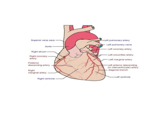
CENTER FOR PHYSIOTHERAPY AND
REHABILITATION SCIENCE
JAMIA MILLIA ISLAMIA
SUBMITTED TO: DR. JAMAL ALI MOIZ
SUBMITTED BY: FARZANA KHATOON
MPT 3RD SEM
coronary angiography technique
in management of coronary
artery disease
Coronary Angiography
• Coronary angiography remains the gold standard for
detecting clinically significant atherosclerotic
coronary artery diseas
• The technique was first performed by Dr. Mason
Sones at the Cleveland Clinic in 1958
• The American College of Cardiology/American Heart
Association Task Force on Assessment of
Cardiovascular Procedures was formed to make
recommendations regarding appropriate utilization of
technology in the diagnosis and treatment of patients
with cardiovascular disease.
• Coronary angiography is one such important technique.
The uses of coronary angiography have undergone an
extraordinary expansion in the recent past, in part
stimulated by the development of improved techniques
and new forms of treatment, particularly for patients
with atherosclerotic coronary heart disease.
• The primary purpose of coronary angiography is
to define the anatomy of the coronary arteries
when such information is needed for patient
management.
• This anatomic definition includes assessment of
the presence, extent and severity of obstructive
atherosclerotic coronary artery disease, coronary
artery size, coronary collateral flow, thrombus
formation, dynamic obstructions (coronary
spasm) or congenital coronary artery anomalies.
• Coronary angiography is used not only in diagnosis but also to
assess the appropriateness and feasibility of various forms of
therapy aimed directly at the coronary arteries, such as
percutaneous coronary angioplasty, coronary artery bypass
surgery, thrombolysis or treatments designed to cause
regression of atherosclerosis.
• Information provided by coronary angiography is useful for
assessing the results of therapy and in helping to formulate
prognosis in patients with coronary artery disease.
• It should be emphasized that coronary angiography does not
provide direct information about the patient's functional
capacity and symptoms or the functional significance of a
given coronary lesion.
Coronary artery anatomy
• The left and right coronary arteries (LCA, RCA) arise from
the anterior aortic root.
• The LCA divides into the left anterior descending (LAD)
and left circumflex (LCX) arteries.
• The LAD gives rise to two 'diagonal' branches. The left
(obtuse) marginal artery arises from the LCX at the
posterior interventricular sulcus.
• The RCA gives off a right marginal artery branch and in
70% of people supplies the posterior descending artery
(PDA). The LCA supplies mainly the left side of the heart.
The RCA supplies the right heart, 20-30% of the left
ventricle (LV) and in the majority of people the sinoatrial
and atrioventricular nodes.
Classification of Applications of
Coronary Angiography
• Class I: Conditions for which there is general
agreement that coronary angiography is justified. A
Class I indication should not be taken to mean that
coronary angiography is the only acceptable diagnostic
procedure.
• Class II: Conditions for which coronary angiography is
frequently performed, but there is a divergence of
opinion with respect to its justification in terms of value
and appropriateness.
• Class III: Conditions for which there is general
agreement that coronary angiography is not ordinarily
justified.
What is a cardiac catheterization
• Coronary artery disease is the narrowing or blockage
of the coronary (heart) arteries, After an
interventional procedure, the coronary artery is
opened, increasing blood flow to the heart.
• Cardiac catheterization (also called cardiac cath or
coronary angiogram) is an invasive imaging
procedure that to evaluate heart function
• Cardiac catheterization is used to:
• Evaluate or conform the presence of coronary artery
disease, valve disease or disease of the aorta
• Evaluate heart muscle function
• Determine the need for further treatment (such as an
interventional procedure or coronary artery bypass
graft, or CABG, surgery)
• During a cardiac catheterization, a long, narrow tube
called a catheter is inserted through a plastic
introducer sheath (a short, hollow tube that is inserted
into a blood vessel in your arm or leg).
• The catheter is guided through the blood vessel to the
coronary arteries with the aid of a special x-ray
machine
• Contrast material is injected through the catheter and
x-ray movies are created as the contrast material
moves through the heart’s chambers, valves and
major vessels. This part of the procedure is called a
coronary angiogram (or coronary angiography)
• The digital photographs of the contrast material are
used to identify the site of the narrowing or blockage
in the coronary artery
• Additional imaging procedures called intra-vascular
ultrasound (IVUS) and fractional flow reserve (FFR),
may be performed along with cardiac catheterization
in some cases to obtain detailed images of the walls
of the blood vessels.
• With IVUS a miniature sound-probe (transducer) is
positioned on the tip of a coronary catheter. The
catheter is threaded through the coronary arteries and,
using high-frequency sound waves produces detailed
images of the inside walls of the arteries. IVUS
produces an accurate picture of the location and
extent of plaque.
• With FFR a special wire is threaded through the
artery and a vasodilator medication is given. This test
is functionally performing a very high quality stress
test for a short segment of the artery.
What is an interventional procedure
• An interventional procedure is a non-surgical
treatment used to open narrowed coronary arteries to
improve blood ow to the heart.
• An interventional procedure can be performed during
a diagnostic cardiac catheterization when a blockage
is identied, or it may be scheduled after a
catheterization has conrmed the presence of coronary
artery disease
• An interventional procedure starts out the same way
as a cardiac catheterization. Once the catheter is in
place, one of these interventional procedures is
performed to open the artery: balloon angioplasty,
stent placement, rotablation or cutting balloon.
Balloon angioplasty with stenting:
• In most cases, balloon angioplasty is performed in
combination with the stenting procedure.
• A stent is a small, metal mesh tube that acts as a
scaffold to provide support inside the coronary artery. A
balloon catheter placed over a guide wire is used to
insert the stent into the narrowed artery.
• Once in place, the balloon is inated and the stent
expands to the size of the artery and holds it open.
• The balloon is deated and removed, and the stent stays
in place permanently. During a period of several weeks,
the artery heals around the stent. In this way, restenosis
is somewhat diminished
• Angioplasty with stenting is most commonly
recommended for patients who have a blockage in
one or two coronary arteries. If there are blockages in
more than two coronary arteries, coronary artery
bypass graft surgery may be recommended
Balloon angioplasty:
• A procedure in which a small balloon at the tip of the
catheter is inserted near the blocked or narrowed area
of the coronary artery.
• The technical name for balloon angioplasty is
percutaneous transluminal coronary angioplasty
(PTCA) or percutaneous coronary intervention (PCI).
• When the balloon is inated, the fatty plaque or blockage
is compressed against the artery walls and the diameter
of the blood vessel is widened (dilated) to increase
blood ow to the heart. This procedure is sometimes
complicated by vessel recoil and restenosis.
Goals
• To visualize coronary arteries, branches, collaterals
and anomalies
• Precise localization relative to major and minor side
branches, thrombi and areas of calcification
• To visualize vessel bifurcations, origin of side
branches and specific lesion characteristics (length,
eccentricity, calcium etc)
INDICATION
• Diagnosis of CAD in clinically suspected pts.
• Providing peri-interventional information for
percutaneous coronary intervention
• Coronary anomalies
• To exclude stenoses before non-coronary cardiac
surgery (valve surgery after 40 yrs of age)
• Determine patency of coronary artery bypass graft
• In patients with non–ST-segment elevation acute
coronary syndromes with high-risk features (e.g.,
ongoing ischemia, heart failure)
• In patients with acute ST-segment elevation
myocardial infarction (STEMI)
• Primary percutaneous intervention (PCI) is usually
performed in the same procedure, immediately after
the diagnostic procedure
CONTRAINDICATIONS
• Coagulopathy
• Decompensate congestive heart failure
• Uncontrolled Hypertension
• CVA
• GI Hemorrhage
• Pregnancy
• Inability for patient cooperation
• Active infection
• Renal Failure
• Contrast medium allergy
Before the Procedure
• After patient is properly identified, the procedure
must be explained before consent can be signed
• Baseline vital signs will be done and as long as these
are within the doctor’s interest, can proceed with the
procedure
• Blood tests must be done including BUN, creatnine,
PTT, INR, insulin/sugar levels
Procedure
Arterial Puncture
• Access is easiest from right side of patient due to
aortic bend
• Puncture is generally done via the femoral artery
• Alternative sites include the radial and brachial
arteries of the arm
• After numbing the groin area, the femoral artery is
palpated and a needle is inserted in that direction
• When blood comes out of needle, the artery has been
accessed
• A small, flexible guidewire is then inserted into the
lumen of the needle
• The needle can then be removed but the wire must
maintain position
• After removing the needle, a flexible plastic tube can
be placed over the wire and introduced into the artery.
This is called a oneway sheath (allows insertion of
catheters and wires without blood escaping)
• The catheter is then inserted over the guidewire but
through the sheet and advanced into placement to the
aorta.
• MC Catheter used is Judkins.
• Movement of catheter is monitored under fluoroscopy
with the cardiologist manipulating its movements
• The fluoroscopic machine is manipulated by a
qualified, scrubbed in, radiologic technologist
• When catheter is in place, wire can be removed and
contrast administered
7th round seminar
7th round seminar

7th round seminar

  • 1.
    CENTER FOR PHYSIOTHERAPYAND REHABILITATION SCIENCE JAMIA MILLIA ISLAMIA SUBMITTED TO: DR. JAMAL ALI MOIZ SUBMITTED BY: FARZANA KHATOON MPT 3RD SEM
  • 2.
    coronary angiography technique inmanagement of coronary artery disease
  • 3.
    Coronary Angiography • Coronaryangiography remains the gold standard for detecting clinically significant atherosclerotic coronary artery diseas • The technique was first performed by Dr. Mason Sones at the Cleveland Clinic in 1958
  • 4.
    • The AmericanCollege of Cardiology/American Heart Association Task Force on Assessment of Cardiovascular Procedures was formed to make recommendations regarding appropriate utilization of technology in the diagnosis and treatment of patients with cardiovascular disease. • Coronary angiography is one such important technique. The uses of coronary angiography have undergone an extraordinary expansion in the recent past, in part stimulated by the development of improved techniques and new forms of treatment, particularly for patients with atherosclerotic coronary heart disease.
  • 5.
    • The primarypurpose of coronary angiography is to define the anatomy of the coronary arteries when such information is needed for patient management. • This anatomic definition includes assessment of the presence, extent and severity of obstructive atherosclerotic coronary artery disease, coronary artery size, coronary collateral flow, thrombus formation, dynamic obstructions (coronary spasm) or congenital coronary artery anomalies.
  • 6.
    • Coronary angiographyis used not only in diagnosis but also to assess the appropriateness and feasibility of various forms of therapy aimed directly at the coronary arteries, such as percutaneous coronary angioplasty, coronary artery bypass surgery, thrombolysis or treatments designed to cause regression of atherosclerosis. • Information provided by coronary angiography is useful for assessing the results of therapy and in helping to formulate prognosis in patients with coronary artery disease. • It should be emphasized that coronary angiography does not provide direct information about the patient's functional capacity and symptoms or the functional significance of a given coronary lesion.
  • 7.
    Coronary artery anatomy •The left and right coronary arteries (LCA, RCA) arise from the anterior aortic root. • The LCA divides into the left anterior descending (LAD) and left circumflex (LCX) arteries. • The LAD gives rise to two 'diagonal' branches. The left (obtuse) marginal artery arises from the LCX at the posterior interventricular sulcus. • The RCA gives off a right marginal artery branch and in 70% of people supplies the posterior descending artery (PDA). The LCA supplies mainly the left side of the heart. The RCA supplies the right heart, 20-30% of the left ventricle (LV) and in the majority of people the sinoatrial and atrioventricular nodes.
  • 9.
    Classification of Applicationsof Coronary Angiography • Class I: Conditions for which there is general agreement that coronary angiography is justified. A Class I indication should not be taken to mean that coronary angiography is the only acceptable diagnostic procedure. • Class II: Conditions for which coronary angiography is frequently performed, but there is a divergence of opinion with respect to its justification in terms of value and appropriateness. • Class III: Conditions for which there is general agreement that coronary angiography is not ordinarily justified.
  • 10.
    What is acardiac catheterization • Coronary artery disease is the narrowing or blockage of the coronary (heart) arteries, After an interventional procedure, the coronary artery is opened, increasing blood flow to the heart. • Cardiac catheterization (also called cardiac cath or coronary angiogram) is an invasive imaging procedure that to evaluate heart function
  • 11.
    • Cardiac catheterizationis used to: • Evaluate or conform the presence of coronary artery disease, valve disease or disease of the aorta • Evaluate heart muscle function • Determine the need for further treatment (such as an interventional procedure or coronary artery bypass graft, or CABG, surgery)
  • 12.
    • During acardiac catheterization, a long, narrow tube called a catheter is inserted through a plastic introducer sheath (a short, hollow tube that is inserted into a blood vessel in your arm or leg). • The catheter is guided through the blood vessel to the coronary arteries with the aid of a special x-ray machine
  • 13.
    • Contrast materialis injected through the catheter and x-ray movies are created as the contrast material moves through the heart’s chambers, valves and major vessels. This part of the procedure is called a coronary angiogram (or coronary angiography) • The digital photographs of the contrast material are used to identify the site of the narrowing or blockage in the coronary artery
  • 14.
    • Additional imagingprocedures called intra-vascular ultrasound (IVUS) and fractional flow reserve (FFR), may be performed along with cardiac catheterization in some cases to obtain detailed images of the walls of the blood vessels.
  • 15.
    • With IVUSa miniature sound-probe (transducer) is positioned on the tip of a coronary catheter. The catheter is threaded through the coronary arteries and, using high-frequency sound waves produces detailed images of the inside walls of the arteries. IVUS produces an accurate picture of the location and extent of plaque. • With FFR a special wire is threaded through the artery and a vasodilator medication is given. This test is functionally performing a very high quality stress test for a short segment of the artery.
  • 16.
    What is aninterventional procedure • An interventional procedure is a non-surgical treatment used to open narrowed coronary arteries to improve blood ow to the heart. • An interventional procedure can be performed during a diagnostic cardiac catheterization when a blockage is identied, or it may be scheduled after a catheterization has conrmed the presence of coronary artery disease
  • 17.
    • An interventionalprocedure starts out the same way as a cardiac catheterization. Once the catheter is in place, one of these interventional procedures is performed to open the artery: balloon angioplasty, stent placement, rotablation or cutting balloon.
  • 18.
    Balloon angioplasty withstenting: • In most cases, balloon angioplasty is performed in combination with the stenting procedure. • A stent is a small, metal mesh tube that acts as a scaffold to provide support inside the coronary artery. A balloon catheter placed over a guide wire is used to insert the stent into the narrowed artery. • Once in place, the balloon is inated and the stent expands to the size of the artery and holds it open. • The balloon is deated and removed, and the stent stays in place permanently. During a period of several weeks, the artery heals around the stent. In this way, restenosis is somewhat diminished
  • 19.
    • Angioplasty withstenting is most commonly recommended for patients who have a blockage in one or two coronary arteries. If there are blockages in more than two coronary arteries, coronary artery bypass graft surgery may be recommended
  • 20.
    Balloon angioplasty: • Aprocedure in which a small balloon at the tip of the catheter is inserted near the blocked or narrowed area of the coronary artery. • The technical name for balloon angioplasty is percutaneous transluminal coronary angioplasty (PTCA) or percutaneous coronary intervention (PCI). • When the balloon is inated, the fatty plaque or blockage is compressed against the artery walls and the diameter of the blood vessel is widened (dilated) to increase blood ow to the heart. This procedure is sometimes complicated by vessel recoil and restenosis.
  • 21.
    Goals • To visualizecoronary arteries, branches, collaterals and anomalies • Precise localization relative to major and minor side branches, thrombi and areas of calcification • To visualize vessel bifurcations, origin of side branches and specific lesion characteristics (length, eccentricity, calcium etc)
  • 22.
    INDICATION • Diagnosis ofCAD in clinically suspected pts. • Providing peri-interventional information for percutaneous coronary intervention • Coronary anomalies • To exclude stenoses before non-coronary cardiac surgery (valve surgery after 40 yrs of age) • Determine patency of coronary artery bypass graft
  • 23.
    • In patientswith non–ST-segment elevation acute coronary syndromes with high-risk features (e.g., ongoing ischemia, heart failure) • In patients with acute ST-segment elevation myocardial infarction (STEMI) • Primary percutaneous intervention (PCI) is usually performed in the same procedure, immediately after the diagnostic procedure
  • 24.
    CONTRAINDICATIONS • Coagulopathy • Decompensatecongestive heart failure • Uncontrolled Hypertension • CVA • GI Hemorrhage • Pregnancy • Inability for patient cooperation • Active infection • Renal Failure • Contrast medium allergy
  • 25.
    Before the Procedure •After patient is properly identified, the procedure must be explained before consent can be signed • Baseline vital signs will be done and as long as these are within the doctor’s interest, can proceed with the procedure • Blood tests must be done including BUN, creatnine, PTT, INR, insulin/sugar levels
  • 26.
    Procedure Arterial Puncture • Accessis easiest from right side of patient due to aortic bend • Puncture is generally done via the femoral artery • Alternative sites include the radial and brachial arteries of the arm
  • 27.
    • After numbingthe groin area, the femoral artery is palpated and a needle is inserted in that direction • When blood comes out of needle, the artery has been accessed • A small, flexible guidewire is then inserted into the lumen of the needle • The needle can then be removed but the wire must maintain position
  • 28.
    • After removingthe needle, a flexible plastic tube can be placed over the wire and introduced into the artery. This is called a oneway sheath (allows insertion of catheters and wires without blood escaping) • The catheter is then inserted over the guidewire but through the sheet and advanced into placement to the aorta. • MC Catheter used is Judkins.
  • 29.
    • Movement ofcatheter is monitored under fluoroscopy with the cardiologist manipulating its movements • The fluoroscopic machine is manipulated by a qualified, scrubbed in, radiologic technologist • When catheter is in place, wire can be removed and contrast administered