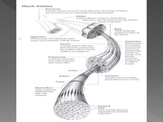
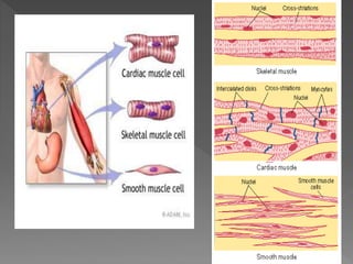
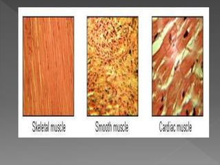
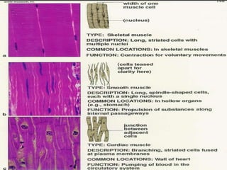
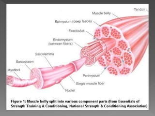
Muscle is a contractile tissue found throughout the body that produces movement when stimulated. There are three main types of muscle tissue: smooth, cardiac, and skeletal muscle. Skeletal muscle is striated, voluntary muscle attached to bones that allows for conscious control of movement. It comprises over 600 muscles in the musculoskeletal system, including axial muscles that control facial expression and posture, and appendicular muscles of the limbs. Muscle fibers contract when stimulated by motor nerves, producing movement through their interaction with bones and tendons.
The human skeleton is the internal framework of the human body. It is composed of around 270 bones at birth – this total decreases to around 206 bones by adulthood after some bones get fused together. The bone mass in the skeleton makes up about 14% of the total body weight and reaches maximum density around age 21
a quick visual understanding of what actually nervous tissue is made up of at cellular level its functions nerve cell types chemical synapse detailed structure of neuron
Skeletal system. anatomy and physiology of skeletal system. appendicular skel...mamtabisht10
SKELETAL SYSTEM
bones, cartilage and ligaments are tightly joined to form a strong, flexible framework called skeletal system
anatomy and physiology of axial and appendicular skeletal system
Axial Skeleton: The axial skeleton includes the skull, spine, ribs and sternum.
Appendicular Skeleton:
The appendicular skeleton includes the appendages of the body, which are the shoulders, arms, hips, and legs.
detail notes on connective tissue..
Connective tissue (CT) is one of the four basic types of animal tissue, along with epithelial tissue, muscle tissue, and nervous tissue. It develops from the mesoderm. Connective tissue is found in between other tissues everywhere in the body, including the nervous system. In the central nervous system, the three outer membranes (the meninges) that envelop the brain and spinal cord are composed of connective tissue.
All connective tissue consists of three main components: fibers (elastic and collagenous fibers), ground substance and cells. Not all authorities include blood or lymph as connective tissue because they lack the fiber component. All are immersed in the body water.
This ppt contains an elaborate description of muscles, focussing on muscles of mastication. both main muscles and accessory muscles have been described along with palpation of each. A brief is given about disorders.
Skeletal system
Divisions of skeletal system,
types of bone,
salient features and functions
of bones of axial and appendicular skeletal system Organization of skeletal muscle,
physiology of muscle contraction,
neuromuscular junction.
Its abt normal developmental milestones of a child from birth till 1 year.... Especially normal motor milestones...
"Because once u dont knw whts normal, u cant knw n differentiate between an abnormal"
2. Muscles are red, fleshy mass of the body
located under deep fascia.
The muscles are also found in heart and in
various organs like stomach, intestine,
urinogenital organs and also in blood
vessels.
3.
4. Muscle is a contractile tissue which
brings about movements.
Muscles can be regarded as motors of
body.
Study of muscle is called General
myology.
10. Smkuellteitnaul c(vleoaltuendt acryyli n&d srtirciaalt esdtr)u mctuusrcelse hfiabveirnsg a are c lear display of longitudinal and cross striations.
Skeletal muscle or "voluntary muscle" is anchored tboybteonndeoannsd( oisr ubsyeda ptoo neeffuercots siskealte ata flemwo pvleamceesn) t such tahsilso pcoosmtuortaiol ncoanntdr oinl i ms gaeinntearianlilny gm paoinsttuarine.e Tdh aosu ag h tsou bccoonnscscioiouus sc roenftlreoxl, ltihkee mnounsc-lpeos srteusrpaol nmsiubslcel erse.a Actn amvuesrcalgee a anddu altn m aavleer aisg me aaddue lut pfe omf a4le2 %is omf asdkee luepta ol f 36% (as a percentage of body mass)
Skeletal muscle fibers is striated, or striped and is under control of the individual’s will.
For this reason, it is often called “voluntary” muscle tissue.
11. Skeletal muscle tissues are usually attached to
bones. That is known as osseous tissues which
is innervated with somatic nerves through
which volitional control is performed.
When muscle fibers are stimulated by an
action of a never fiber, the fibers contract
and relax.
This interaction between muscle and nerve
fibers produces movement.
It can perform works of rapid, powerful
contraction as well as that of prolonged slow
sustained tonic contraction.
12.
13. Musculoskeletal system: comprises
those muscles that have an attachment
to skeletal system.
There are over 600 muscles in the
musculoskeletal system.
Axial skeleton muscles: control facial
expression; mastication; eye; tongue
and neck movements; respiration; the
abdominal wall; pelvic region;
movement of the vertebral column.
Appendicular skeletal muscles: include
the pectoral girdle, pelvic girdle, and
upper and lower extremities.
14.
15.
16. 1. Muscle Belly: The fleshy bulging, central part of a muscle.
2. Muscle fiber: A cylindrical, multinucleate cell composed of numerous myofibrils that
contracts when stimulated.
3. Myofibrils: A myofibril is a basic unit of a muscle. Muscles are composed of tubular cells
called myocytes or myofibers. Myofibers are composed of tubular myofibrils. Myofibrils
are composed of long proteins such as actin, myosin, and titin, and other proteins that
hold them together.
Myofilaments: Myofilaments, the filaments of myofibrils constructed from proteins. The
principal types of muscle are striated muscle, obliquely striated muscle and smooth
muscle. Various arrangements of myofilaments create different muscles. S has transverse
bands of filaments. In obliquely striated muscle, the filaments are staggered, and
smooth muscle has irregular arrangements of filaments.
There are three different types of myofilaments: thick, thin, and elastic filaments.
Thick filaments consist primarily of the protein myosin. Each thick filament are
approximately 15 nm in diameter, and each is made of several hundred molecules
of myosin.
Thin filaments, 7 nm in diameter, consist primarily of the protein actin. All thin filaments
are attached the Z disc.
Elastic filaments, 1 nm in diameter, are made of titin, a large springy protein. They flank
each thick filament and anchor it to the Z disc, the end point of a sarcomere.
1. Origin
2. Insertion
3. Tendon
4. Aponeurosis
17.
18.
19. 9. Epimysium
10. Perimysium
11. Endomysium
12. Sarcomere
13. Sarcolemma
14. Sarcoplasm
15. Sarcoplasmic reticulum
16. Motor nerve
17. Motor end-plate
18. Neurovascular hilum
19. Motor point
20. Myotome
21. Arrangement of fibers Shape
• The arrangement of muscle fibers varies according to the
direction, force and range of habitual movement at a
particular joint.
• The force of movement is directly proportional to the
number and size of muscle fibers and range of movement
is directly proportional to length of muscle fibers.
22. The muscles can be classified according to
arrangement of their fasciculi into the
following groups:
1. Parallel: muscles whose fibers run parallel
to one another.
Quadrilateral (thyrohyoid)
Strap (sternohyoid and Sartorius)
Strap-like with tendinous intersections
(rectus abdominis)
Fusiform (biceps brachii and digastric)
23.
24. 2. Oblique: feather-shaped muscles with
oblique fibers and a central tendon.
Unipennate (flexor pollicis longus, extensor
digitorum longus, peroneus tertius and
palmar interossei)
Bipennate (rectus femoris, dorsal interossei,
peroneus longus and flexor hallucis longus)
Multipennate (deltoid and subscapularis)
Triangular (adductor longus and
temporalis)
Circumpennate (tibialis anterior)
25.
26. 3. Circular: muscles whose
fibers encompass an
opening.
Sphincter radial
4. Convergent: fan-shaped
muscle fibers at
the origin that converge
at the insertion point.
tricipital