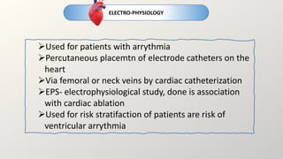
The topics for the day
CORONARY
ANGIOGRAPHY
ELECTRO-
PHYSIOLOGY
RADIO-
NUCLEOTIDE
IMAGING
CARDIAC
CATHERTERISATION
CARDIAC CATHERTERISATION
 An invasive technique of passing a specialised catheter through peripheral artery or vein into the
heart under X-Ray guidance
 Usually performed in conjunction with coronary angiography
 Gold standard diagnostic test for assessing heart and its vasculature
CONTRAINDICATIONS
1. Acute Decompensated heart failure
2. CKD- contast induced AKI
3. Bacteremia- lead to sepsis
4. Active GI bleeding
5. Contrast allergy- anaphylactic
reaction
6. On anticoagulant therapy- can lead
to vascular site bleeding
7. Electrolyte imbalance
CARDIAC CATHERTERISATION
COMPLICATIONS OF CATHETERIZATION
1. Tachy/brady-arrythmia
2. Vascular site bleeding- most common
complications. 1.5 to 2%
3. Contrast induced AKI- increase in serum
creatinine >0.5mg/dl. Seen in patients with risk
factors- DM, CKD, CHF, Anemia
4. Contrast allergy- IgE type hypersensitivity
reaction. If history is suggestive, treat with
antihistamines 24 hours prior
5. Myocardial infarction, stroke and death-
very rare complications. Less than 1%
POSTOPERATIVE CARE
• Remove vascular sheath, achieve
homeostasis
• Bed rest- 2 hours
• Advise adequate fluid intake
• Avoid strenuous activity
• Discharge same day, overnight
hospitalization only in high rise
cases(comorbities present)
• Patient with radiation exposure,
clinical follow up after 1 month
CARDIAC CATHERTERISATION
 Patient must be fasted for 6 hours prior procedure
 IV conscious sedation, but remain awake
 In suspected CAD, administer 325 mg aspirin
 Stop all anticoagulants 2-3 days prior to surgery- maintain INR <1.7
 No prophylactic Abx- sterile procedure
 Vascular Access: cardiac catheter is introduced percutaneously
under local anesthetic
 Via femoral or radial artery- Left heart catheterization
 Via femoral, radial or internal jugular vein- Right heart
catherterization
 Left heart catheterization: pass the guide wire through the artery
to aorta, inject dye for coronary angiography/ measure
hemodynamics/ ventriculography/ aortography
 Right heart catheterization: not performed as routine
catheterization. Indicated in unexplained dyspnea, PAH, CHD. A
balloon tiped SWAN GANZ catheter is used for measuring PCWP
CARDIAC CATHERTERISATION
A: x ray source
B: movable patient table
C: Lead shield
D: Video display
CARDIAC CATHERTERISATION
DIAGNOSTIC USES OF CARDIAC CATHETERISATION
HEMODYNAMICS
• Detect pressure and volumes in all chambers of the heart in systole and diastole
• AS- systolic pressure gradient between LA and LV
• MS- Diastolic pressure gradient between LA and LV
• HOCM- brackenborough-braunwald sign: post PVC increase in aortic-LV pressure and decrease in aortic PP
• Regurigitant volume in MR and TR
• To differentiate between CCP, CT and RCM- RAP, Y descent, square root sign
CARDIAC OUTPUT
• Using FICK’S PRINCIPLE- total oxygen consumption is equal to the product of cardiac output and
arteriovenous oxygen difference.
• 𝐶𝑂 =
𝑣𝑂2
𝐶𝑎−𝐶𝑣
• THERMODILUTION- injection of 10ml NS into right atrium(room temperature)measuring the temperature
deviations in pulmonary artery using a thermistor tipped catheter
CARDIAC CATHERTERISATION
DIAGNOSTIC USES OF CARDIAC CATHETERISATION
VASCULAR RESISTANCE
• Using ohms law. Resistance(for e.g. systemic) is equal to mean pressure(mean aortic pressure- mean RAP) and mean
flow(cardiac output)
• Measured in Wood units(x 80 to dynes-s-cm2)
• SVR 11-15 woods or 900-1200 dynes-s-cm2 – increased in stress, low output syndrome, hypertension
• PVR less than 2 woods or 50-250 dynes-s-cm2- PAH
VALVE AREA
• Gorlin formula and modified Hakki formula
• <1 cm2 severe AS
• <1.5 cm2 severe MS
INTRACARDIAC SHUNTS
• Congenital heart disease
• Using oxygen saturation. Step up saturationleft to right shunt. Step down right to left shunt
• Shunt ratio ratio of pulmonary blood flow by systemic blood flow(difference in mean arterial and venous oxygen
content
• Shunt ratio >1.5 ASD
CARDIAC CATHERTERISATION
DIAGNOSTIC USES OF CARDIAC CATHETERISATION
VENTRICULOGRAPHY
• Assessment of left ventricular function. Pigtail cathetercontrastvisualise cardiac cycle
• Hypokinetic movements, akinetic or dyskinetic(paradoxical)
• Can detect MVP, severity of MR(contrast reflux)-1+ tp 4+, LV aneurysm, VSD
AORTOGRAPHY
• Aneursym, coarctation and PDA
CINEFLOUROGRAPHY OF PROSTHETIC VALVES
• Detect any prosthetic valve dysfuntion)(immobility or fixity), thrombus or pannus
INTERVENTIONAL USES
• Coronary artery balloon angioplasty
• Stent placement
• Transcatheter abortive valve replacement(TAVR)
• Ablation
• Biopsy
CARDIAC CATHERTERISATION
CORONARY ANGIOGRAPHY
INJECTION OF RADIOOPAQUE CONTRAST IN THE CORONARY
ARTERIES WITH SERIAL RADIO IMAGING
• 2 coronary ostia,3 main coronary arteries- RCA, LAD and
LCX
• Ramus intermedius- posterior interventricular branch
directly from main LCA
• RCA- supplies SA node(60%) and AV node(90%), posterior
descending branch(right dominant in
90%)inferoposterior LV, Right AV groove RA and RV.
• LAD- anterior septal perforators, anterior, later and apical
LV
• LCX- lateral, posterior and inferior parts of LV, Posterior
descending branch(left dominance in 10%)
• 1-2% have coronary anomalies; separate ostia for LAD and
LCXmost common(0.41%)
CORONARY ANGIOGRAPHY
 Coronary circulation is visualized in multiple orthogonal
projections by angiography
 Luminal narrowings are visualised indicating coronary artery
stenosis.
 Narrowings are compared to “normal” segment and expressed
as percentage
 >50% stenosis  significant
 Myocardial bridge a kink in the LAD normal present, mistaken
for stenosis. It is due to the dip of the vessel from epicardium to
myocardium consequent contraction in systole
compression. In contrast to stenosis, returns to normal in
diastole
 Thrombolysis in Myocardial Infaction(TIMI) flow grade- relative
duration of the time taken for contrast to fill the artery
 TIMI grade 1 minimal filling. TIMI grade 2 gradual filling.
These suggest severe CAD
RADIO-
NUCLEOTIDE IMAGING
Used for evaluating cardiac function but MRI have replaced due to superior quality
and lesser radiation exposure
Myocardial perfusion scanning:
• IV injection of technetium
• Induce stress(exercise or dopamine)
• Scintiscan using gamma camera of myocardium
• Assess myocardial metabolism/function
Blood pool imaging:
• Injection of radio-isotope labelled RBCs
• Using gamma camera evaluate the heart 4 to 5 min after injection
• Shows ejection fractions, size and shape of cardiac chambers
ELECTRO-PHYSIOLOGY
Used for patients with arrythmia
Percutaneous placemtn of electrode catheters on the
heart
Via femoral or neck veins by cardiac catheterization
EPS- electrophysiological study, done is association
with cardiac ablation
Used for risk stratifaction of patients are risk of
ventricular arrythmia
heart.pptx

heart.pptx

  • 2.
    The topics forthe day CORONARY ANGIOGRAPHY ELECTRO- PHYSIOLOGY RADIO- NUCLEOTIDE IMAGING CARDIAC CATHERTERISATION
  • 3.
    CARDIAC CATHERTERISATION  Aninvasive technique of passing a specialised catheter through peripheral artery or vein into the heart under X-Ray guidance  Usually performed in conjunction with coronary angiography  Gold standard diagnostic test for assessing heart and its vasculature CONTRAINDICATIONS 1. Acute Decompensated heart failure 2. CKD- contast induced AKI 3. Bacteremia- lead to sepsis 4. Active GI bleeding 5. Contrast allergy- anaphylactic reaction 6. On anticoagulant therapy- can lead to vascular site bleeding 7. Electrolyte imbalance
  • 4.
    CARDIAC CATHERTERISATION COMPLICATIONS OFCATHETERIZATION 1. Tachy/brady-arrythmia 2. Vascular site bleeding- most common complications. 1.5 to 2% 3. Contrast induced AKI- increase in serum creatinine >0.5mg/dl. Seen in patients with risk factors- DM, CKD, CHF, Anemia 4. Contrast allergy- IgE type hypersensitivity reaction. If history is suggestive, treat with antihistamines 24 hours prior 5. Myocardial infarction, stroke and death- very rare complications. Less than 1% POSTOPERATIVE CARE • Remove vascular sheath, achieve homeostasis • Bed rest- 2 hours • Advise adequate fluid intake • Avoid strenuous activity • Discharge same day, overnight hospitalization only in high rise cases(comorbities present) • Patient with radiation exposure, clinical follow up after 1 month
  • 5.
    CARDIAC CATHERTERISATION  Patientmust be fasted for 6 hours prior procedure  IV conscious sedation, but remain awake  In suspected CAD, administer 325 mg aspirin  Stop all anticoagulants 2-3 days prior to surgery- maintain INR <1.7  No prophylactic Abx- sterile procedure  Vascular Access: cardiac catheter is introduced percutaneously under local anesthetic  Via femoral or radial artery- Left heart catheterization  Via femoral, radial or internal jugular vein- Right heart catherterization  Left heart catheterization: pass the guide wire through the artery to aorta, inject dye for coronary angiography/ measure hemodynamics/ ventriculography/ aortography  Right heart catheterization: not performed as routine catheterization. Indicated in unexplained dyspnea, PAH, CHD. A balloon tiped SWAN GANZ catheter is used for measuring PCWP
  • 6.
    CARDIAC CATHERTERISATION  Patientmust be fasted for 6 hours prior procedure  IV conscious sedation, but remain awake  In suspected CAD, administer 325 mg aspirin  Stop all anticoagulants 2-3 days prior to surgery- maintain INR <1.7  No prophylactic Abx- sterile procedure  Vascular Access: cardiac catheter is introduced percutaneously under local anesthetic  Via femoral or radial artery- Left heart catheterization  Via femoral, radial or internal jugular vein- Right heart catherterization  Left heart catheterization: pass the guide wire through the artery to aorta, inject dye for coronary angiography/ measure hemodynamics/ ventriculography/ aortography  Right heart catheterization: not performed as routine catheterization. Indicated in unexplained dyspnea, PAH, CHD. A balloon tiped SWAN GANZ catheter is used for measuring PCWP
  • 7.
    CARDIAC CATHERTERISATION A: xray source B: movable patient table C: Lead shield D: Video display
  • 8.
    CARDIAC CATHERTERISATION DIAGNOSTIC USESOF CARDIAC CATHETERISATION HEMODYNAMICS • Detect pressure and volumes in all chambers of the heart in systole and diastole • AS- systolic pressure gradient between LA and LV • MS- Diastolic pressure gradient between LA and LV • HOCM- brackenborough-braunwald sign: post PVC increase in aortic-LV pressure and decrease in aortic PP • Regurigitant volume in MR and TR • To differentiate between CCP, CT and RCM- RAP, Y descent, square root sign CARDIAC OUTPUT • Using FICK’S PRINCIPLE- total oxygen consumption is equal to the product of cardiac output and arteriovenous oxygen difference. • 𝐶𝑂 = 𝑣𝑂2 𝐶𝑎−𝐶𝑣 • THERMODILUTION- injection of 10ml NS into right atrium(room temperature)measuring the temperature deviations in pulmonary artery using a thermistor tipped catheter
  • 9.
    CARDIAC CATHERTERISATION DIAGNOSTIC USESOF CARDIAC CATHETERISATION VASCULAR RESISTANCE • Using ohms law. Resistance(for e.g. systemic) is equal to mean pressure(mean aortic pressure- mean RAP) and mean flow(cardiac output) • Measured in Wood units(x 80 to dynes-s-cm2) • SVR 11-15 woods or 900-1200 dynes-s-cm2 – increased in stress, low output syndrome, hypertension • PVR less than 2 woods or 50-250 dynes-s-cm2- PAH VALVE AREA • Gorlin formula and modified Hakki formula • <1 cm2 severe AS • <1.5 cm2 severe MS INTRACARDIAC SHUNTS • Congenital heart disease • Using oxygen saturation. Step up saturationleft to right shunt. Step down right to left shunt • Shunt ratio ratio of pulmonary blood flow by systemic blood flow(difference in mean arterial and venous oxygen content • Shunt ratio >1.5 ASD
  • 10.
    CARDIAC CATHERTERISATION DIAGNOSTIC USESOF CARDIAC CATHETERISATION VENTRICULOGRAPHY • Assessment of left ventricular function. Pigtail cathetercontrastvisualise cardiac cycle • Hypokinetic movements, akinetic or dyskinetic(paradoxical) • Can detect MVP, severity of MR(contrast reflux)-1+ tp 4+, LV aneurysm, VSD AORTOGRAPHY • Aneursym, coarctation and PDA CINEFLOUROGRAPHY OF PROSTHETIC VALVES • Detect any prosthetic valve dysfuntion)(immobility or fixity), thrombus or pannus INTERVENTIONAL USES • Coronary artery balloon angioplasty • Stent placement • Transcatheter abortive valve replacement(TAVR) • Ablation • Biopsy
  • 11.
  • 12.
    CORONARY ANGIOGRAPHY INJECTION OFRADIOOPAQUE CONTRAST IN THE CORONARY ARTERIES WITH SERIAL RADIO IMAGING • 2 coronary ostia,3 main coronary arteries- RCA, LAD and LCX • Ramus intermedius- posterior interventricular branch directly from main LCA • RCA- supplies SA node(60%) and AV node(90%), posterior descending branch(right dominant in 90%)inferoposterior LV, Right AV groove RA and RV. • LAD- anterior septal perforators, anterior, later and apical LV • LCX- lateral, posterior and inferior parts of LV, Posterior descending branch(left dominance in 10%) • 1-2% have coronary anomalies; separate ostia for LAD and LCXmost common(0.41%)
  • 13.
    CORONARY ANGIOGRAPHY INJECTION OFRADIOOPAQUE CONTRAST IN THE CORONARY ARTERIES WITH SERIAL RADIO IMAGING • 2 coronary ostia,3 main coronary arteries- RCA, LAD and LCX • Ramus intermedius- posterior interventricular branch directly from main LCA • RCA- supplies SA node(60%) and AV node(90%), posterior descending branch(right dominant in 90%)inferoposterior LV, Right AV groove RA and RV. • LAD- anterior septal perforators, anterior, later and apical LV • LCX- lateral, posterior and inferior parts of LV, Posterior descending branch(left dominance in 10%) • 1-2% have coronary anomalies; separate ostia for LAD and LCXmost common(0.41%)
  • 14.
    CORONARY ANGIOGRAPHY  Coronarycirculation is visualized in multiple orthogonal projections by angiography  Luminal narrowings are visualised indicating coronary artery stenosis.  Narrowings are compared to “normal” segment and expressed as percentage  >50% stenosis  significant  Myocardial bridge a kink in the LAD normal present, mistaken for stenosis. It is due to the dip of the vessel from epicardium to myocardium consequent contraction in systole compression. In contrast to stenosis, returns to normal in diastole  Thrombolysis in Myocardial Infaction(TIMI) flow grade- relative duration of the time taken for contrast to fill the artery  TIMI grade 1 minimal filling. TIMI grade 2 gradual filling. These suggest severe CAD
  • 15.
    RADIO- NUCLEOTIDE IMAGING Used forevaluating cardiac function but MRI have replaced due to superior quality and lesser radiation exposure Myocardial perfusion scanning: • IV injection of technetium • Induce stress(exercise or dopamine) • Scintiscan using gamma camera of myocardium • Assess myocardial metabolism/function Blood pool imaging: • Injection of radio-isotope labelled RBCs • Using gamma camera evaluate the heart 4 to 5 min after injection • Shows ejection fractions, size and shape of cardiac chambers
  • 16.
    ELECTRO-PHYSIOLOGY Used for patientswith arrythmia Percutaneous placemtn of electrode catheters on the heart Via femoral or neck veins by cardiac catheterization EPS- electrophysiological study, done is association with cardiac ablation Used for risk stratifaction of patients are risk of ventricular arrythmia