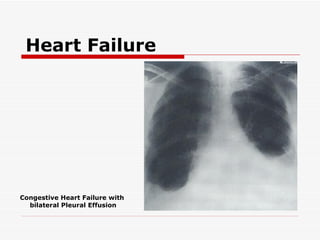
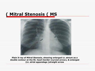
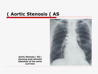
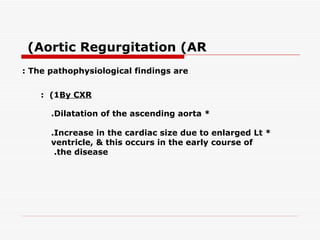
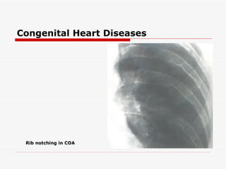
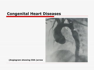
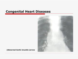
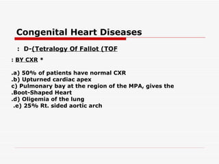
Heart Failure
plain X-ray findings: include the followings The *

a( Enlarged cardiac shadow +/- specific chamber
.enlargement
b( Evidence of pulmonary venous hypertension
 .(zoneenlargement of the vessels in the upper(
.c( Evidence of pulmonary edema
d( Pleural Effusion : It is usually bilateral, often larger on
the Rt. than on the Lt. side; but if it is unilateral it is
 .almost always on the Rt. side


 : Note
 Acute Lt. ventricular failure, small effusion is seen at the costo-phrenic
 angle, running up the lateral chest wall; (this fluid may, in fact, be edema of
 .(the lungs rather than true pleural effusion
Heart Failure


Congestive Heart Failure with
  bilateral Pleural Effusion
Valvular Heart Diseases

   Mitral Valve Diseases
   : They Include

   ( -1M itralS tenosis (MS
    ( -2M itralR egurgitation (MR
( Mitral Stenosis ( MS
: The pathophysiological findings are
    : (1By CXR

                  .Lt. atrial enlargement + normal cardiac size *

                                            .Mitral calcification *

                              .Pulmonary venous hypertension *

       .Pulmonary edema *

       Pulmonary arterial hypertension, will lead to enlarged *
       .(cardiac size (Rt. ventricle is enlarged
( Mitral Stenosis ( MS


  Plain X-ray of Mitral Stenosis, showing enlarged Lt. atrium as a
 double contour at the Rt. heart border (curved arrow(, & enlarged
               (Lt. atrial appendage (straight arrow
(Mitral Regurgitation (MR
: The pathophysiological findings are


    : (1By CXR

     Lt. atrium & Lt. ventricle are enlarged, so cardiac size *
     will be enlarged in its Lt. ventricular .configuration

     .Pulmonary venous hypertension *

      .Pulmonary edema *

  : Note
    Lt. atrial enlargement & pulmonary venous hypertension are the
    important signs of MR, which differs from MS by Lt. ventricular
    .enlargement
Valvular Heart Diseases

  Aortic Valve Diseases
   : They Include

   ( -1A orticS tenosis (AS
    ( -2A orticR egurgitation (AR
( Aortic Stenosis ( AS
: The pathophysiological findings are

    : (1By CXR

     .Aortic valve calcification *
     Post stenotic dilatation of the ascending aorta (the *
     .(major feature
     .(Lt. ventricular enlargement (Late feature *
     .(Increase pulmonary venous pressure (Late feature *

     .Both late features will lead to Lt. ventricular failure -
( Aortic Stenosis ( AS


    Aortic Stenosis ( AS (
    showing post-stenotic
    dilatation of the aorta
           ((arrows
(Aortic Regurgitation (AR
: The pathophysiological findings are


    : (1By CXR

      .Dilatation of the ascending aorta *

      .Increase in the cardiac size due to enlarged Lt *
      ventricle, & this occurs in the early course of
       .the disease
& Lt. Atrial Myxoma
Other Intra-cardiac Masses
.Intracardiac tumors are extremely rare *

Lt. atrial myxoma is the most frequently encountered, it *
is a benign tumor which arises : from
.a( Intra-atrial septum
.b( Lt. atrial walls

.As it enlarges, it becomes pedunculated to float in the Lt *
atrial cavity, & therefore it will interfere with mitral valve
function & mimic MS or MR in both ways (clinically & by
.(CXR

It can be differentiated from other intra-cardiac masses *
by MRI & Echo., & the only differential Dx is Lt. atrial
.thrombus in patient with Rheumatic MS
& Lt. Atrial Myxoma
Other Intra-cardiac Masses


      Lt. Atrial Myxoma shown by 2-dimentional
         echocardiography– modified apical 4-
                     chamber view
Congenital Heart Diseases

   : A-(Lt. to Rt. Shunt (as in ASD, VSD, & PDA

CXR findings will When the shunt is 2/1 or more, the following *
: be seen
.(a( Enlarged cardiac size (cardiomegally
b( Enlarged Mean Pulmonary Artery (MPA(, hilar pulmonary
.arteries
.c( Pulmonary plethora

Absent of Lt. ventricular enlargement in the presence of increase *
.pulmonary flow is mainly indicate ASD
Congenital Heart Diseases


    VSD in a child
Congenital Heart Diseases
: B-(Pulmonary Stenosis (PS


BY CXR: : there will be *

.a( Normal heart size
b( Enlargement of MPA Lt. pulmonary artery
.(Post-Stenotic Dilatation(
Congenital Heart Diseases
: C-(Coarctation Of Aorta (COA

 It is an abnormal aortic arch due to presence of *
 narrowing, just distal to the origin of the Lt. subclavian
  .artery

CXR findings: include *
  .a( Indentation of the aortic arch
  & ,(b( Dilatation above (dilatation of the Lt. subclavian artery
  below (post-stenotic .(dilatation
  c( Enlarged cardiac size & ascending aorta due to long standing
  .hypertension
  d( Rib notching is due to enlargement of the intercostal arteries
  which act as a collateral, & there will small cortical indentation
  on the inferior margin of the posterior halves of the ribs from
  .the 3rd or 4th rib, & downward

  .Note : COA itself can be seen by angiography or MRI
Congenital Heart Diseases


 Rib notching in COA
Congenital Heart Diseases


(Angiogram showing COA (arrow
Congenital Heart Diseases


(Abnormal Aortic knuckle (arrow
Congenital Heart Diseases
: D-(Tetralogy Of Fallot (TOF
 : they are & *
 .a( VSD
 b( Rt. ventricular outflow obstruction (valvular or
 .(subvalvular
 .c( Rt. ventricular hypertrophy
  .d( Overriding of aorta over the VSD
Congenital Heart Diseases
 : D-(Tetralogy Of Fallot (TOF
: BY CXR *

.a( 50% of patients have normal CXR
.b( Upturned cardiac apex
c( Pulmonary bay at the region of the MPA, gives the
.Boot-Shaped Heart
.d( Oligemia of the lung
 .e( 25% Rt. sided aortic arch
Thank You

radilology. CVS lec 4.(dr.abeer)

  • 1.
    Heart Failure plain X-rayfindings: include the followings The * a( Enlarged cardiac shadow +/- specific chamber .enlargement b( Evidence of pulmonary venous hypertension .(zoneenlargement of the vessels in the upper( .c( Evidence of pulmonary edema d( Pleural Effusion : It is usually bilateral, often larger on the Rt. than on the Lt. side; but if it is unilateral it is .almost always on the Rt. side : Note Acute Lt. ventricular failure, small effusion is seen at the costo-phrenic angle, running up the lateral chest wall; (this fluid may, in fact, be edema of .(the lungs rather than true pleural effusion
  • 2.
    Heart Failure Congestive HeartFailure with bilateral Pleural Effusion
  • 3.
    Valvular Heart Diseases Mitral Valve Diseases : They Include ( -1M itralS tenosis (MS ( -2M itralR egurgitation (MR
  • 4.
    ( Mitral Stenosis( MS : The pathophysiological findings are : (1By CXR .Lt. atrial enlargement + normal cardiac size * .Mitral calcification * .Pulmonary venous hypertension * .Pulmonary edema * Pulmonary arterial hypertension, will lead to enlarged * .(cardiac size (Rt. ventricle is enlarged
  • 5.
    ( Mitral Stenosis( MS Plain X-ray of Mitral Stenosis, showing enlarged Lt. atrium as a double contour at the Rt. heart border (curved arrow(, & enlarged (Lt. atrial appendage (straight arrow
  • 6.
    (Mitral Regurgitation (MR :The pathophysiological findings are : (1By CXR Lt. atrium & Lt. ventricle are enlarged, so cardiac size * will be enlarged in its Lt. ventricular .configuration .Pulmonary venous hypertension * .Pulmonary edema * : Note Lt. atrial enlargement & pulmonary venous hypertension are the important signs of MR, which differs from MS by Lt. ventricular .enlargement
  • 7.
    Valvular Heart Diseases Aortic Valve Diseases : They Include ( -1A orticS tenosis (AS ( -2A orticR egurgitation (AR
  • 8.
    ( Aortic Stenosis( AS : The pathophysiological findings are : (1By CXR .Aortic valve calcification * Post stenotic dilatation of the ascending aorta (the * .(major feature .(Lt. ventricular enlargement (Late feature * .(Increase pulmonary venous pressure (Late feature * .Both late features will lead to Lt. ventricular failure -
  • 9.
    ( Aortic Stenosis( AS Aortic Stenosis ( AS ( showing post-stenotic dilatation of the aorta ((arrows
  • 10.
    (Aortic Regurgitation (AR :The pathophysiological findings are : (1By CXR .Dilatation of the ascending aorta * .Increase in the cardiac size due to enlarged Lt * ventricle, & this occurs in the early course of .the disease
  • 11.
    & Lt. AtrialMyxoma Other Intra-cardiac Masses .Intracardiac tumors are extremely rare * Lt. atrial myxoma is the most frequently encountered, it * is a benign tumor which arises : from .a( Intra-atrial septum .b( Lt. atrial walls .As it enlarges, it becomes pedunculated to float in the Lt * atrial cavity, & therefore it will interfere with mitral valve function & mimic MS or MR in both ways (clinically & by .(CXR It can be differentiated from other intra-cardiac masses * by MRI & Echo., & the only differential Dx is Lt. atrial .thrombus in patient with Rheumatic MS
  • 12.
    & Lt. AtrialMyxoma Other Intra-cardiac Masses Lt. Atrial Myxoma shown by 2-dimentional echocardiography– modified apical 4- chamber view
  • 13.
    Congenital Heart Diseases : A-(Lt. to Rt. Shunt (as in ASD, VSD, & PDA CXR findings will When the shunt is 2/1 or more, the following * : be seen .(a( Enlarged cardiac size (cardiomegally b( Enlarged Mean Pulmonary Artery (MPA(, hilar pulmonary .arteries .c( Pulmonary plethora Absent of Lt. ventricular enlargement in the presence of increase * .pulmonary flow is mainly indicate ASD
  • 14.
  • 15.
    Congenital Heart Diseases :B-(Pulmonary Stenosis (PS BY CXR: : there will be * .a( Normal heart size b( Enlargement of MPA Lt. pulmonary artery .(Post-Stenotic Dilatation(
  • 16.
    Congenital Heart Diseases :C-(Coarctation Of Aorta (COA It is an abnormal aortic arch due to presence of * narrowing, just distal to the origin of the Lt. subclavian .artery CXR findings: include * .a( Indentation of the aortic arch & ,(b( Dilatation above (dilatation of the Lt. subclavian artery below (post-stenotic .(dilatation c( Enlarged cardiac size & ascending aorta due to long standing .hypertension d( Rib notching is due to enlargement of the intercostal arteries which act as a collateral, & there will small cortical indentation on the inferior margin of the posterior halves of the ribs from .the 3rd or 4th rib, & downward .Note : COA itself can be seen by angiography or MRI
  • 17.
    Congenital Heart Diseases Rib notching in COA
  • 18.
  • 19.
  • 20.
    Congenital Heart Diseases :D-(Tetralogy Of Fallot (TOF : they are & * .a( VSD b( Rt. ventricular outflow obstruction (valvular or .(subvalvular .c( Rt. ventricular hypertrophy .d( Overriding of aorta over the VSD
  • 21.
    Congenital Heart Diseases : D-(Tetralogy Of Fallot (TOF : BY CXR * .a( 50% of patients have normal CXR .b( Upturned cardiac apex c( Pulmonary bay at the region of the MPA, gives the .Boot-Shaped Heart .d( Oligemia of the lung .e( 25% Rt. sided aortic arch
  • 22.