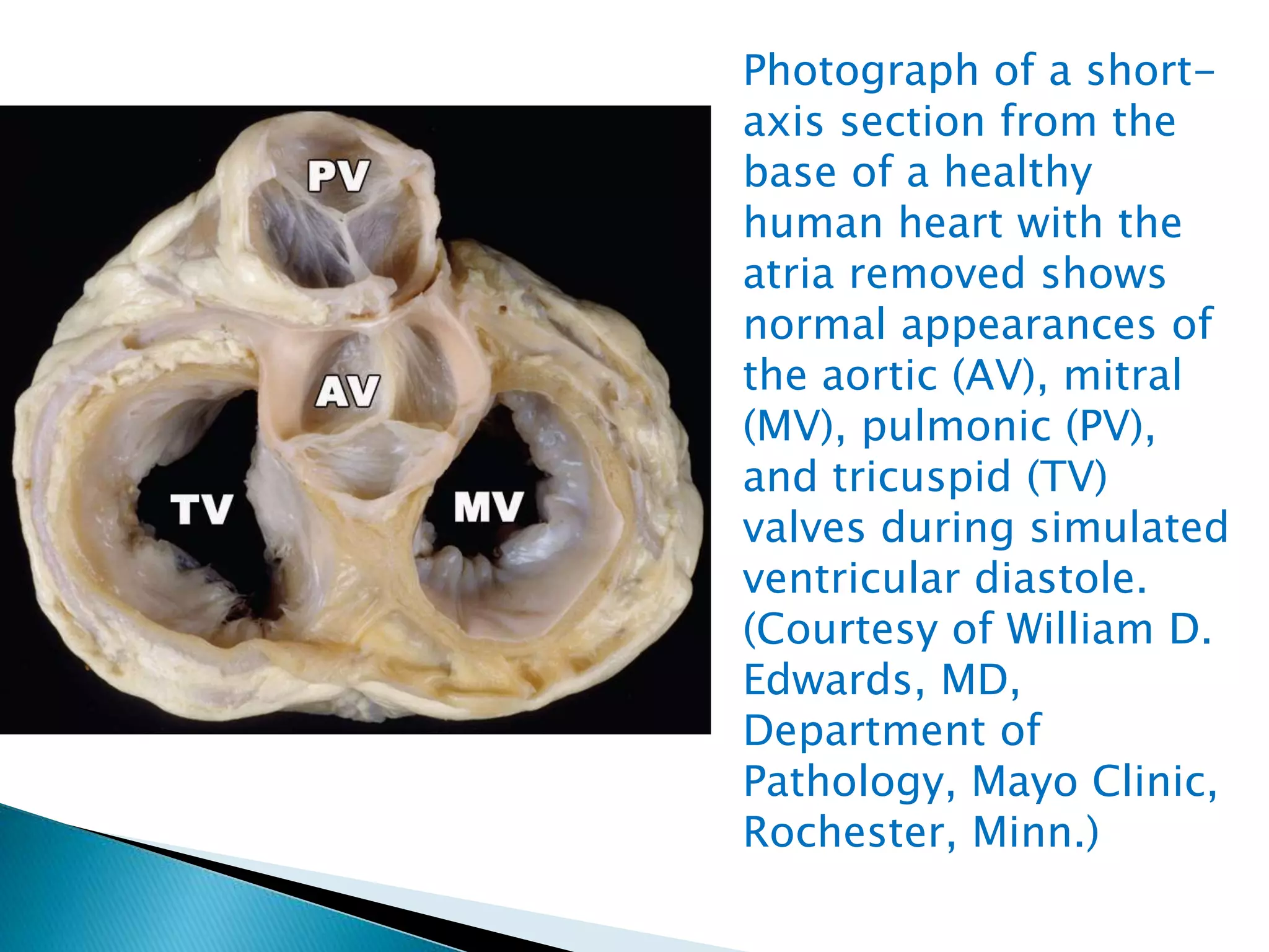
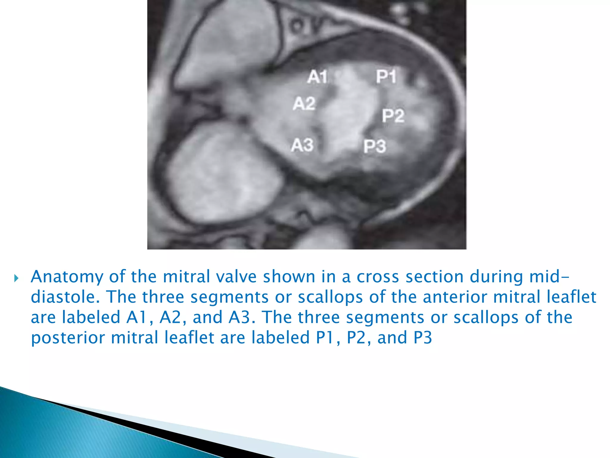
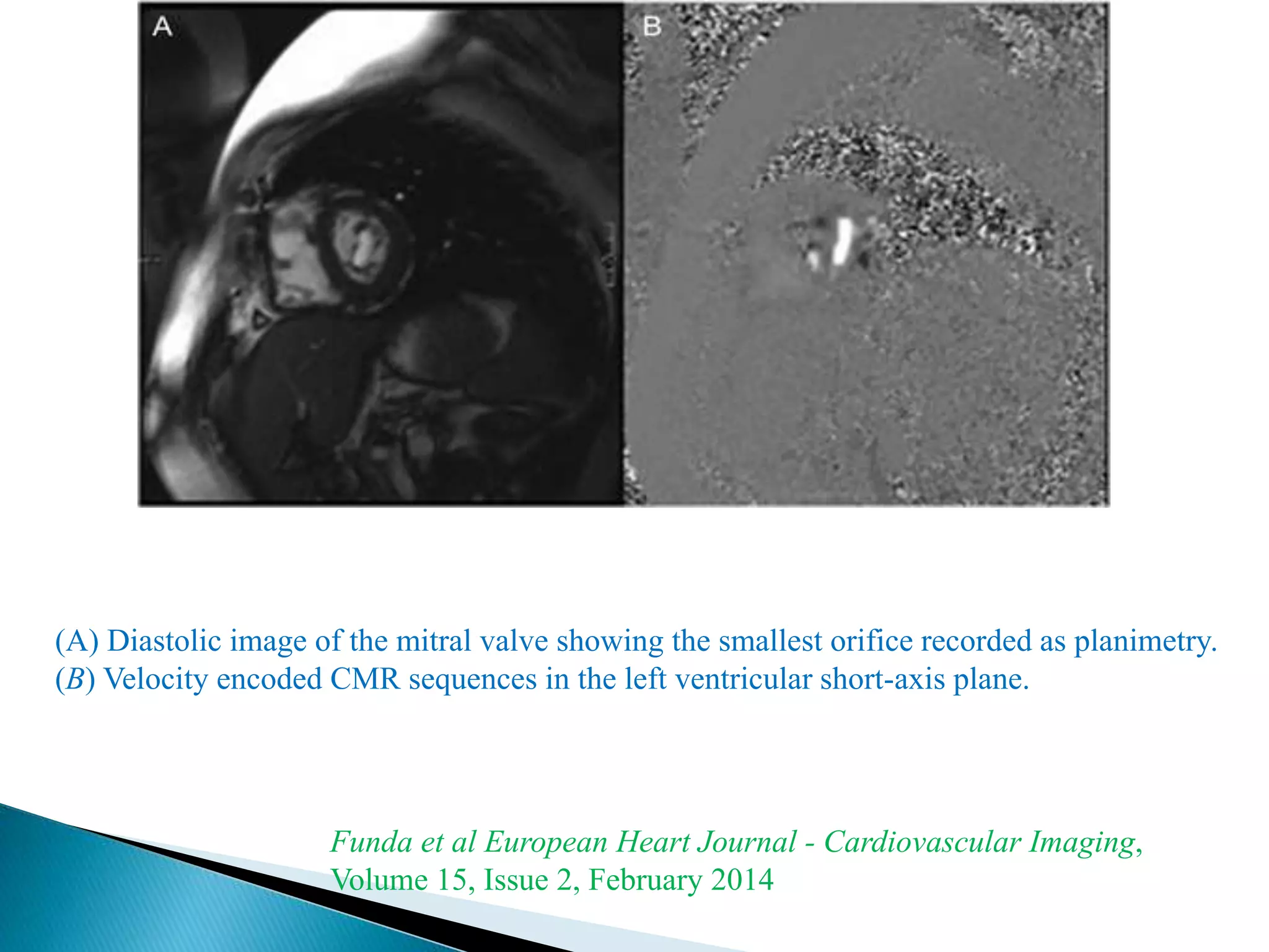
The mitral valve develops between the 5th and 15th weeks of gestation. It consists of an annulus, two leaflets, chordae tendineae and papillary muscles. Rheumatic fever is a leading cause of mitral stenosis, which results from repeated bouts of inflammation damaging the mitral valve over time. Mitral stenosis causes elevated left atrial pressure and left atrial enlargement, often resulting in pulmonary hypertension. Echocardiography is the primary imaging method used to evaluate the mitral valve anatomy and measure the severity of mitral stenosis.
This document provides an overview of echocardiographic assessment of mitral regurgitation. It describes the anatomy of the mitral valve including the leaflets, annulus, chordae, and papillary muscles. It discusses Carpentier's functional classification system for describing the mechanism of mitral valve dysfunction. Methods for assessing severity are covered, including color flow imaging, continuous wave Doppler, vena contracta width, proximal isovelocity surface area, and volumetric assessment. Key points are made about evaluating jet direction, duration, and velocity in context of blood pressure. The importance of assessing left ventricular and left atrial size and function is also highlighted.
ECHOCARDIOGRAPHIC EVALUATION OF MITRAL VALVE DISEASEPraveen Nagula
MITRAL VALVE ANATOMY , M MODE FINDINGS IN MITRAL STENOSIS,EVALUATION OF THE SEVERITY OF LESION,CALCIFIC MS,CCMA,CONGENITAL LESIONS,GUIDELINES ALL IN DETAIL....
1) Transthoracic and transesophageal echocardiography are important modalities for assessing atrial septal defects (ASDs). TTE can identify RV volume overload and septal flattening, while TEE precisely measures defect size and evaluates rim morphology.
2) The four main types of ASDs - ostium secundum, ostium primum, sinus venosus, and coronary sinus defects - have distinguishing echo features. Doppler can demonstrate shunt direction and magnitude.
3) Echocardiography guides percutaneous ASD closure by assessing defect and rim anatomy, device sizing, and post-procedure result. Understanding echo features is key to ensuring procedure success.
This document discusses the anatomy, embryology, and management of L-TGA (transposition of the great arteries). Some key points:
- In L-TGA, the ventricles are inverted such that the morphologic right ventricle is on the left and pumps blood to the lungs, while the morphologic left ventricle is on the right and pumps blood to the body.
- Embryologically, abnormal leftward looping of the heart tube during development results in the inverted ventricles. The conduction system and coronary arteries also have abnormal anatomy.
- Clinical features may include congenital heart block, progressive tricuspid regurgitation, pulmonary stenosis, and heart failure. Diagn
Tissue Doppler echocardiography allows assessment of myocardial motion using Doppler ultrasound. It uses frequency shifts of ultrasound waves to calculate myocardial velocity, focusing on lower velocities than blood flow Doppler. There are two techniques: pulsed TDE uses a sample volume gate while color-coded TDE uses autocorrelation to display multigated velocity data superimposed on images. TDE is useful for evaluating systolic and diastolic left ventricular function by measuring velocities of the mitral annulus, and can help distinguish conditions like constrictive pericarditis from restrictive cardiomyopathy.
A lecture on the echocardiographic evaluation of hypertrophic cardiomyopathy. Starts with an overview of the topic then a systematic approach to diagnosis and then a differential diagnosis followed by take-home messages and conclusion.
This document discusses the use of echocardiography in evaluating various types of cardiomyopathies. It provides echocardiographic features of dilated cardiomyopathy including dilated chambers, normal wall thickness, and complications like mitral regurgitation. Hypertrophic cardiomyopathy features include unexplained hypertrophy, diastolic dysfunction, and left ventricular outflow tract obstruction. Restrictive cardiomyopathies show hypertrophy, enlarged atria, restricted filling, and elevated pressures. Left ventricular non-compaction and arrhythmogenic right ventricular cardiomyopathy also have distinct echocardiographic characteristics described.
This ECG shows a 58-year-old female patient who presented with chest pain. The ECG reveals sinus rhythm with 5:4 second-degree sinoatrial exit block, where every fifth impulse is blocked at the SA junction. This type of SA block is characterized by no change in P-R interval before the pause and a pause interval that equals approximately two to four times the normal P-P cycle length. SA node dysfunction can be intrinsic, due to degenerative causes like fibrosis, or extrinsic, due to autonomic influences or drugs that suppress the SA node. Diagnosis is usually clinical or via ECG and pacemaker implantation may be indicated for symptomatic patients.
This document summarizes the history and classification of sinus of Valsalva aneurysm (SOVA). Some key points:
- SOVA was first described in 1839 and the first successful repair was in 1956 using cardiopulmonary bypass.
- SOVAs can be congenital or acquired due to various connective tissue/inflammatory disorders.
- The majority originate from the right coronary cusp (77%) and most commonly rupture into the right ventricle (67.9%).
- The classic Sakakibara classification categorizes SOVAs arising from the right coronary cusp into three types based on location of rupture/protrusion. A modified classification exists for non-cor
1) The document describes methods for quantifying mitral regurgitation (MR), including Carpentier's classification of MR types and echocardiographic parameters for assessing MR severity.
2) Proximal isovelocity surface area (PISA) uses the conservation of mass principle to calculate regurgitant volume and orifice area based on measurements of the PISA radius and aliasing velocity.
3) Several limitations of PISA are discussed, but it provides a quantitative assessment of MR with acceptable reproducibility when used appropriately.
1) Complete transposition of the great arteries (d-TGA) is a congenital heart defect where the aorta arises from the right ventricle and the pulmonary artery arises from the left ventricle, causing ventriculoarterial discordance.
2) In d-TGA, the systemic and pulmonary circulations are arranged in parallel rather than in series, requiring blood flow between the circuits through connections like an atrial or ventricular septal defect.
3) Echocardiography is useful for diagnosing d-TGA by demonstrating the aorta originating from the right ventricle and pulmonary artery from the left ventricle, as well as identifying the origins of the coronary arteries.
The document discusses assessment of mechanical dyssynchrony for cardiac resynchronization therapy. It defines electrical and mechanical dyssynchrony, and describes the deleterious hemodynamic effects of left ventricular dyssynchrony. It then summarizes various echocardiographic tools for assessing atrioventricular dyssynchrony, interventricular dyssynchrony, and intraventricular dyssynchrony, including M-mode, tissue Doppler imaging, and three-dimensional echocardiography. Measurement techniques for different dyssynchrony parameters such as septal-posterior wall motion delay, lateral wall postsystolic displacement, and time to peak systolic velocity are outlined.
Hypertrophic cardiomyopathy (HCM) is defined by a thickened left ventricular wall without an identifiable cause. It can range from asymptomatic to causing heart failure, arrhythmias, or sudden cardiac death. Treatment depends on whether the left ventricular outflow tract (LVOT) is obstructed. For symptomatic patients with LVOT obstruction despite maximum medical therapy, septal reduction procedures like alcohol septal ablation or surgical myectomy are recommended. Alcohol septal ablation involves injecting alcohol into a septal perforator artery to ablate tissue and reduce the gradient. Surgical myectomy directly resects septal muscle. Both procedures significantly reduce gradients and improve symptoms but surgical myectomy provides better gradient and symptom reduction with a lower risk of
Echo assessment of aortic valve diseaseNizam Uddin
This document discusses the echocardiographic assessment of aortic valve diseases. It describes how aortic stenosis is classified based on its location as valvular, subvalvular, or supravalvular. It outlines the etiology of valvular aortic stenosis and discusses echocardiographic methods for assessing the severity of aortic stenosis including peak transvalvular velocity, mean transvalvular gradient, and aortic valve area using the continuity equation. The document also discusses the assessment of aortic regurgitation severity using measurements such as vena contracta width, regurgitant jet width and area, pressure half time, diastolic flow reversal, and regurgitant volume and fraction. Methods for
ECG localization of accessory pathways slideshareCardiology
This presentation is simplified view of accessory pathways in heart and their localization with help of algorithms and ECG examples. Try to read this PPT in power point to see full effects and animations.
This document discusses techniques for localizing the site of origin of ventricular tachycardia based on electrocardiogram characteristics. It describes that right ventricular outflow tract tachycardias typically present with left bundle branch block morphology while left ventricular sites may present with either right or left bundle branch block depending on exit site. Specific leads are discussed that can provide clues about anterior vs posterior, septal vs free wall origin within the outflow tracts. Other areas like fascicles, papillary muscles and mitral/tricuspid annuli are also summarized.
This document discusses percutaneous mitral valve interventions for mitral regurgitation. It begins by describing the anatomy of the mitral valve and causes of mitral regurgitation. It then discusses the natural history of mitral regurgitation and indications for surgery. Current percutaneous options are described including the MitraClip device, which is the only FDA approved one. The MitraClip procedure involves grasping the leaflets edges to reduce regurgitation. Early results show high rates of procedural success for MitraClip in patients at high risk for surgery. Complications are usually low at 15-19% and include bleeding, partial clip detachment, and stroke.
This document discusses the use of echocardiography in evaluating congenital heart diseases in adults. It outlines the indications for echocardiography and describes how to perform the examination and interpret findings. Key abnormalities that can be identified include atrial septal defects, ventricular septal defects, atrioventricular septal defects, anomalies of venous inflow, and abnormalities of ventricular morphology. Echocardiography is well-suited for diagnosing and monitoring these congenital heart conditions in adulthood.
This document summarizes the echocardiographic assessment of mitral stenosis (MS). It describes the anatomy of the mitral valve and causes of MS. Methods for assessing MS severity include measuring the pressure gradient, mitral valve area using planimetry and pressure half-time, and pulmonary artery pressure. Suitability for percutaneous transvenous mitral commissurotomy is evaluated. Concomitant valve lesions are also identified. Stress echocardiography may be used when symptoms are equivocal. Transesophageal echocardiography is recommended in some cases.
This document discusses various echocardiographic scoring systems used to assess mitral valve anatomy and predict outcomes of percutaneous balloon mitral valvuloplasty (PBMV). The Wilkins score and Commissural Calcification score are described in detail. The Wilkins score grades leaflet thickening, mobility, calcification and subvalvular involvement on a scale of 4-16. A score ≤8 indicates favorable anatomy for PBMV. The Commissural Calcification score quantifies calcification at each commissure. Other discussed scores include the Cormier score, RT-3DE score, Chen score, Reid score and Nobuyoshi score. Limitations of the scoring systems and ideas for an ideal future scoring
This document discusses the echocardiographic evaluation of mitral valve prolapse (MVP). It describes the use of M-mode, 2D, and 3D echocardiography to diagnose MVP and assess mitral regurgitation severity. Measurement of the vena contracta and use of the proximal isovelocity surface area method are emphasized for accurate regurgitant quantification. Surgical indications and repair techniques are also summarized.
Three sentences:
The document provides details on the anatomy and evaluation of aortic stenosis using echocardiography. It describes the normal aortic valve anatomy and how various types of aortic stenosis like calcific, rheumatic, bicuspid and subvalvular present on echo. Quantitative assessment of aortic stenosis severity is done using Doppler ultrasound to measure the maximum jet velocity and calculate the pressure gradient across the stenotic valve.
The document discusses electrocardiogram (ECG) patterns associated with cardiac chamber enlargement, specifically right atrial enlargement (RAE) and left atrial enlargement (LAE). RAE is suggested by a tall, peaked P wave in leads II, III, AVF and a positive P wave in V1. LAE results in prolongation of the left atrial component of the P wave, increased posterior deviation of the left atrial vector, and left axis deviation of the P wave. The diagnostic accuracy of ECG findings for chamber enlargement is limited but can provide clues when correlated with imaging studies.
The document discusses various coronary artery anomalies including anomalies of origination, course, and intrinsic anatomy. Some key points include:
- Coronary artery anomalies have a global incidence of 5.64% and incidence of sudden death is 0.6%
- Anomalous origination of the left main coronary artery from the pulmonary artery (ALCAPA) is a rare but serious anomaly if left untreated
- Certain anomalous coronary artery courses, such as between the aorta and pulmonary artery, are associated with higher risks of sudden cardiac death
- Other anomalies discussed include single coronary arteries, coronary hypoplasia, ectasia/aneurysms, and intramural coronary arteries
Anatomy of mitral valve echo evaluationmadhusiva03
The document discusses the anatomy and function of the mitral valve complex. It notes that the mitral valve has a triple function regulating blood flow between the left atrium and ventricle. The mitral valve complex relies on normal morphology and function of the annulus, leaflets, chordae tendineae, papillary muscles, and left ventricle. Echocardiography is useful for evaluating each of these structures and identifying abnormalities that can cause mitral dysfunction. Detailed assessment of the leaflet segments, called scallops, aids in characterizing valvular lesions.
This document provides an overview of standard transthoracic echocardiography (TTE) and transesophageal echocardiography (TEE) views. It describes the imaging windows, planes, and positions for obtaining basic and modified TTE views such as parasternal, apical, and subcostal views. It also outlines TEE imaging levels and how to manipulate the probe to obtain standard midesophageal and transgastric views, including 4-chamber, 2-chamber, aortic valve, and left ventricular views. The document aims to guide practitioners in performing comprehensive TTE and TEE exams through appropriate patient positioning and transducer manipulation.
This document discusses various imaging approaches for evaluating acute chest pain and thoracic abnormalities. It begins by outlining how cardiac CT angiography can be used to assess coronary artery disease and causes of non-cardiac chest pain such as aortic disorders, pulmonary embolism, and other thoracic issues. Examples of CT and MRI images are provided to illustrate different pathologies. The document then focuses on specific conditions like aortic dissection, pulmonary embolism, pneumothorax, and pleural effusions. Imaging findings and diagnostic criteria for each condition are summarized.
This document summarizes various congenital heart diseases including obstructive, acyanotic, and cyanotic lesions. It describes defects that cause increased, decreased, or mixed blood flow patterns. Specific conditions covered include coarctation of the aorta, atrial and ventricular septal defects, patent ductus arteriosus, tetralogy of Fallot, transposition of the great arteries, and Ebstein's anomaly. Diagrams and descriptions of characteristic radiographic findings are provided for each condition.
The document discusses imaging of congenital heart diseases, describing the main types of defects such as atrial septal defects (ASD), ventricular septal defects (VSD), and patent ductus arteriosus (PDA). It provides details on the anatomy, classifications, imaging findings, and clinical presentations of each type of defect. Examples of echocardiograms and chest x-rays are shown to illustrate the imaging appearance of various congenital heart abnormalities.
The document discusses several types of congenital heart diseases that can present in adults, including atrial septal defects, ventricular septal defects, patent ductus arteriosus, coarctation of the aorta, tetralogy of Fallot, Ebstein's anomaly, and transposition of the great arteries. It provides details on the anatomy, clinical presentation, diagnostic workup, and treatment options for each condition.
Congenitally corrected transposition of great arteriesDheeraj Sharma
This document provides an overview of congenitally corrected transposition of the great arteries (CCTGA). Key points include:
- CCTGA is a rare congenital heart defect where the ventricles are transposed but the atria are connected to the physically opposite ventricles, resulting in circulatory pathways in series.
- Patients may be asymptomatic for years but eventually develop right ventricular failure or left ventricular outflow tract obstruction. Diagnosis is made through physical exam, chest x-ray, and electrocardiogram showing right ventricular hypertrophy.
- Associated anomalies include ventricular septal defects, pulmonary stenosis, Ebstein's anomaly of the tricuspid valve, and heart block. Surgical
This document presents two medical case studies involving imaging findings and diagnoses. The first case involves a woman with cough and dyspnea, and imaging shows a normal left lung but small right pulmonary artery and hypoplastic right lung. The most likely diagnosis is Swyer James Syndrome. The second case involves a woman with IV drug abuse presenting with fevers and leg pain, and imaging shows a filling defect across the iliac arteries. The most likely diagnosis is an embolism.
Radiographic Presentation of Congenital Heart DiseaseTarique Ajij
1. The document discusses the radiographic presentation of various congenital heart diseases including atrial septal defects, ventricular septal defects, patent ductus arteriosus, atrioventricular septal defects, pulmonic stenosis, aortic stenosis, coarctation of the aorta, tetralogy of Fallot, Ebstein's anomaly, transposition of the great arteries, truncus arteriosus, and total anomalous pulmonary venous connection.
2. Key findings on chest x-rays are described such as enlargement of specific heart chambers, changes in pulmonary vascularity, positioning of the great vessels, and rib notching.
3. Diagnosis is made through precordial examination, echocard
1. An atrial septal defect is an opening in the septum between the left and right atria, allowing blood to shunt from the left to the right side of the heart.
2. It is one of the most common congenital heart defects found in adults.
3. Symptoms range from none in small defects to fatigue and shortness of breath from right heart strain in large defects that cause significant shunting of blood from the left to the right atrium.
Disorders of the heart valves commonly result from infective endocarditis, rheumatic fever, ischemia, trauma, or congenital defects. The two major types are stenosis, where the valve opening narrows, and regurgitation, where the valve does not close properly. Aortic stenosis causes the left ventricle to work harder and hypertrophy. Mitral stenosis obstructs blood flow from the left atrium to the left ventricle. Aortic and mitral regurgitation cause the heart to pump blood twice per cycle, leading to dilation and failure over time. Imaging like echocardiography is used to diagnose valve disorders while treatment involves managing symptoms medically or replacing defective valves surgically.
This document describes a case report of a 26-year-old adult with a ventricular septal defect (VSD). It provides background on VSD, including that it is a structural abnormality characterized by a defect in the septum separating the two heart chambers. The document discusses the patient's condition, including definitions, causes, symptoms, diagnostic tests and their results, differential diagnosis, and treatment options for VSD.
Tricuspid valve disease involves the valve on the right side of the heart. It can be caused by structural abnormalities present from birth (congenital) or acquired later in life. The two main types are tricuspid stenosis, where the valve does not open fully, and tricuspid regurgitation, where the valve does not close properly and allows blood to flow backward. Symptoms include fatigue, swelling, and pain. Diagnosis involves echocardiography and Doppler ultrasound of the heart. Treatment options include medical management or surgical repair or replacement of the tricuspid valve.
This document provides an overview of congenital heart disease, including prevalence, circulatory adjustments at birth, hemodynamic classifications, and descriptions of specific conditions like atrial septal defect (ASD) and ventricular septal defect (VSD). It notes that congenital heart defects affect 6-8 per 1000 live births and can range widely in severity. Diagnosis typically occurs by 1 week or 1 month of age. After birth, clamping of the umbilical cord and expansion of the lungs cause pressure changes and closure of passages between circulations. Conditions are classified as acyanotic or cyanotic depending on oxygen saturation levels. ASD and VSD are both described in detail including typical clinical features, imaging findings, and management
Врожд. пороки сердца у взрослых Heart deferts(англ).pptBHARGAVKINI
The document discusses several types of congenital heart disease that can present in adults, including atrial septal defects, ventricular septal defects, patent ductus arteriosus, coarctation of the aorta, and bicuspid aortic valve. It provides details on the pathophysiology, clinical presentation, diagnostic workup, and treatment options for each condition. The focus is on how these congenital heart defects are managed when patients reach adulthood rather than during childhood.
Valvular heart disease affects the mitral, aortic, tricuspid, and pulmonary valves. Imaging plays an important role in assessing valve stenosis and regurgitation, effects on ventricular function, and associated pulmonary pathologies. Echocardiography is the main imaging modality and can evaluate valve structure and function, ventricular size, and pressures. Chest x-ray is also useful and can show valve calcification and chamber enlargement. Imaging is used to assess disease severity and guide management.
The document describes a case of a 3-week-old infant referred for tachypnea. Chest x-ray showed cardiomegaly and normal pulmonary vascularity. Aortogram showed normal aorta but retrograde filling of the left coronary artery from collaterals, with the proximal left coronary draining into the pulmonary artery rather than the aorta. The most likely diagnosis is Bland-White-Garland Syndrome, a rare condition where one coronary artery, usually the left, originates from the pulmonary artery rather than the aorta.
Congenital heart disease is an abnormality present at birth that affects the structure or function of the heart. The most common types are acyanotic conditions like atrial septal defects, ventricular septal defects, and patent ductus arteriosus which allow blood to flow from the left to the right side of the heart. Cyanotic conditions like tetralogy of Fallot and transposition of the great arteries prevent oxygenated blood from reaching the body. Abnormal development during embryogenesis can disrupt the normal partitioning of the heart, leading to these defects.
A ventricular septal defect (VSD) is a hole in the septum separating the left and right ventricles of the heart. VSDs are the most common type of congenital heart defect, occurring in about 2 out of every 1000 live births. They can range from small to large in size. Echocardiography is the primary way to diagnose a VSD and determine its location and size. Small VSDs may close on their own, but larger defects often require surgery to repair.
The document discusses various congenital cardiac defects including:
1) Ventricular septal defects which allow left-to-right shunting of blood and can cause pressure/volume changes in the ventricles.
2) Transposition of the great arteries where the aorta arises from the right ventricle and pulmonary artery from the left ventricle, causing right-to-left shunting.
3) Tetralogy of Fallot, a condition characterized by four defects that cause deoxygenated blood to bypass the lungs and mix with oxygenated blood.
The document discusses mitral stenosis and mitral regurgitation. For mitral stenosis, rheumatic heart disease is the leading cause and results in thickening and fusion of the mitral valve leaflets. This narrowing of the valve orifice leads to elevated left atrial pressures and pulmonary hypertension. Symptoms include dyspnea and palpitations. Mitral regurgitation can be acute or chronic, and has various etiologies such as rheumatic heart disease. Chronic mitral regurgitation results in left ventricular and left atrial enlargement, while acute mitral regurgitation can cause pulmonary edema due to a sudden rise in left atrial pressures. Echocardiography is important for evaluating
This document provides information on various types of acyanotic congenital heart defects, including their anatomy, physiology, clinical features, diagnosis, treatment and prognosis. It discusses atrial septal defects (ASD), ventricular septal defects (VSD), and patent ductus arteriosus (PDA). ASDs are classified based on their location. VSDs account for one-quarter of all congenital heart defects and result in left-to-right shunting. PDA causes left-to-right shunting between the aorta and pulmonary artery. Surgical or catheterization closure is often recommended for larger defects.
New microsoft office power point presentationRiyadhWaheed
This document discusses the role of 3D ultrasound in evaluating uterine diseases and anomalies. It begins by reviewing female reproductive tract embryology. It then describes the American Fertility Society's 7-class system for classifying Müllerian duct anomalies, including uterine agenesis, unicornuate uterus, bicornuate uterus, septate uterus, and DES exposure anomalies. It discusses how 3D ultrasound and MRI can be used to distinguish between septate and bicornuate anomalies. The document also discusses how 3D ultrasound can be used to measure endometrial volume and vascularity, which are important indicators of endometrial receptivity and pregnancy potential.
This document discusses renal hypertension and techniques for imaging renal artery stenosis. It begins by describing renal artery anatomy and notes that hypertension affects about 20% of the adult population, with 5-20% due to secondary causes such as renal artery stenosis. Renal artery stenosis is most commonly caused by atherosclerosis or fibromuscular dysplasia. Imaging techniques for evaluating renal artery stenosis include ultrasound, CT angiography, MRI angiography, and catheter angiography. Each modality has advantages and limitations for assessing the renal arteries and branches.
Doppler in obstetric power point presentation (4)RiyadhWaheed
Doppler ultrasound is used in obstetrics to evaluate fetal growth and well-being. It assesses blood flow in the umbilical artery (placental circulation), middle cerebral artery (fetal circulation), and uterine arteries (maternal circulation). Abnormal Doppler readings include increased resistance and absent/reversed end diastolic flow in the umbilical artery, which indicate placental insufficiency and fetal growth restriction. The middle cerebral artery Doppler shows the brain-sparing effect in hypoxic fetuses. Together, Doppler ultrasound provides important information about the fetus's condition and helps time delivery.
Doppler ultrasound uses blood flow signals to evaluate arteries and veins. It can detect abnormalities in flow patterns caused by stenosis or occlusion. The key parameters measured include peak systolic velocity (PSV), end diastolic velocity (EDV), and ratios of PSV in different vessel segments. Proper Doppler technique optimizes settings for sample volume placement, angle correction, and velocity scales. Common carotid artery (CCA) and internal carotid artery (ICA) waveforms are analyzed and compared bilaterally to detect asymmetries indicating stenosis. Degree of stenosis can be estimated from increased PSV ratios at and above areas of narrowing. Ulcerated or heterogeneous plaques on ultrasound also suggest unstable lesions.
Trans-Cranial Doppler (TCD) is a non-invasive ultrasound technique used to evaluate cerebral blood flow velocities. There are two main types of TCD devices - non-duplex devices which identify arteries "blindly" based on Doppler shift and duplex devices which combine Doppler with B-mode imaging to directly visualize arteries. TCD allows evaluation of intracranial steno-occlusive disease, vasospasm, aneurysms, and other conditions. It can detect elevated velocities indicative of stenosis but has limitations including operator dependence and inability to image distal arteries. TCD is useful for monitoring conditions like sickle cell disease where elevated velocities increase stroke risk.
This document discusses several types of congenital heart diseases that cause cyanosis, including transposition of the great arteries, truncus arteriosus, total anomalous pulmonary venous connection, single ventricle, and double outlet right ventricle. Imaging modalities like CT and MRI play an important role in the diagnosis and surgical planning of these conditions by precisely demonstrating vascular anatomy and associated anomalies.
Classical cong.h disease 5th power point presentation (3)RiyadhWaheed
This document discusses the classic imaging signs of several congenital cardiovascular abnormalities seen on radiographs. It describes signs such as the "egg on a string" appearance of transposition of the great arteries, the "snowman" sign of total anomalous pulmonary venous return, the "scimitar sign" of anomalous pulmonary venous drainage, the "gooseneck deformity" of endocardial cushion defect, the "boot shape" of tetralogy of Fallot, the "box shape" of Ebstein's anomaly, and the "figure 3 sign" of aortic coarctation. Understanding these classic signs is essential for radiologists and physicians to identify congenital heart abnormalities.
This document provides an overview of tricuspid and pulmonary valve diseases. It discusses the anatomy of the right atrium, right ventricle, and tricuspid valve. It then covers tricuspid stenosis, including causes, chest X-ray findings, echocardiography findings, and severity assessment. Tricuspid regurgitation is also discussed, including causes, chest X-ray findings, and echocardiography findings. MRI and CT imaging of tricuspid valve disease is briefly mentioned. The document concludes by stating that Ebstein anomaly is a myopathy.
1) Mitral regurgitation occurs when a portion of the left ventricular blood volume is directed backwards into the left atrium during systole instead of forwards through the aorta.
2) Causes of mitral regurgitation include myxomatous degeneration, rheumatic fever, hypertrophic cardiomyopathy, and regional wall motion abnormalities.
3) Cardiac MRI and CT are useful for evaluating mitral regurgitation through assessment of valve morphology, ventricular volumes and function, visualization of regurgitant jets, and quantification of regurgitant volumes. Phase contrast imaging allows for measurement of forward flow and calculation of regurgitant fraction.
TEST BANK For Community Health Nursing A Canadian Perspective, 5th Edition by...Donc Test
TEST BANK For Community Health Nursing A Canadian Perspective, 5th Edition by Stamler, Verified Chapters 1 - 33, Complete Newest Version Community Health Nursing A Canadian Perspective, 5th Edition by Stamler, Verified Chapters 1 - 33, Complete Newest Version Community Health Nursing A Canadian Perspective, 5th Edition by Stamler Community Health Nursing A Canadian Perspective, 5th Edition TEST BANK by Stamler Test Bank For Community Health Nursing A Canadian Perspective, 5th Edition Pdf Chapters Download Test Bank For Community Health Nursing A Canadian Perspective, 5th Edition Pdf Download Stuvia Test Bank For Community Health Nursing A Canadian Perspective, 5th Edition Study Guide Test Bank For Community Health Nursing A Canadian Perspective, 5th Edition Ebook Download Stuvia Test Bank For Community Health Nursing A Canadian Perspective, 5th Edition Questions and Answers Quizlet Test Bank For Community Health Nursing A Canadian Perspective, 5th Edition Studocu Test Bank For Community Health Nursing A Canadian Perspective, 5th Edition Quizlet Test Bank For Community Health Nursing A Canadian Perspective, 5th Edition Stuvia Community Health Nursing A Canadian Perspective, 5th Edition Pdf Chapters Download Community Health Nursing A Canadian Perspective, 5th Edition Pdf Download Course Hero Community Health Nursing A Canadian Perspective, 5th Edition Answers Quizlet Community Health Nursing A Canadian Perspective, 5th Edition Ebook Download Course hero Community Health Nursing A Canadian Perspective, 5th Edition Questions and Answers Community Health Nursing A Canadian Perspective, 5th Edition Studocu Community Health Nursing A Canadian Perspective, 5th Edition Quizlet Community Health Nursing A Canadian Perspective, 5th Edition Stuvia Community Health Nursing A Canadian Perspective, 5th Edition Test Bank Pdf Chapters Download Community Health Nursing A Canadian Perspective, 5th Edition Test Bank Pdf Download Stuvia Community Health Nursing A Canadian Perspective, 5th Edition Test Bank Study Guide Questions and Answers Community Health Nursing A Canadian Perspective, 5th Edition Test Bank Ebook Download Stuvia Community Health Nursing A Canadian Perspective, 5th Edition Test Bank Questions Quizlet Community Health Nursing A Canadian Perspective, 5th Edition Test Bank Studocu Community Health Nursing A Canadian Perspective, 5th Edition Test Bank Quizlet Community Health Nursing A Canadian Perspective, 5th Edition Test Bank Stuvia
Basavarajeeyam is a Sreshta Sangraha grantha (Compiled book ), written by Neelkanta kotturu Basavaraja Virachita. It contains 25 Prakaranas, First 24 Chapters related to Rogas& 25th to Rasadravyas.
Rasamanikya is a excellent preparation in the field of Rasashastra, it is used in various Kushtha Roga, Shwasa, Vicharchika, Bhagandara, Vatarakta, and Phiranga Roga. In this article Preparation& Comparative analytical profile for both Formulationon i.e Rasamanikya prepared by Kushmanda swarasa & Churnodhaka Shodita Haratala. The study aims to provide insights into the comparative efficacy and analytical aspects of these formulations for enhanced therapeutic outcomes.
TEST BANK For Basic and Clinical Pharmacology, 14th Edition by Bertram G. Kat...rightmanforbloodline
TEST BANK For Basic and Clinical Pharmacology, 14th Edition by Bertram G. Katzung, Verified Chapters 1 - 66, Complete Newest Version.
share - Lions, tigers, AI and health misinformation, oh my!.pptxTina Purnat
• Pitfalls and pivots needed to use AI effectively in public health
• Evidence-based strategies to address health misinformation effectively
• Building trust with communities online and offline
• Equipping health professionals to address questions, concerns and health misinformation
• Assessing risk and mitigating harm from adverse health narratives in communities, health workforce and health system
TEST BANK For An Introduction to Brain and Behavior, 7th Edition by Bryan Kol...rightmanforbloodline
TEST BANK For An Introduction to Brain and Behavior, 7th Edition by Bryan Kolb, Ian Q. Whishaw, Verified Chapters 1 - 16, Complete Newest Versio
TEST BANK For An Introduction to Brain and Behavior, 7th Edition by Bryan Kolb, Ian Q. Whishaw, Verified Chapters 1 - 16, Complete Newest Version
Muktapishti is a traditional Ayurvedic preparation made from Shoditha Mukta (Purified Pearl), is believed to help regulate thyroid function and reduce symptoms of hyperthyroidism due to its cooling and balancing properties. Clinical evidence on its efficacy remains limited, necessitating further research to validate its therapeutic benefits.
Histololgy of Female Reproductive System.pptxAyeshaZaid1
Dive into an in-depth exploration of the histological structure of female reproductive system with this comprehensive lecture. Presented by Dr. Ayesha Irfan, Assistant Professor of Anatomy, this presentation covers the Gross anatomy and functional histology of the female reproductive organs. Ideal for students, educators, and anyone interested in medical science, this lecture provides clear explanations, detailed diagrams, and valuable insights into female reproductive system. Enhance your knowledge and understanding of this essential aspect of human biology.
2. The embryologic development of the mitral
valve is a complex process that unfolds in
the endocardial cushion between the 5th
and 15th weeks of gestation .
The fully developed mitral apparatus
consists of an annulus, two valve leaflets,
chordae tendineae, and papillary muscles .
3. Normal anatomy of the mitral valve. Photographs of the left heart
from a 16-year-old boy in long-axis (two-chamber) view (a) and of
the mitral valve removed and cut open along the mid P2 segment
(b). All the components of the valve are visible: the annulus (curved
arrow in a), anterior leaflet (# in b), posterior leaflet (* in b), chordae
tendineae (straight arrows), and papillary muscles (arrowheads).
(Case courtesy of William D. Edwards, MD, Department of Pathology,
Mayo Clinic, Rochester, Minn.)
4. Photograph of a short-
axis section from the
base of a healthy
human heart with the
atria removed shows
normal appearances of
the aortic (AV), mitral
(MV), pulmonic (PV),
and tricuspid (TV)
valves during simulated
ventricular diastole.
(Courtesy of William D.
Edwards, MD,
Department of
Pathology, Mayo Clinic,
Rochester, Minn.)
5. Anatomy of the mitral valve shown in a cross section during mid-
diastole. The three segments or scallops of the anterior mitral leaflet
are labeled A1, A2, and A3. The three segments or scallops of the
posterior mitral leaflet are labeled P1, P2, and P3
6. CMR interrogation of the mitral valve. Using a cross-sectional view of the mitral valve as a
reference point (A), serial long-axis views are prescribed through the A1 to P1 scallops (B), the A2
to P2 scallops (C), or the A3 to P3 scallops (D) to produce long-axis cine views interrogating the
individual scallops and coaptation points of the mitral valve. In this example, there is adequate
coaptation of the A1 to P1 scallops (B) and the A3 to P3 scallops (D) but impaired coaptation of
the A2 to P2 scallops, demonstrating a flail P2 scallop (C). AO: aorta; LV: left ventricle.
Juan et al, Methodist Debakey Cardiovasc J. 2013 Jul-Sep; 9(3): 142–148
7. normal anterior (A1, A2, A3) and
posterior (P1, P2, P3) mitral valve
leaflets, which are composed of
lateral (A1, P1), middle (A2, P2),
and medial (A3, P3) segments. (5)
Axial diastolic images from
contrast-enhanced
Normal appearances of the
anterior (white arrow in a) and
posterior (arrowhead in a) mitral
leaflets, left ventricular outflow
tract (black arrow in a), and
anterolateral (arrow in b) and
posteromedial (arrowhead in b)
papillary muscles.
CT normal anatomy
8. Mitral stenosis (MS) is the most common valvular
heart disease encountered in developing countries.
The cause of MS is almost always chronic rheumatic
heart disease.
In developed countries, mitral stenosis is relatively
uncommon.
In MS , increase left atrial pressure is necessary to
move blood across the stenotic mitral valve and into
the left ventricle. Chronic elevation of left atrial
pressure causes atrial dilatation and pulmonary
vascular hypertension.
.
9. Atrial fibrillation due to atrial dilatation and
dyspnea due to pulmonary vascular
hypertension are common symptoms of mitral
stenosis.
Prolonged pulmonary vascular hypertension
may also lead to right ventricular dilatation
and failure, as well as tricuspid regurgitation
10. The incidence of isolated MS is about
25%. Combined MS and mitral
regurgitation (MR) account for 40% of
cases. Associated aortic valve involvement
is seen in 35% of cases.
11. Patient susally remain asymptomatic until the stenosis
is < 2 cm2 area.
Breathlessness: reduced lung compliance , due to
chronic
pulmonary venous congestion.
Fatigue- low cardiac output.
Oedema and ascites (right heart failure).
Palpitation ( Atrial fibrillation)
Haemoptysis (pulmonary congestion, pulmonary
hypertension).
Cough ( pulmonary congestion).
Chest pain ( pulmonary hypertension).
Thromboembolic complications ( e.g; stroke, ischemic
limb).
12. *Atrial fibrillation
*Mitral facies (calcification)
*Auscultation
-Loud 1st heart sound
- Opening snap: may be audible and move closer to
S2 with increase in severity.
*Mid-diastolic murmur: turbulent flow produces
characteristic low pitched.
-Accentuated by exercise
- Inaudible if the valve is heavily calcified.
*Crepitating, pulmonary oedema, effusion (raised
pulmonary capillary pressure).
14. Rheumatic mitral stenosis : characterized by
* Restricted opening of the thickened valve
from commissural fusion.
*Valve calcification, or both results in a “fish-
mouth” appearance on short-axis images.
*Bowing of a thickened and fibrotic anterior
leaflet during diastole results in a “hockey-
stick” appearance that is best seen on two- or
four-chamber images .
16. Acute phase subsides
Fibrosis alters leaflet and cusp structure
Results in leaflet or cuspal thickening along valvular
margins of closure.
Valves affected
Most often mitral valve alone.
Then most often mitral and aortic together.
Lastly aortic alone.
18. Mitral orifice becomes smaller
Two circulatory changes
*To maintain LV filling across narrowed valve,
left atrial pressure ↑
Blood flow across mitral valve is ↓which to
↓cardiac output
19. Clinical
chest X ray *
CT&MRI ***
Angiography *
Echocardiography ****.
20. Chest x ray findings on the heart
*Usually normal heart or slightly enlarged heart.
*Straightening of left heart border.
*Convexity along left heart border secondary to enlarged
left atrial appendage.
*small aortic knob from decreased COP.
*Double density of left atrial enlargement.
*Rarely , right atrial enlargement from tricuspid
insufficiency.
24. -Calcification of the valve seen best in lateral view.
-Rarely calcification of left atrial wall due to fibrosis from
long standing disease.
-Rarely calcification of pulmonary arteries from PAH.
25. Calcification of mitral annulus does not signify presence of mitral valve disease, Occurs
in older women, Usually asymptomatic, Rarely associated with Mitral Stenosis
26. -Cephalization
-Elevation of left main stem bronchus
-Enlargement of main pulmonary artery
secondary to PAH in severe, chronic disease.
-Multiple small hemorrhage in the lung,
Pulmonary hemosiderosis and ossification.
27. ↑pulmonary venous and capillary pressure
Normal 5-10 mm Hg
Cephalization 10-15 mm Hg
Kerley B Lines 15-20 mmHg
Pulmonary Interstitial Edema 20-25 mmHg
Pulmonary Alveolar Edema > 25 mmHg
30. Enlarged MPA and straightening of left heart border due to enlarged left
atrium
31. PA chest X-ray of a patient with mitral stenosis in which a double right contour , an increase of the carina
angle, elevated left main bronchus, a peribronchial cuffing and bilateral perihilar haze (arrow head) are
also observed secondary to an acute pulmonary edema in intertitial stage B. Axial chest CT at mediastinal
window of another patient shows a calcification of the mitral valve (arrowhead) and cardiomegaly in a
patient with severe mitral stenosis.
32. *Assessment of mitral valve leaflets
*Detection of calcification.
*Valve area planimetry.
*Assessment of papillary muscles.
*Assessment of cardiac chambers.
*Detection of valve masses, thrombosis.
33. Mitral valve area
planimetry.
A- left ventricle
along horizontal and
vertical (B) long
heart axis and cross
sections (C and D)
along short heart
axis at level of mitral
valve leaflets.
Arrowheads indicate
minor stair-step
artifacts due to
arrhythmia in atrial
fibrillation.
56 y o with heavily calcified , severely stenosed MV
Alexander et al, 2011.AJR
34. 14.MDCT short axis for Middle age woman with rheumatic mitral stenosis shows thickening of
the mitral valve leaflets (arrows) with commissural fusion and calcification (arrowhead), features
that produce a characteristic “fish-mouth” appearance. (15) Photograph of a short-axis section
from the base of the heart of a 44-year-old woman with rheumatic mitral stenosis shows diffuse
fibrous leaflet thickening (arrow) and commissural fusion (arrowhead) that cause the valve to
resemble a fish mouth. A large antero septal myocardial infarction with associated mural
attenuation also is depicted. (Fig 15 courtesy of William D. Edwards, MD, Department of
Pathology, Mayo Clinic, Rochester, Minn.)
35. Axial diastolic image from contrast-enhanced ECG-gated dual-source
64- channel CT in an 80-year-old woman shows leaflet calcifications
(arrows), a cause of mitral stenosis.
36. Mitral annular calcification on CT.
Courtesy of M. Urena-Alcazar.
39. (16)Two-chamber diastolic view from ECG-gated 1.5-T SSFP MR imaging in a 74-year-old
woman with rheumatic mitral stenosis shows anterior leaflet thickening and bowing (arrow),
which produces a “hockey-stick” appearance. The posterior leaflet is also thickened (arrowhead).
(17) Photograph of a long-axis section (three-chamber view) from the heart of a 55-year-old
woman with rheumatic mitral stenosis shows prominent thickening of the mitral leaflets
and chordae, with associated bowing of the anterior leaflet (arrow) producing the
characteristic hockey-stick appearance.
40. Quantifiable parameters of the severity of mitral
stenosis include the valve area measured with
planimetry and the mean diastolic gradient across the
valve on velocity-encoded phase-contrast cine images
Short-axis diastolic view from
ECG-gated 1.5-T SSFP MR
imaging performed for planimetry
in a 67-year-old woman shows mild
mitral stenosis.
41. Caseous degeneration of the mitral annulus in a 72-year-old woman. Axial double-
inversion-recovery image (a) and two-chamber SSFP image (b) from ECG-gated
1.5-T MR imaging show a low-signal-intensity mass in the posterior aspect of the
annulus (arrow).
42. Three-chamber view from
ECG-gated 1.5-T SSFP MR
imaging in a 20-year-old
woman shows all the chordae
arising from the
posteromedial papillary
muscle (arrow), a finding that
represents a parachute mitral
valve.
43. Cine MRI in MS
Observable features include;
Mitral leaflet thickening.
Reduced diastolic opening.
Abnormal valve motion toward the left ventricular
outflow tract.
45. Phase contrast images
VEC-MRI
Velocity-encoded cine-magnetic resonance imaging (VEC-
MRI) is a relatively new method for quantitation of blood
flow with the potential to measure high-velocity jets
across stenotic valves.
46. SSFP images of abnormal mitral and aortic valves with the abnormal valve leaflets (A) closed and (B) at maximal
opening. Rheumatic mitral stenosis is shown in the 3-chamber and short-axis image plane. Note significant
thickening and calcification of both the mitral valve leaflets and chordae tendineae, along with chordal fusion, as
well as severe left atrial enlargement. At maximal opening of the mitral valve leaflets, the signal void seen within the
left ventricle represents turbulent diastolic filling of the left ventricle (arrow). A bicuspid aortic valve with significant
aortic stenosis is shown in a short-axis image plane. MR images courtesy of Raymond J. Kim, MD, Duke
Cardiovascular Magnetic Resonance Center, Durham, NC.
Peter et al, circulation 2009
47. (A) Diastolic image of the mitral valve showing the smallest orifice recorded as planimetry.
(B) Velocity encoded CMR sequences in the left ventricular short-axis plane.
Funda et al European Heart Journal - Cardiovascular Imaging,
Volume 15, Issue 2, February 2014
48. Degree of
stenosis
Valve area cm2 Mean gradient
mmHg
Normal 4-6 0
Mild 1.6-3.9 <5
Moderate 1.0-1.5 5-10
Severe <1.0 >10
49. Exists as isolated abnormality 25% of time.
Coexists with VSD 30% of time.
Coexists with another form of left ventricular outflow
obstruction 40% of time —SHONE’S Syndrome.
50. A parachute mitral valve is a valvular congenital abnormality usually
identified in infants or young children, though it can present later, in
adulthood.
Pathology
Parachute mitral valves occur when all the chorda tendiae are attached to a
single papillary muscle origin, this single origin means the valve has
limited opening, thus causing a relative obstruction. When presenting in
infancy, the condition usually progresses to mitral stenosis.
Associations
It may occur as a single anomaly but is recognized as part of the Shone
complex.
Other recognized associations include:
Supravalvular ring.
Subaortic stenosis.
Aortic Coarcatation.
51. Analysis of mitral valve should include
–leaflet thickening, calcification, leaflet mobility,
subvalvular invovement.
Diagnosis of mitral stenosis done by 2D echo , demonstrate
bowing, elbowing or hockey stick appearance of anterior
leaflet.
52. The following parameters need to be assessed about the
valve morphology.:
*Thickening.
*Mobility.
*Subvalvar fusion.
*Commissural fusion.
*Calcification.
54. Mitral valve area (MVA) measured by planimetry in short-axis
view of mitral valve correlates best with explanted valves and is
the reference standard.
This measurement is not affected by flow conditions, compliance
of LA, and presence of associated valve lesions.
This method is a very familiar technique by 2D and 3D
echocardiography en-face view of mitral valve.
Smallest orifice is the maximum opening in mid-diastole at the
tips of mitral leaflets. This is identified while scanning from LA
to left ventricular (LV) apex and frozen for planimetry in short
axis of mitral valve
56. Echographic analysis of mitral stenosis. Transthoracic
echocardiography. Parasternal long-axis (left) and short-axis (right)
views showing MS. Note on the short-axis view the bilateral
commissural fusion without calcification. LA, left atrium; LAX,
long-axis view; LV, left ventricle; SAX, short-axis view.
Courtesy of Dr E. Brochet.
57. Parasternal short axis view of the mitral valve at the level of the tips to measure mitral valve
area (MVA) by planimetry. (A) Prior to percutaneous balloon mitral valvuloplasty (PBMV),
showing fused both commissures with MVA = 1.2 cm2. (B) Same patient after PBMV, showing
complete opening of the anterolateral commissure and partial opening of the posteromedial
commissure. MVA = 2.0 cm2.
A S OMRAN et al J Saudi Heart Assoc. 2011 Jan;
23(1): 51–58
59. 3D TEE zoom-mode of mitral valve with severe rheumatic
MS.
A S OMRAN et al J Saudi Heart Assoc. 2011 Jan;
23(1): 51–58
60. Calculation of mitral valve area (MVA) by QLAB software implemented in the 3D echo
machine. (A) Two orthogonal views of mitral valve are derived from a 3D zoom-mode
acquisition of the mitral valve. After proper alignment of lines representing x, y and z axis,
mitral valve orifice will appear and MVA can be traced. (B) MVA was 1.1 cm2. this software
still needs validation.
A S OMRAN et al J Saudi Heart Assoc. 2011 Jan;
23(1): 51–58
62. leaflets separation is measured in PLAX view and apical
four-chamber view. The distance between the tips of both
leaflets when widely separated in diastole is measured for
at least three cardiac cycles, and then, the average is taken.
An index of 0.8 cm or less predicts severe MS. 1.1–1.2 or
more indicates mild MS.
63. Parasternal long axis view in diastole, showing diastolic doming (hockey-stick
shape) of anterior mitral valve leaflet (AMVL) and thickened, restricted posterior
mitral valve leaflet (PMVL). RV = right ventricle, LV = left ventricle, LA = left
atrium.
A S OMRAN et al J Saudi Heart Assoc. 2011 Jan;
23(1): 51–58
64. (a) Measuring thickness of the tip of anterior leaflet in diastole. (b)
Objective measurement of leaflet mobility (ab/xy)
Gnanavelu Ganesan 2017
C- MV leaflet thickness and mobility
65. The normal thickness of mitral leaflet is 2–4 mm. Usually,
thickness of mitral leaflets increases at the margins in MS
and extend toward body and whole leaflet is thickened in
severe cases. Depending on the thickness, four grades are
given in Wilkins score.
Mitral leaflet thickness can be compared to posterior aortic
wall thickness, and the ratio gives an objective assessment.
Normally, the ratio of valve thickness/posterior aortic wall
thickness is <1.4. The ratio between 1.4 and 2.0 indicates
mild thickening, the ratio between 2 and 5 indicates
moderate thickening, and ratio >5 indicates severe
thickening.
66. Objective grading of mobility of mitral valve - Reid system
H/L ratio ab/xy Grade Score
<0.25 Mild 0
0.25-0.44 Moderate 1
>0.44 Severe 2
68. Calcification is identified by bright echogenic spots over the
leaflets. The presence of calcium over commissures is an
absolute contraindication for BMV; however, some
experienced operators do perform BMV when only one
commissure is calcified. Calcium restricted to the body of the
leaflets is not a contraindication for BMV.
69. A-Doppler gradient
Continuous wave Doppler and color-guided parallel alignment of
Doppler beam in apical four-chamber view are necessary to
achieve maximum velocity across mitral valve. Pulse wave
Doppler or high pulse repetition frequency can be of value and
give better spectral Doppler waveform because of better signal to
noise ratio.
Maximum and mean gradients are calculated by tracing the
diastolic flow waveform. Mean gradient is hemodynamically more
relevant than peak gradient because maximal gradient depends on
LA compliance, LV diastolic function, and associated MR. Mean
gradient more than 10 mmHg indicates severe MS.
70. (a) Measuring peak, mean gradients, and
pressure half time. Mean gradient measures 14
mmHg and P1/2t of 264 ms suggesting severe
mitral stenosis. (b) In this spectral Doppler
waveform, only gradients can be measured.
P1/2 t cannot be measured.
71. B-Pressure half time
Pressure half time (P1/2t) is the time interval between the
maximum mitral gradient in early diastole and the time point
where the gradient becomes half of the peak initial value,
expressed in milliseconds.
Normal P1/2time is 20-40 ms
Valve area is inversely related to the decline of the velocity of
diastolic transmitral blood flow. MVA is derived using an
empirical formula: MVA = 220/P1/2t cm2. P1/2t is derived by
tracing the slope of deceleration of E wave on Doppler spectral
display of transmitral flow, and the valve area is calculated
automatically by the software.
Grades of MS according to PHT(ms), mild (<150), moderate (150-
220), severe (>220).
72. Calculation of the mitral valve area (MVA) by the method of
pressure half-time (P1/2t).
A S OMRAN et al J Saudi Heart Assoc. 2011 Jan;
23(1): 51–58
73. C- Mean pressure gradient across the mitral valve can be
measured in apical views. Modal Doppler (most dense portion
of the Doppler curve) should be used for calculation .
The gradient can be measured by tracing the dense outline of
mitral diastolic inflow and the mean pressure gradient is
automatically calculated.
The severity can be assessed as mild (<5), moderate (5–10)
and severe (>10).
74. Continuous wave Doppler parallel to the mitral inflow in apical 4 chamber view to
measure mean peak gradient (Mean PG) across the mitral valve. Measurements should be
done in 3–5 consecutive beats and averaged.
A S OMRAN et al J Saudi Heart Assoc. 2011 Jan;
23(1): 51–58
75. D- MVA by continuity equation
Continuity equation is based on the law of conservation of
mass and assumes that volume of blood flow through the
mitral annulus should be equal to flow across the mitral
orifice. LV outflow tract (LVOT) can be substituted for
mitral annulus. This substitution is valid only if there is no
significant AR.
76. Flow across LVOT =
LVOT area (LVOT
diameter2 × 0.785) ×
LVOT velocity time
integral (VTI).
Then MVA = LVOT
flow/MS VTI.
77.
78. Mitral valve area by continuity equation (a) parasternal
long-axis view to measure the left ventricular outflow
tract diameter (b) A4C view to measure mitral stenosis
velocity time integral (c) A5C view to measure left
ventricular outflow tract velocity time integral. Left
ventricular outflow tract diameter = 1.96 cm, left
ventricular outflow tract velocity time integral = 17.4
cm, mitral stenosis velocity time integral = 74.9 cm,
mitral valve area = 1.96 × 1.96 × 0.785 × 17.4/74.9 =
0.7 cm2
79. LA size should be assessed in PLAX view. The widest
dimension antero-posteriorly is measured. Although the
American Society of Echocardiography does not
recommend this dimension as a standard measure of LA, it
is an important parameter for BMV. LA size <5 cm predicts
better procedural success of BMV. Aneurysmally dilated LA
(>6 cm), however, predicts unfavorable results after BMV
and procedural failure.
Spontaneous echo contrast (SEC) may be present within
LA. Objective assessment of SEC is available and may be
used. LA should be carefully examined along its free wall,
roof, near appendage, and near pulmonary veins for the
presence of thrombus.
80. Very dense SEC in both atria imaged on transesophageal
echocardiography from a patient with prior mitral valve replacement
surgery. LA, left atrium; PV, prosthetic valve; RA, right atrium.
81. Mitral stenosis as demonstrated with 2-dimensional (2D)
echocardiography.
82. (a) Ball valve thrombus (Type V). (b) Transesophageal echocardiography mid esophageal 90° 2 chamber view –
pectinate muscle in the left atrial appendage. (c) Transesophageal echocardiography mid esophageal 60° short-
axis view showing clear left atrial appendage. (d) Transesophageal echocardiography mid esophageal 60° short-
axis view showing spontaneous echo contrast in the left atrial appendage. (e) Transesophageal echocardiography
mid esophageal 60° short-axis view showing Type IIb thrombus in the left atrial appendage
83. Interatrial septum should be carefully assessed for the
presence of patent foramen ovale or ASD (Lutembacher's
syndrome). Interatrial septal aneurysm or bulging septum
toward the right atrium should be noted and reported because
this feature may give rise to difficulty in septal puncture. The
presence of thrombus over interatrial septum is a
contraindication to BMV.
86. Estimation of pulmonary artery systolic pressure and the
right ventricular systolic pressure (RVSP) is necessary.
It can be measured from tricuspid regurgitation velocity by
Bernoulli equation . RVSP can also be assessed during
exercise in borderline cases.
87. (A) Estimation of pulmonary artery systolic pressure (right ventricular systolic
pressure) using TR velocity and gradient in short axis view in a patient with severe MS
and severe pulmonary hypertension (B) Calculation of left atrial volume using method
of discs (MOD) in apical 4 chamber view in same patient, showing severe increase of
LA volume.
A S OMRAN et al J Saudi Heart Assoc. 2011 Jan;
23(1): 51–58
88. In grading the severity of MS based on echocardiographic
evaluation, the following recommendations by the EAE/ASE
(Baumgartber et al., 2009) are the used standards of practice.
89. mild moderate severe
Specific finding
Valve area cm2 >1.5 1.0-1.5 <1.0
Supportive finding
Mean pressure gradient (mmHg)a <5 5-10 >10
PHT ms <150 150-220 >220
Pulmonary artery pressure
(mmHg)
<30 30-50 >50
a -At heart rates 60-80 bpm in regular rhythm
90. 3D echocardiography, especially 3D TEE is a merging tool and is
very promising to assess anatomy of mitral valve due to excellent
location of mitral valve in relation with esophagus. 3D TEE can
show en-face view of the mitral valve from left atrial and left
ventricular side.
Morphology of the mitral valve, degree of fusion of the
commissures, area of mitral valve by QLAB software, result of
the balloon valvuloplasty and mechanism of possible post
balloon mitral regurgitation are the information which can be
driven from 3D TEE.
This technique can be used in Catheterization Laboratory during
PBMV and in the operation room during surgical correction of
mitral stenosis.
91. 3D TEE zoom-mode acquisition of a patient with severe rheumatic mitral stenosis,
viewing from LV side. (A) Prior to balloon valvuloplasty, mitral valve area (MVA)
calculated by the grid and 3D QLAB was 0.7–0.8 cm2. (B) Same view, 2 days after
successful PBMV, both commissures are fully split. MVA calculated by same methods
showed increasing to 2.1 cm2.
A S OMRAN et al J Saudi Heart Assoc. 2011 Jan;
23(1): 51–58
93. M mode changes in MS
Posterior mitral leaflet has a less exaggerated independent pattern
of motion with a W shape.
In rheumatic MS there is a distinct and easily recognizable
Distortion of this M mode pattern.
*Thickening of leaflets
*Delay in amplitude and slope of the E wave (delayed valve
opening)
*A slow descent or flattening of the E-F slope (increased in LV
filling pressure).
*Decrease in amplitude of the A wave (decreased atrial
contraction).
94. DE amplitude , normal 17-30 mm
DE slope =240-380 mm/s
EF slope= 50-180 mm/s
95. On M mode in patient with MS specifically rheumatic
origin , PML moves anteriorly and in parallel with the
AML rather than in usual posterior direction, this is highly
specific for MS.
Segal et al Echocardiography clinical application in mitral stenosis JAMA 1966, Ticzon et
al 1975.
96. M mode echo of mitral valve in normal, MS and false MS
97. E- F slope
The slower and flatter the slope of E wave, the more severe the MS.
A slow slope of 10-20 mm/s with a valve area less than 1.0 cm2.
Flattening of E-F slope is due to;
*increase in LV filling pressure.
*poor LV compliance.
*pulmonary hypertension.
A wave is absent in atrial fibrillation.
98. Grading of MS according to E-F slope
E-F slope MS
>35 mm/s Normal
26-35 mm/s mild
15-26 mm/s Moderate
<15 mm/s severe
Winter and associates emphasized that a correlation between E-F slope and
valve area could only be seen with an amplitude greater than 10 mm.
103. Elucidating the B bump on the mitral valve echogram in patients with severe left
Ventricular systolic dysfunction.
1-mitral B bump is essentially a late diastolic phenomina in which the leaflets
Keep a semi-opened without LV inflow effectiveness.
2- the resultant LA pressure which prolongates the duration of AR
Wave beyond A wave, analogously work over mitral leaflets , pushing them
toward LV generated the bump.
3-DR is caused by LVEDP higher than LA pressure and coexists with B bump
without a cause effect relationship.
May 2004, 96:1:7-12
104.
105.
106. Mitral Masses; causes
1-Nonneoplastic causes.
Mitral annular calcification.
Vegetation (usually infective).
Caseous degeneration of mitral annulus.
Thrombus.
2-Benign neoplasms.
Papillary fibroelastoma
Myxoma
3-Malignant neoplasms
Lymphoma
Sarcoma
Metastasis
CT SCAN IS SUPERIOR DUE
TO HIGH SPATIAL
RESOLUTION AND
LESS ARTIFACTS
107. (19) Axial diastolic image from contrast-enhanced 16-channel CT in a 71- year-old woman
shows a left atrial myxoma (arrow) obstructing mitral inflow. (20) Photograph of a long-
axis cardiac section (three-chamber view) shows a large myxoma arising from the left
atrial septum and blocking the mitral orifice (arrow). (Fig 20 courtesy of William D.
Edwards, MD, Department of Pathology, Mayo Clinic, Rochester, Minn.)
108. Mitral vegetation in a 78-year-
old man with infective
endocarditis. (a) Axial image
from contrast-enhanced 64-
channel CT shows a hypo
attenuating mass (arrow)
adhering to the mitral valve. (b)
Intraoperative photograph shows
a large vegetation (arrow)
adhering to the posterior mitral
leaflet (arrowhead).
109. Caseous degeneration of the mitral
annulus in an 86-year-old woman.
Four-chamber view from contrast-
enhanced ECG-gated 64- channel
multidetector CT shows a mass in the
posterior aspect of the annulus
(arrow). The mass is demarcated by a
peripheral enhancing rim of
calcification and contains a central
region of lesser hyper attenuation.
110. Great thanks for your attention
Great thanks for your attention