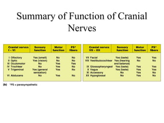
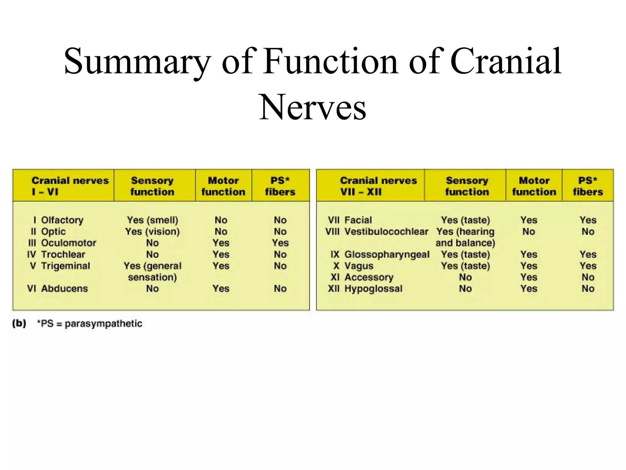
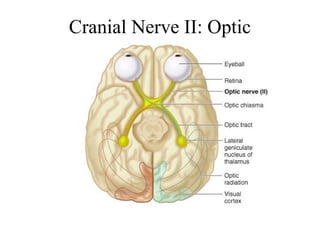
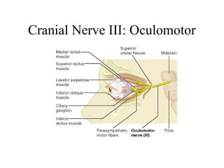
Summary of Function of Cranial 
Nerves
Cranial Nerve I: Olfactory 
• Arises from the olfactory epithelium 
• Passes through the cribriform plate of the 
ethmoid bone 
• Fibers run through the olfactory bulb and 
terminate in the primary olfactory cortex 
• Functions solely by carrying afferent 
impulses for the sense of smell
Cranial Nerve I: Olfactory
Cranial Nerve II: Optic 
• Arises from the retina of the eye 
• Optic nerves pass through the optic canals and 
converge at the optic chiasm 
• They continue to the thalamus where they synapse 
• From there, the optic radiation fibers run to the 
visual cortex 
• Functions solely by carrying afferent impulses for 
vision
Cranial Nerve II: Optic
Cranial Nerve III: Oculomotor 
• Fibers extend from the ventral midbrain, pass 
through the superior orbital fissure, and go to the 
extrinsic eye muscles 
• Functions in raising the eyelid, directing the 
eyeball, constricting the iris, and controlling lens 
shape 
• The latter 2 functions are parasympathetically 
controlled 
• Parasympathetic cell bodies are in the ciliary 
ganglia
Cranial Nerve III: Oculomotor
Cranial Nerve IV: Trochlear 
• Fibers emerge from the dorsal midbrain and 
enter the orbits via the superior orbital 
fissures; innervate the superior oblique 
muscle 
• Primarily a motor nerve that directs the 
eyeball
Cranial Nerve IV: Trochlear
Cranial Nerve V: Trigeminal 
• Composed of three divisions 
– Ophthalmic (V1) 
– Maxillary (V2) 
– Mandibular (V3) 
• Fibers run from the face to the pons via the superior orbital fissure 
(V1), the foramen rotundum (V2), and the foramen ovale (V3) 
• Conveys sensory impulses from various areas of the face (V1) and 
(V2), and supplies motor fibers (V3) for mastication 
• Tic douloureux or trigeminal neuralgia 
- Most excruciating pain known (?) 
- Caused by inflammation of nerve 
- In severe cases, nerve is cut; relieves agony but results in loss 
of sensation on that side of the face
Cranial Nerve V: Trigeminal
Cranial Nerve VI: Abducens 
• Fibers leave the inferior pons and enter the orbit 
via the superior orbital fissure 
• Primarily a motor nerve innervating the lateral 
rectus muscle (abducts the eye; thus the name 
abducens)
Cranial Nerve VII: Facial 
• Fibers leave the pons, travel through the internal acoustic 
meatus, and emerge through the stylomastoid foramen to the 
lateral aspect of the face 
• Motor functions include; 
– Facial expression 
– Transmittal of parasympathetic impulses to lacrimal and 
salivary glands (submandibular and sublingual glands) 
• Sensory function is taste from taste buds of anterior two-thirds 
of the tongue
Cranial Nerve VII: Facial
Facial Nerve (CN VII) 
• Bell’s palsy: paralysis of facial muscles on 
affected side and loss of taste sensation 
• Caused by herpes simplex I virus 
• Lower eyelid droops 
• Corner of mouth sags 
• Tears drip continuously and eye cannot be 
completely closed (dry eye may occur) 
• Condition my disappear spontaneously without 
treatment
Cranial Nerve VIII: 
Vestibulocochlear 
• Fibers arise from the hearing and equilibrium 
apparatus of the inner ear, pass through the 
internal acoustic meatus, and enter the brainstem 
at the pons-medulla border 
• Two divisions – cochlear (hearing) and vestibular 
(balance) 
• Functions are solely sensory – equilibrium and 
hearing
Cranial Nerve VIII: Vestibulocochlear
Cranial Nerve IX: 
Glossopharyngeal 
• Fibers emerge from the medulla, leave the skull 
via the jugular foramen, and run to the throat 
• Nerve IX is a mixed nerve with motor and sensory 
functions 
• Motor – innervates part of the tongue and 
pharynx, and provides motor fibers to the parotid 
salivary gland 
• Sensory – fibers conduct taste and general sensory 
impulses from the tongue and pharynx
Cranial Nerve IX: Glossopharyngeal
Cranial Nerve X: Vagus 
• The only cranial nerve that extends beyond the head 
and neck 
• Fibers emerge from the medulla via the jugular 
foramen 
• The vagus is a mixed nerve 
• Most motor fibers are parasympathetic fibers to the 
heart, lungs, and visceral organs 
• Its sensory function is in taste 
• Paralysis leads to hoarseness 
• Total destruction incompatible with life
Cranial Nerve X: Vagus
Cranial Nerve XI: Accessory 
• Formed from a cranial root emerging from the 
medulla and a spinal root arising from the superior 
region of the spinal cord 
• The spinal root passes upward into the cranium via 
the foramen magnum 
• The accessory nerve leaves the cranium via the 
jugular foramen 
• Primarily a motor nerve 
– Supplies fibers to the larynx, pharynx, and soft palate 
– Innervates the trapezius and sternocleidomastoid, 
which move the head and neck
Cranial Nerve XI: Accessory
Cranial Nerve XII: Hypoglossal 
• Fibers arise from the medulla and exit the 
skull via the hypoglossal canal 
• Innervates both extrinsic and intrinsic 
muscles of the tongue, which contribute to 
swallowing and speech 
• If damaged, difficulties in speech and 
swallowing; inability to protrude tongue
Cranial Nerve XII: Hypoglossal

Cranial nerves

  • 1.
    Summary of Functionof Cranial Nerves
  • 2.
    Cranial Nerve I:Olfactory • Arises from the olfactory epithelium • Passes through the cribriform plate of the ethmoid bone • Fibers run through the olfactory bulb and terminate in the primary olfactory cortex • Functions solely by carrying afferent impulses for the sense of smell
  • 3.
  • 4.
    Cranial Nerve II:Optic • Arises from the retina of the eye • Optic nerves pass through the optic canals and converge at the optic chiasm • They continue to the thalamus where they synapse • From there, the optic radiation fibers run to the visual cortex • Functions solely by carrying afferent impulses for vision
  • 5.
  • 6.
    Cranial Nerve III:Oculomotor • Fibers extend from the ventral midbrain, pass through the superior orbital fissure, and go to the extrinsic eye muscles • Functions in raising the eyelid, directing the eyeball, constricting the iris, and controlling lens shape • The latter 2 functions are parasympathetically controlled • Parasympathetic cell bodies are in the ciliary ganglia
  • 7.
  • 8.
    Cranial Nerve IV:Trochlear • Fibers emerge from the dorsal midbrain and enter the orbits via the superior orbital fissures; innervate the superior oblique muscle • Primarily a motor nerve that directs the eyeball
  • 9.
  • 10.
    Cranial Nerve V:Trigeminal • Composed of three divisions – Ophthalmic (V1) – Maxillary (V2) – Mandibular (V3) • Fibers run from the face to the pons via the superior orbital fissure (V1), the foramen rotundum (V2), and the foramen ovale (V3) • Conveys sensory impulses from various areas of the face (V1) and (V2), and supplies motor fibers (V3) for mastication • Tic douloureux or trigeminal neuralgia - Most excruciating pain known (?) - Caused by inflammation of nerve - In severe cases, nerve is cut; relieves agony but results in loss of sensation on that side of the face
  • 11.
    Cranial Nerve V:Trigeminal
  • 12.
    Cranial Nerve VI:Abducens • Fibers leave the inferior pons and enter the orbit via the superior orbital fissure • Primarily a motor nerve innervating the lateral rectus muscle (abducts the eye; thus the name abducens)
  • 13.
    Cranial Nerve VII:Facial • Fibers leave the pons, travel through the internal acoustic meatus, and emerge through the stylomastoid foramen to the lateral aspect of the face • Motor functions include; – Facial expression – Transmittal of parasympathetic impulses to lacrimal and salivary glands (submandibular and sublingual glands) • Sensory function is taste from taste buds of anterior two-thirds of the tongue
  • 14.
  • 15.
    Facial Nerve (CNVII) • Bell’s palsy: paralysis of facial muscles on affected side and loss of taste sensation • Caused by herpes simplex I virus • Lower eyelid droops • Corner of mouth sags • Tears drip continuously and eye cannot be completely closed (dry eye may occur) • Condition my disappear spontaneously without treatment
  • 16.
    Cranial Nerve VIII: Vestibulocochlear • Fibers arise from the hearing and equilibrium apparatus of the inner ear, pass through the internal acoustic meatus, and enter the brainstem at the pons-medulla border • Two divisions – cochlear (hearing) and vestibular (balance) • Functions are solely sensory – equilibrium and hearing
  • 17.
    Cranial Nerve VIII:Vestibulocochlear
  • 18.
    Cranial Nerve IX: Glossopharyngeal • Fibers emerge from the medulla, leave the skull via the jugular foramen, and run to the throat • Nerve IX is a mixed nerve with motor and sensory functions • Motor – innervates part of the tongue and pharynx, and provides motor fibers to the parotid salivary gland • Sensory – fibers conduct taste and general sensory impulses from the tongue and pharynx
  • 19.
    Cranial Nerve IX:Glossopharyngeal
  • 20.
    Cranial Nerve X:Vagus • The only cranial nerve that extends beyond the head and neck • Fibers emerge from the medulla via the jugular foramen • The vagus is a mixed nerve • Most motor fibers are parasympathetic fibers to the heart, lungs, and visceral organs • Its sensory function is in taste • Paralysis leads to hoarseness • Total destruction incompatible with life
  • 21.
  • 22.
    Cranial Nerve XI:Accessory • Formed from a cranial root emerging from the medulla and a spinal root arising from the superior region of the spinal cord • The spinal root passes upward into the cranium via the foramen magnum • The accessory nerve leaves the cranium via the jugular foramen • Primarily a motor nerve – Supplies fibers to the larynx, pharynx, and soft palate – Innervates the trapezius and sternocleidomastoid, which move the head and neck
  • 23.
  • 24.
    Cranial Nerve XII:Hypoglossal • Fibers arise from the medulla and exit the skull via the hypoglossal canal • Innervates both extrinsic and intrinsic muscles of the tongue, which contribute to swallowing and speech • If damaged, difficulties in speech and swallowing; inability to protrude tongue
  • 25.
    Cranial Nerve XII:Hypoglossal