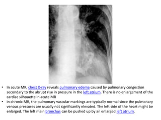
This document provides an overview of acquired mitral valve disease, including:
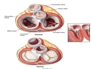
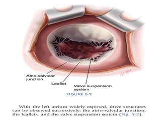
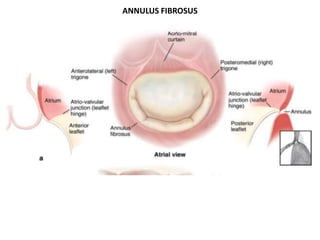
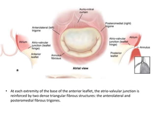
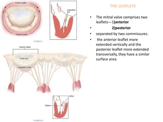
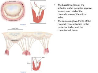
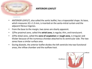
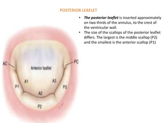
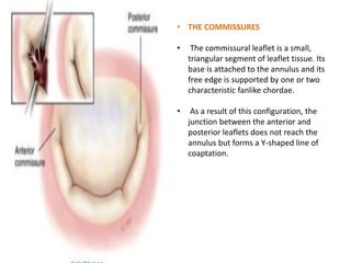
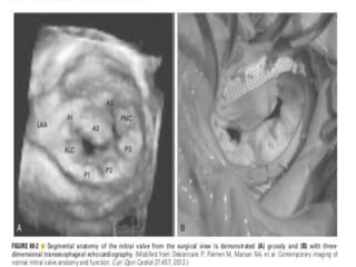
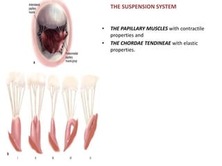
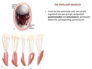
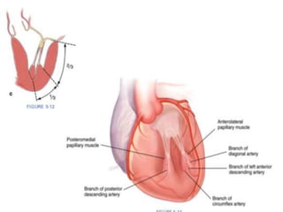
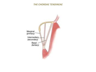
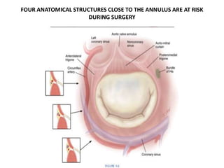
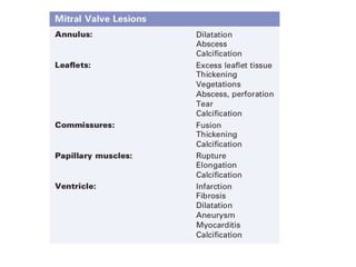
1) It describes the anatomy of the mitral valve, including the annulus, leaflets, commissures, papillary muscles, and chordae tendineae.
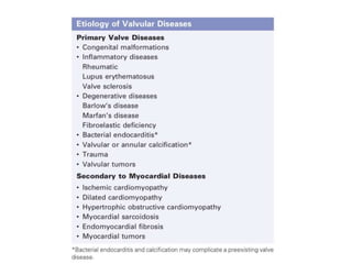
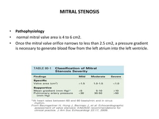
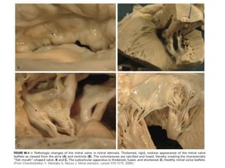
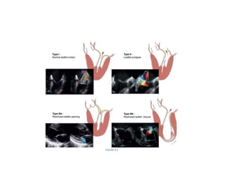
2) It discusses mitral stenosis, including the pathophysiology of reduced orifice area leading to elevated left atrial and pulmonary pressures. Rheumatic fever is the most common cause in developed nations.
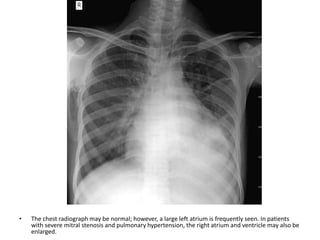
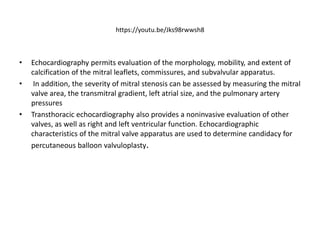
3) It outlines the clinical diagnosis of mitral stenosis through history, examination, ECG, echo, and hemodynamics. Severe untreated mitral stenosis progresses over 10 years from symptom onset to death.