Introduction,
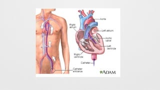
Left Heart Catheterization,
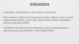
Indications,
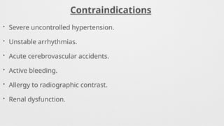
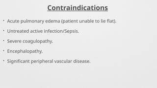
Contraindications,
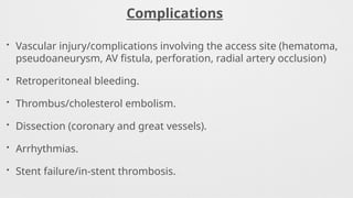
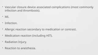
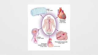
Complications,
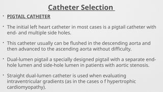
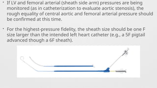
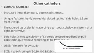
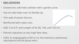
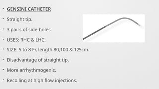
Catheters Used,
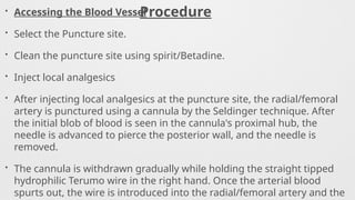
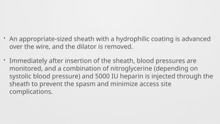
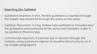
Procedure,
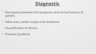
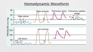
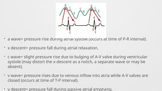
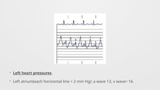
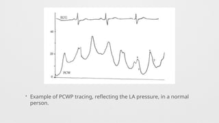
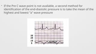
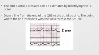
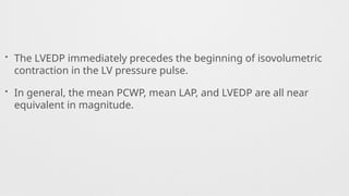
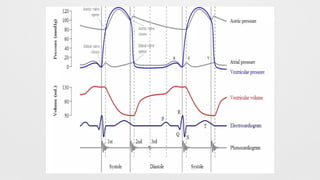
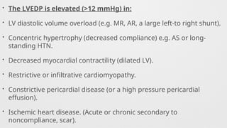
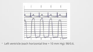
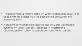
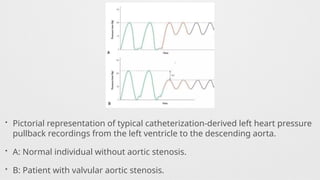
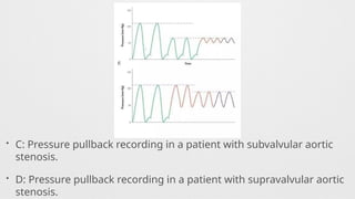
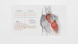
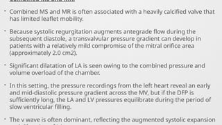
Hemodynamics,
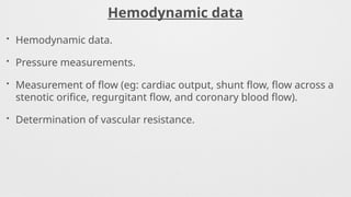
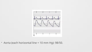
Hemodynamics data,
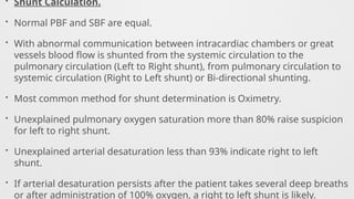
Shunt Calculation,
Oxymetry,
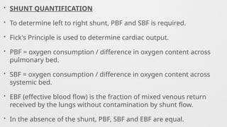
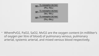
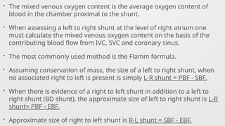
Shunt Quantification,
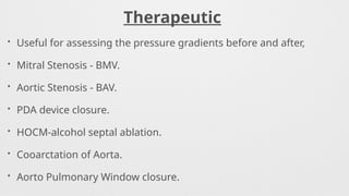
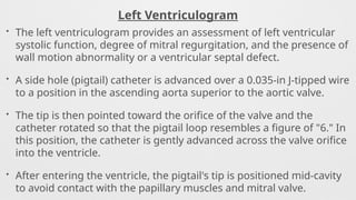
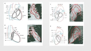
Left Ventriculogram,
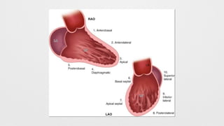
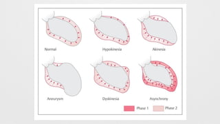
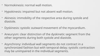
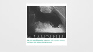
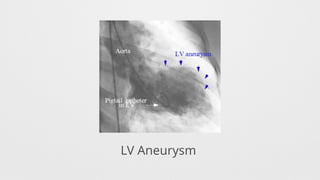
LV Wall motion abnormalities,
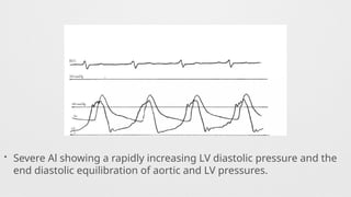
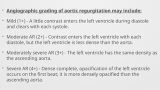
Angiographic Aortic Regurgitation,
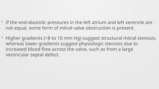
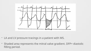
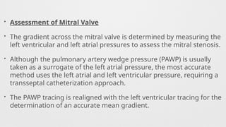
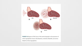
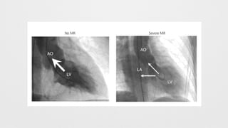
Angiographic Mitral Regurgitation


![Invasive_Cardio-Devices_procedures[1].pdf](https://cdn.slidesharecdn.com/ss_thumbnails/invasivecardio-devicesprocedures1-240129085722-eb86cfb0-thumbnail.jpg?width=640&height=640&fit=bounds)


