Absorbance and Emission
A tool to understand and characterize
the system
Tuhin Kumar Maji
JRF, SNBNCBS
Under supervision of Prof. Samir Kumar Pal
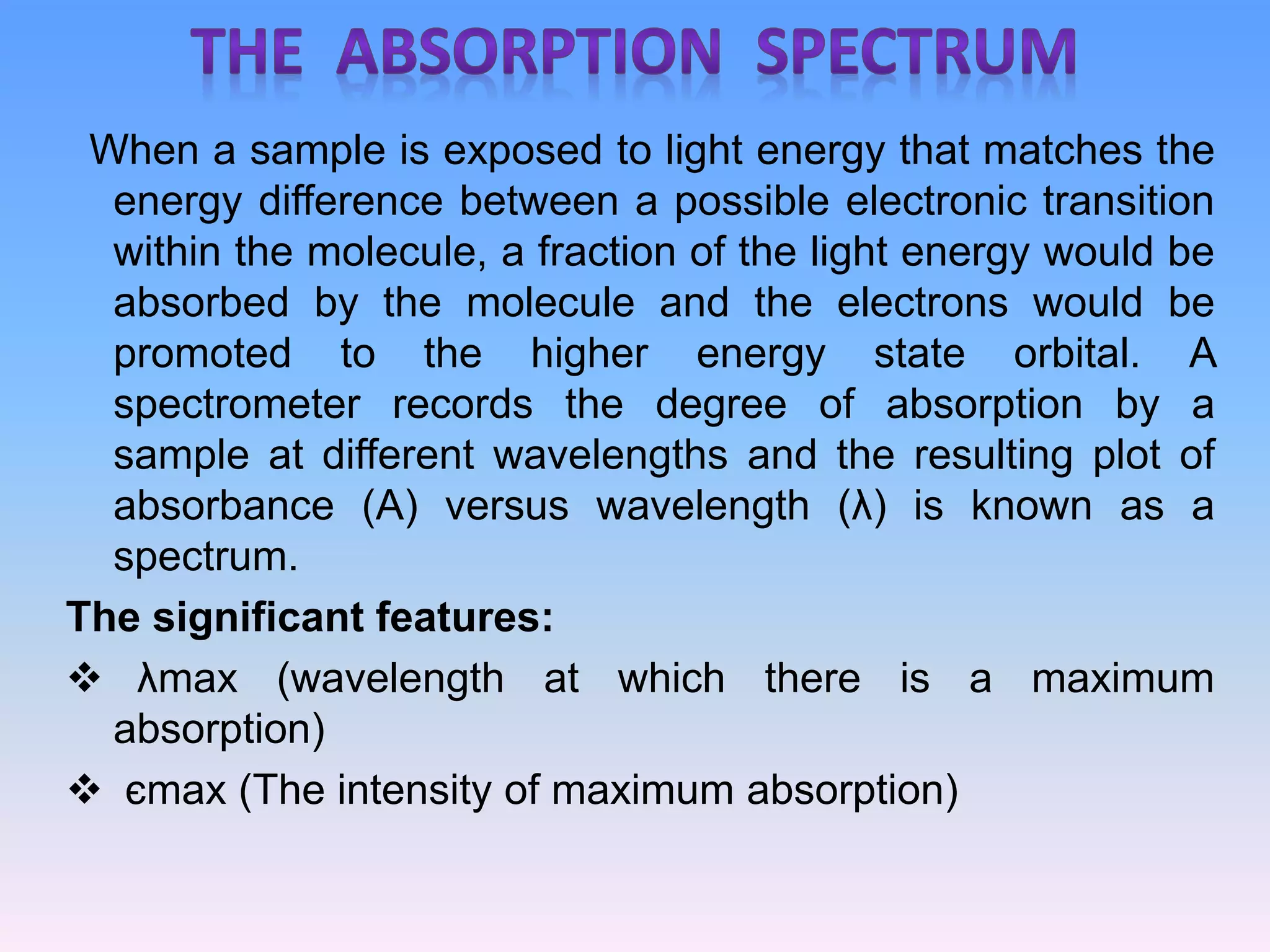
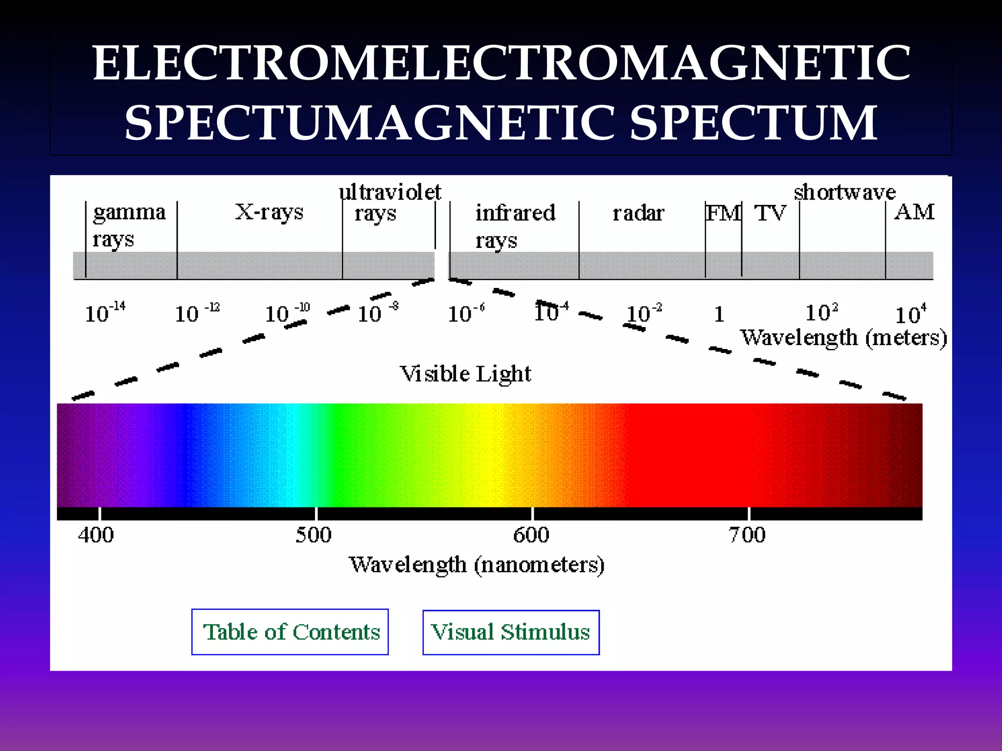
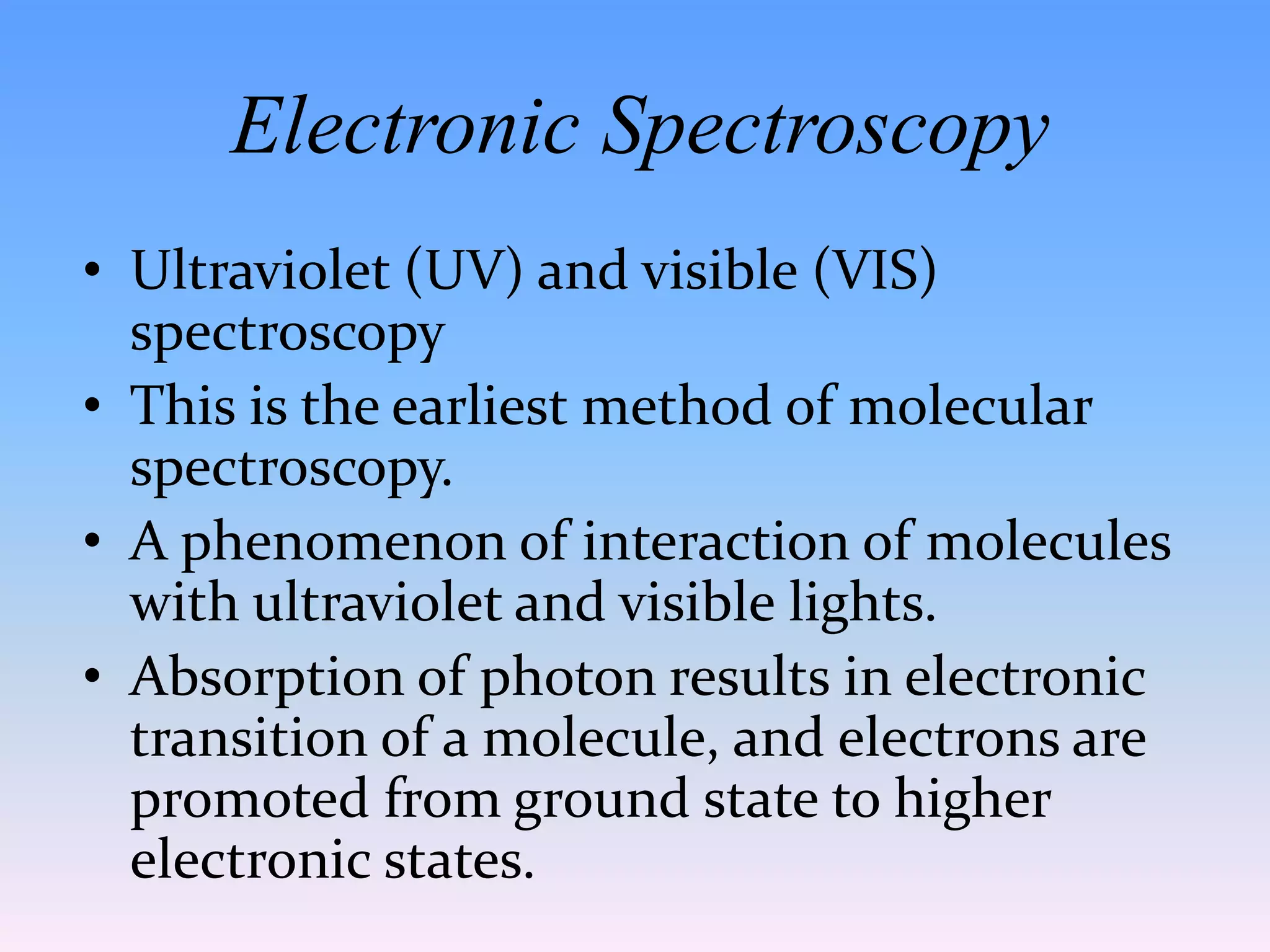
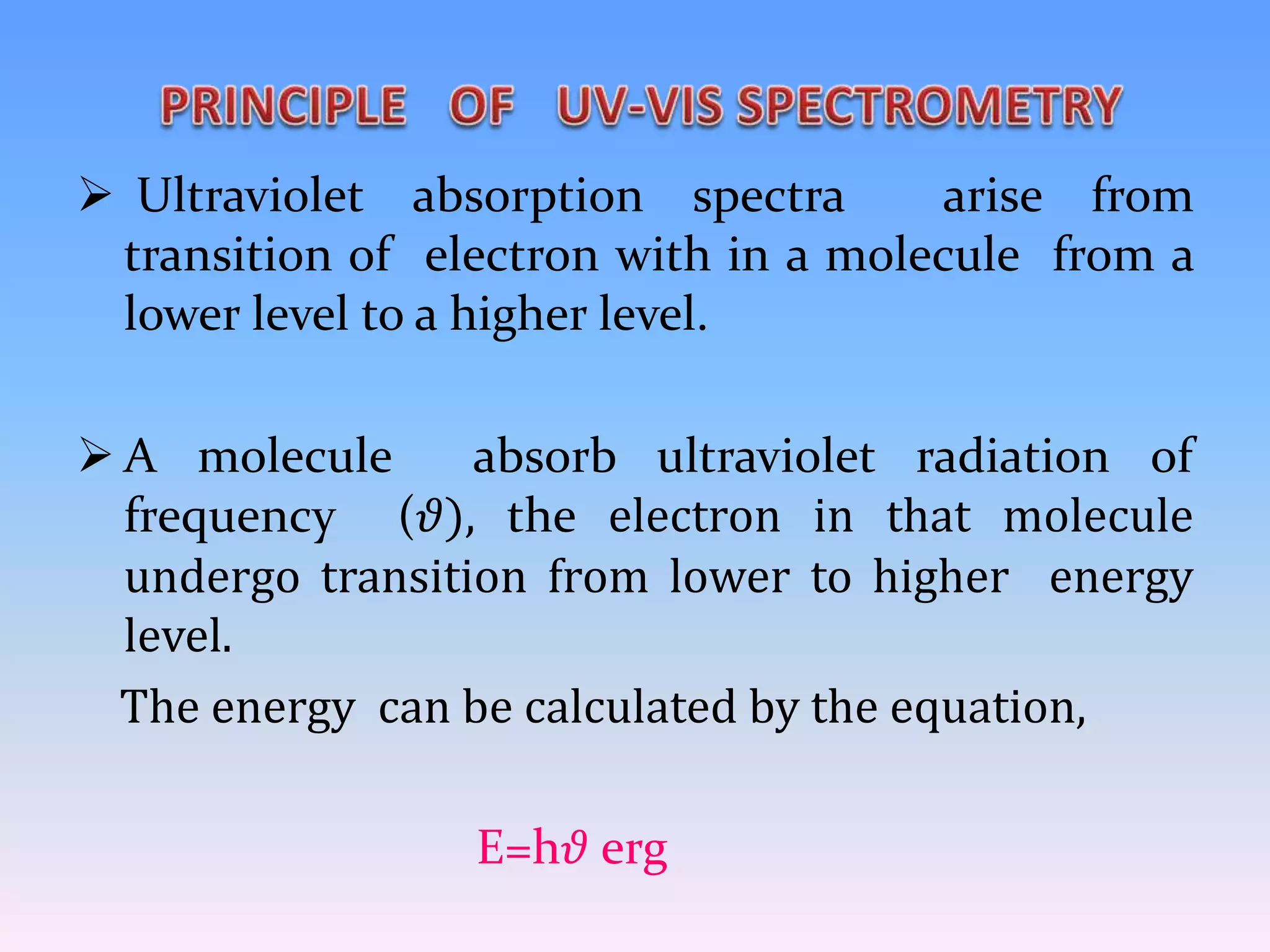
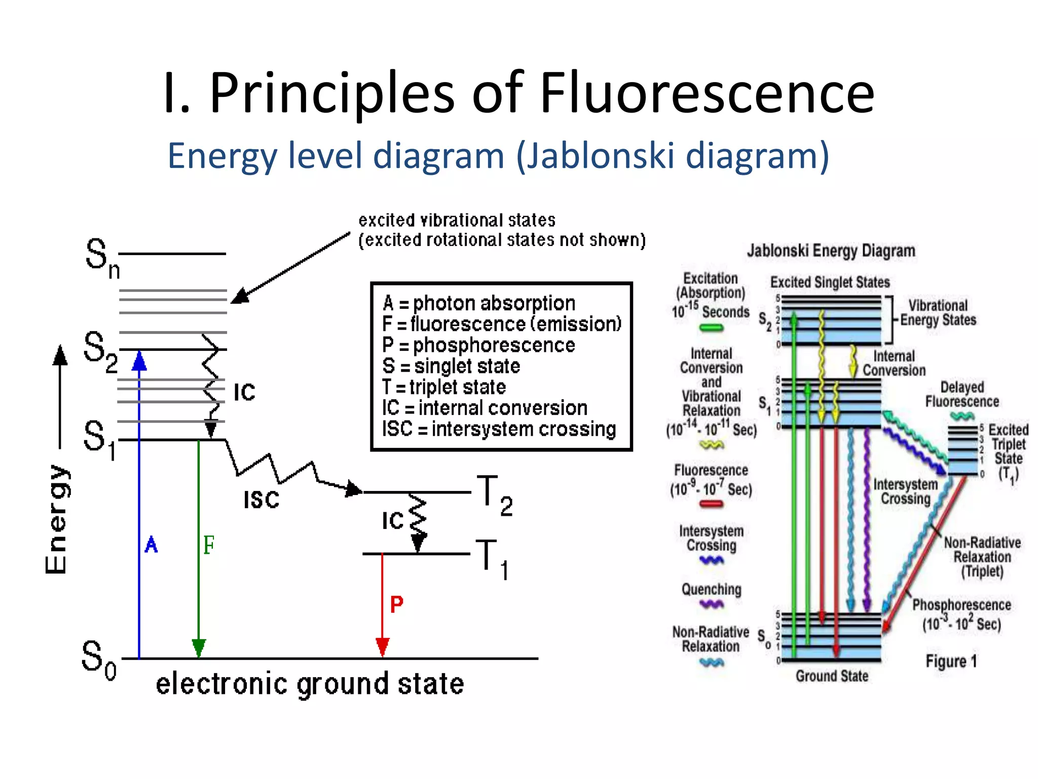
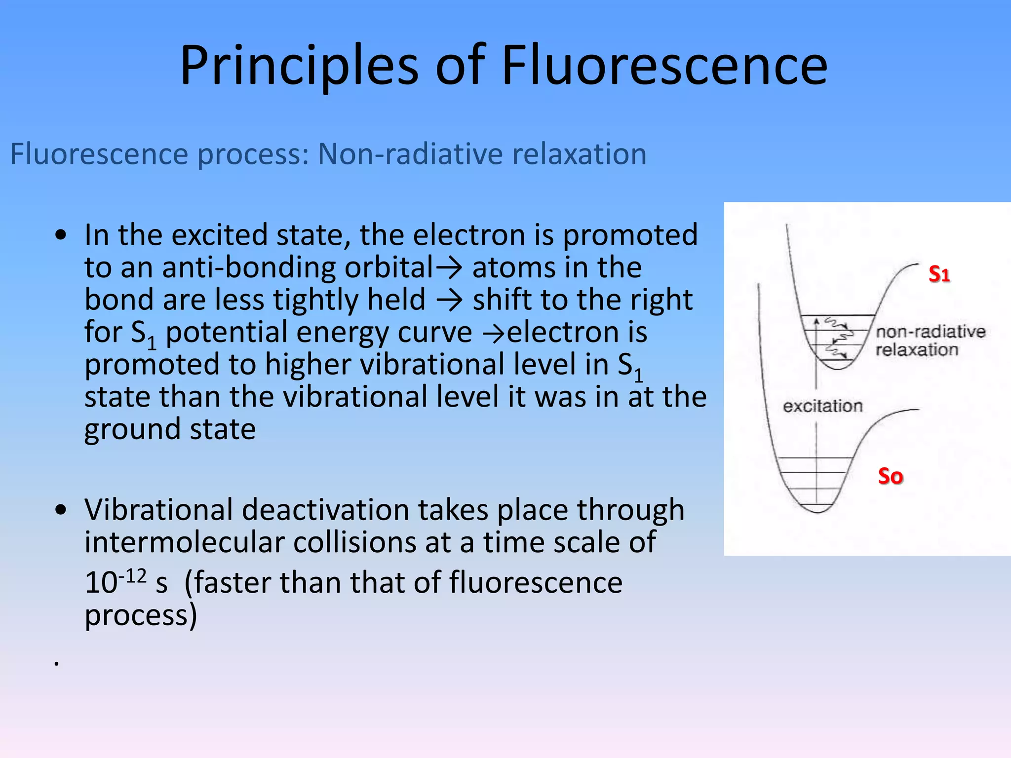
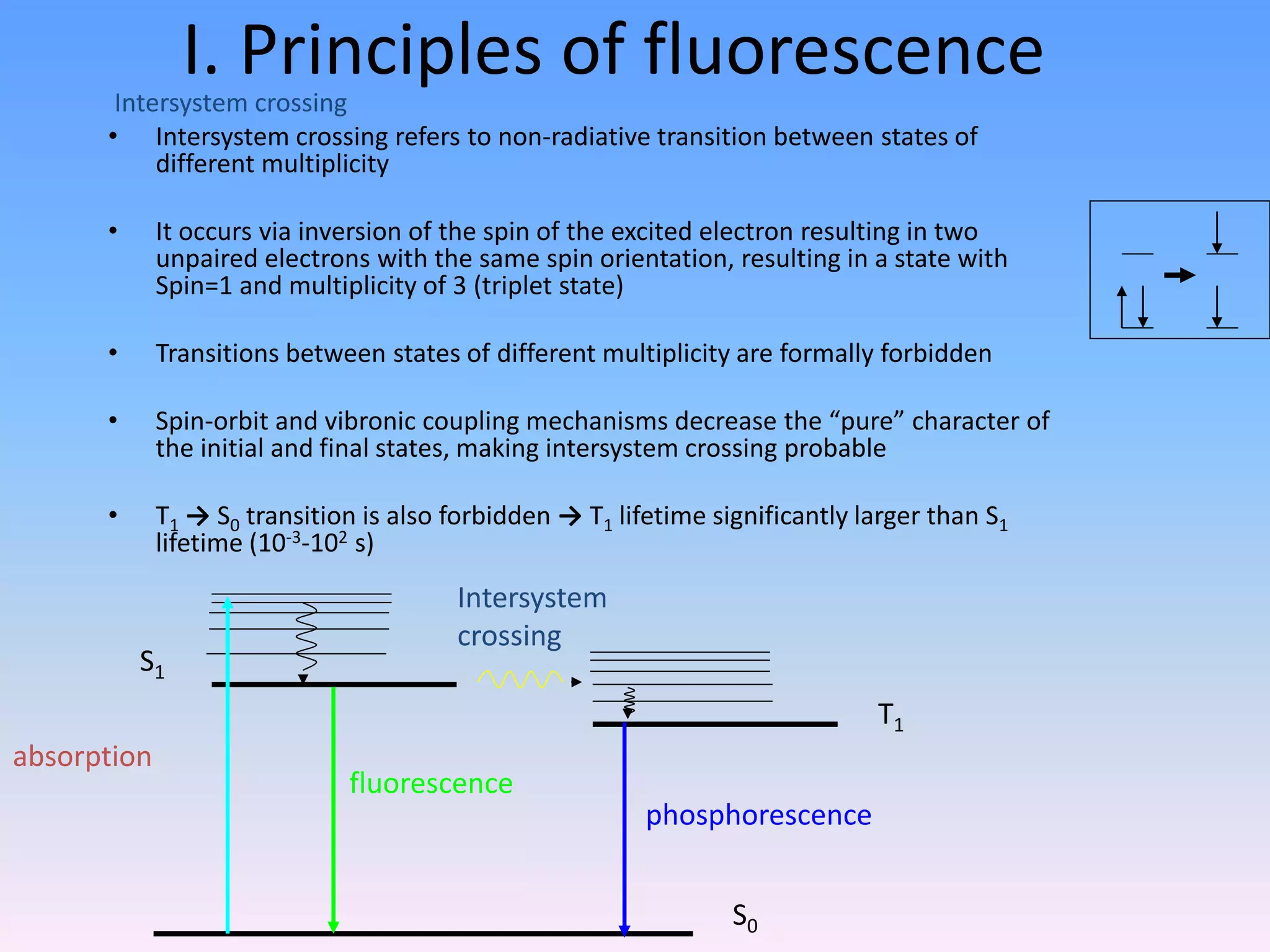
Ultraviolet and visible (UV-Vis) absorption
spectroscopy is the measurement of the
attenuation of a beam of light after it passes
through a sample or after reflection from a
sample surface. Absorption measurements can
be at a single wavelength or over an extended
spectral range.
When a sample is exposed to light energy that matches the
energy difference between a possible electronic transition
within the molecule, a fraction of the light energy would be
absorbed by the molecule and the electrons would be
promoted to the higher energy state orbital. A
spectrometer records the degree of absorption by a
sample at different wavelengths and the resulting plot of
absorbance (A) versus wavelength (λ) is known as a
spectrum.
The significant features:
 λmax (wavelength at which there is a maximum
absorption)
 єmax (The intensity of maximum absorption)
ELECTROMELECTROMAGNETIC
SPECTUMAGNETIC SPECTUM
Electronic Spectroscopy
• Ultraviolet (UV) and visible (VIS)
spectroscopy
• This is the earliest method of molecular
spectroscopy.
• A phenomenon of interaction of molecules
with ultraviolet and visible lights.
• Absorption of photon results in electronic
transition of a molecule, and electrons are
promoted from ground state to higher
electronic states.
 Ultraviolet absorption spectra arise from
transition of electron with in a molecule from a
lower level to a higher level.
 A molecule absorb ultraviolet radiation of
frequency (𝜗), the electron in that molecule
undergo transition from lower to higher energy
level.
The energy can be calculated by the equation,
E=h𝜗 erg
E₁-Eₒ= h𝜗
Etotal=Eelectronic + Evibrotional + Erotational
The energies decreases in the following order:
Electronic ⪢Vibrational ⪢ Rotational
In U.V spectroscopy molecule undergo
electronic transition involving σ, π and n electrons.
 Four types of electronic transition are possible.
i. σ ⇾ σ* transition
ii. n ⇾ σ* transition
iii. n ⇾ π* transition
iv. π ⇾ π* transition
8
BEER’S LAW
“ The intensity of a beam of monochromatic light decrease
exponentially with the increase in concentration of the
absorbing substance” .
Arithmetically;
- dI/ dc ᾱ I
I= Io. exp(-kc) ---------------------eq (1)
ABSORBANCE LAWS
“ When a beam of light is allowed to pass through a
transparent medium, the rate of decrease of
intensity with the thickness of medium is directly
proportional to the intensity of the light”
mathematically;
-dI/ dt ᾱ I
-In . I = kt+b ----------------eq(2)
the combination of eq 1 & 2 we will get
A= Kct
A= ℇct (K=ℇ)
LAMBERT’S LAW
 The real limitation of the beer’s law is successfully
in describing the absorption behavior of dilute
solution only.
 In this regarding it may be considered as a
limiting law.
Know Our Instrument
Know Our Instrument
Light source:
UV - Hydrogen lamp ( hydrogen stored under
pressure) , Deuterium lamp and Xenon lamp-
it is not regularly used because of unstability
and also the radiation of UV causes the
generation of ozone by ionization of the
oxygen molecule.
VIS – Tungsten filament lamp , Tungsten
halogen lamp and carbon arc lamp.
Advantage of double beam spectrophotometer
The ratio of the powers of the sample & reference is constantly
obtained.
It has rapid scanning over the wide wavelength region because of the
above factor
DESCRIPTION OF UV- SPECTROPHOTOMETER
samplereference
detector
I
0
I
0
I
0
I
log(I0/I) =
A
20
0 l,
nm
monochromator/
beam splitter
optics
UV-VIS
sources
Fluorescence spectroscopy
and basic principle
Luminescence
• Emission of photons from electronically
excited states
• Two types of luminescence:
1.Relaxation from singlet excited state
2.Relaxation from triplet excited state
I. Principles of Fluorescence
Singlet and Triplet states
• Ground state – two electrons per orbital; electrons have
opposite spin and are paired
• Singlet excited state
Electron in higher energy orbital has the same spin orientation
with respect to electron in the lower orbital
• Triplet excited state
The excited valence electron may spontaneously reverse its spin
(spin flip). This process is called intersystem crossing.
I. Principles of Fluorescence
Energy level diagram (Jablonski diagram)
Principles of Fluorescence
Fluorescence process: Non-radiative relaxation
• In the excited state, the electron is promoted
to an anti-bonding orbital→ atoms in the
bond are less tightly held → shift to the right
for S1 potential energy curve →electron is
promoted to higher vibrational level in S1
state than the vibrational level it was in at the
ground state
• Vibrational deactivation takes place through
intermolecular collisions at a time scale of
10-12 s (faster than that of fluorescence
process)
.
So
S1
Principles of Fluorescence
Fluorescence process: Emission
• The molecule relaxes from the
lowest vibrational energy level
of the excited state to a vibrational
energy level of the ground state
(10-9 s)
• Relaxation to ground state occurs faster than
time scale of molecular vibration → “vertical”
transition
• The energy of the emitted photon
is lower than that of the incident
photons
So
S1
I. Principles of fluorescenceIntersystem crossing
• Intersystem crossing refers to non-radiative transition between states of
different multiplicity
• It occurs via inversion of the spin of the excited electron resulting in two
unpaired electrons with the same spin orientation, resulting in a state with
Spin=1 and multiplicity of 3 (triplet state)
• Transitions between states of different multiplicity are formally forbidden
• Spin-orbit and vibronic coupling mechanisms decrease the “pure” character of
the initial and final states, making intersystem crossing probable
• T1 → S0 transition is also forbidden → T1 lifetime significantly larger than S1
lifetime (10-3-102 s)
S0
S1
T1
absorption
fluorescence
phosphorescence
Intersystem
crossing
II. Quantum yield
• Quantum yield of fluorescence, Ff, is defined as:
• In practice, is measured by comparative measurements with reference
compound for which has been determined with high degree of accuracy.
absorbedphotonsofnumber
emittedphotonsofnumber
F f
Quantum yield of fluorescence
Know your Instrument
Fluorescence Measurements
Typical fluorescence emission spectrum at 340 nm
excitation (the different components)
0
500000
1000000
1500000
2000000
2500000
3000000
300 350 400 450 500 550 600
Wavelength (nm)
FluorescenceIntensity(a.u.)
Raman
Rayleigh (lexc = lemm)
Fluorescence
Applications in Biological Systems
Absorbance spectrum of
(a) different DNA bases,
(b) single and double
standard DNA
Absorbance spectrum of amino
acids tryptophan, tyrosine and
phenylalanine and a
representative protein BSA
Biological Fluorophores
–Endogenous Fluorophores
amino acids
structural proteins
enzymes and co-enzymes
vitamins
lipids
porphyrins
–Exogenous Fluorophores
Cyanine dyes
Photosensitizers
Molecular markers – GFP, etc.
I. Principles of fluorescence
Wavelength
Absorbance
DONOR
Absorbance
Fluorescence Fluorescence
ACCEPTOR
Molecule 1 Molecule 2
• Fluorescence energy transfer (FRET)
Wavelength
Absorbance
DONOR
Absorbance
Fluorescence Fluorescence
ACCEPTOR
Molecule 1 Molecule 2
Non radiative energy transfer – a quantum mechanical process of resonance between
transition dipoles
Effective between 10-100 Å only
Emission and excitation spectrum must significantly overlap
Donor transfers non-radiatively to the acceptor
Any question ???

Absorbance and emission

  • 1.
    Absorbance and Emission Atool to understand and characterize the system Tuhin Kumar Maji JRF, SNBNCBS Under supervision of Prof. Samir Kumar Pal
  • 2.
    Ultraviolet and visible(UV-Vis) absorption spectroscopy is the measurement of the attenuation of a beam of light after it passes through a sample or after reflection from a sample surface. Absorption measurements can be at a single wavelength or over an extended spectral range.
  • 3.
    When a sampleis exposed to light energy that matches the energy difference between a possible electronic transition within the molecule, a fraction of the light energy would be absorbed by the molecule and the electrons would be promoted to the higher energy state orbital. A spectrometer records the degree of absorption by a sample at different wavelengths and the resulting plot of absorbance (A) versus wavelength (λ) is known as a spectrum. The significant features:  λmax (wavelength at which there is a maximum absorption)  єmax (The intensity of maximum absorption)
  • 4.
  • 5.
    Electronic Spectroscopy • Ultraviolet(UV) and visible (VIS) spectroscopy • This is the earliest method of molecular spectroscopy. • A phenomenon of interaction of molecules with ultraviolet and visible lights. • Absorption of photon results in electronic transition of a molecule, and electrons are promoted from ground state to higher electronic states.
  • 6.
     Ultraviolet absorptionspectra arise from transition of electron with in a molecule from a lower level to a higher level.  A molecule absorb ultraviolet radiation of frequency (𝜗), the electron in that molecule undergo transition from lower to higher energy level. The energy can be calculated by the equation, E=h𝜗 erg
  • 7.
    E₁-Eₒ= h𝜗 Etotal=Eelectronic +Evibrotional + Erotational The energies decreases in the following order: Electronic ⪢Vibrational ⪢ Rotational
  • 8.
    In U.V spectroscopymolecule undergo electronic transition involving σ, π and n electrons.  Four types of electronic transition are possible. i. σ ⇾ σ* transition ii. n ⇾ σ* transition iii. n ⇾ π* transition iv. π ⇾ π* transition 8
  • 9.
    BEER’S LAW “ Theintensity of a beam of monochromatic light decrease exponentially with the increase in concentration of the absorbing substance” . Arithmetically; - dI/ dc ᾱ I I= Io. exp(-kc) ---------------------eq (1) ABSORBANCE LAWS
  • 10.
    “ When abeam of light is allowed to pass through a transparent medium, the rate of decrease of intensity with the thickness of medium is directly proportional to the intensity of the light” mathematically; -dI/ dt ᾱ I -In . I = kt+b ----------------eq(2) the combination of eq 1 & 2 we will get A= Kct A= ℇct (K=ℇ) LAMBERT’S LAW
  • 11.
     The reallimitation of the beer’s law is successfully in describing the absorption behavior of dilute solution only.  In this regarding it may be considered as a limiting law.
  • 12.
  • 13.
    Know Our Instrument Lightsource: UV - Hydrogen lamp ( hydrogen stored under pressure) , Deuterium lamp and Xenon lamp- it is not regularly used because of unstability and also the radiation of UV causes the generation of ozone by ionization of the oxygen molecule. VIS – Tungsten filament lamp , Tungsten halogen lamp and carbon arc lamp.
  • 14.
    Advantage of doublebeam spectrophotometer The ratio of the powers of the sample & reference is constantly obtained. It has rapid scanning over the wide wavelength region because of the above factor DESCRIPTION OF UV- SPECTROPHOTOMETER samplereference detector I 0 I 0 I 0 I log(I0/I) = A 20 0 l, nm monochromator/ beam splitter optics UV-VIS sources
  • 15.
    Fluorescence spectroscopy and basicprinciple Luminescence • Emission of photons from electronically excited states • Two types of luminescence: 1.Relaxation from singlet excited state 2.Relaxation from triplet excited state
  • 16.
    I. Principles ofFluorescence Singlet and Triplet states • Ground state – two electrons per orbital; electrons have opposite spin and are paired • Singlet excited state Electron in higher energy orbital has the same spin orientation with respect to electron in the lower orbital • Triplet excited state The excited valence electron may spontaneously reverse its spin (spin flip). This process is called intersystem crossing.
  • 17.
    I. Principles ofFluorescence Energy level diagram (Jablonski diagram)
  • 18.
    Principles of Fluorescence Fluorescenceprocess: Non-radiative relaxation • In the excited state, the electron is promoted to an anti-bonding orbital→ atoms in the bond are less tightly held → shift to the right for S1 potential energy curve →electron is promoted to higher vibrational level in S1 state than the vibrational level it was in at the ground state • Vibrational deactivation takes place through intermolecular collisions at a time scale of 10-12 s (faster than that of fluorescence process) . So S1
  • 19.
    Principles of Fluorescence Fluorescenceprocess: Emission • The molecule relaxes from the lowest vibrational energy level of the excited state to a vibrational energy level of the ground state (10-9 s) • Relaxation to ground state occurs faster than time scale of molecular vibration → “vertical” transition • The energy of the emitted photon is lower than that of the incident photons So S1
  • 20.
    I. Principles offluorescenceIntersystem crossing • Intersystem crossing refers to non-radiative transition between states of different multiplicity • It occurs via inversion of the spin of the excited electron resulting in two unpaired electrons with the same spin orientation, resulting in a state with Spin=1 and multiplicity of 3 (triplet state) • Transitions between states of different multiplicity are formally forbidden • Spin-orbit and vibronic coupling mechanisms decrease the “pure” character of the initial and final states, making intersystem crossing probable • T1 → S0 transition is also forbidden → T1 lifetime significantly larger than S1 lifetime (10-3-102 s) S0 S1 T1 absorption fluorescence phosphorescence Intersystem crossing
  • 21.
    II. Quantum yield •Quantum yield of fluorescence, Ff, is defined as: • In practice, is measured by comparative measurements with reference compound for which has been determined with high degree of accuracy. absorbedphotonsofnumber emittedphotonsofnumber F f Quantum yield of fluorescence
  • 22.
  • 23.
    Fluorescence Measurements Typical fluorescenceemission spectrum at 340 nm excitation (the different components) 0 500000 1000000 1500000 2000000 2500000 3000000 300 350 400 450 500 550 600 Wavelength (nm) FluorescenceIntensity(a.u.) Raman Rayleigh (lexc = lemm) Fluorescence
  • 24.
    Applications in BiologicalSystems Absorbance spectrum of (a) different DNA bases, (b) single and double standard DNA Absorbance spectrum of amino acids tryptophan, tyrosine and phenylalanine and a representative protein BSA
  • 26.
    Biological Fluorophores –Endogenous Fluorophores aminoacids structural proteins enzymes and co-enzymes vitamins lipids porphyrins –Exogenous Fluorophores Cyanine dyes Photosensitizers Molecular markers – GFP, etc.
  • 27.
    I. Principles offluorescence Wavelength Absorbance DONOR Absorbance Fluorescence Fluorescence ACCEPTOR Molecule 1 Molecule 2 • Fluorescence energy transfer (FRET) Wavelength Absorbance DONOR Absorbance Fluorescence Fluorescence ACCEPTOR Molecule 1 Molecule 2 Non radiative energy transfer – a quantum mechanical process of resonance between transition dipoles Effective between 10-100 Å only Emission and excitation spectrum must significantly overlap Donor transfers non-radiatively to the acceptor
  • 28.