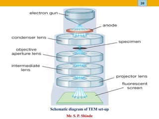
Scanning Electron Microscopy (SEM)
&
Transmission Electron Microscopy (TEM)
Mr. Sanket P. Shinde
Assistant Professor
Pune-Maharashtra.
2
Mr. S. P. Shinde
Introduction
Scanning Electron Microscopy (SEM)
o It is a technique that provides information such as topography, composition
and crystallographic information of an object.
o Topography describes the surface features of an object;
o Composition denotes the elements and compounds of an object and their
relative amounts;
o Crystallography explains the arrangement of atoms in the object.
o The combination of high magnification, large depth of focus, good
resolution, and the ease of observation makes the SEM one of the most
widely uses equipments.
o Scanning electron microscopes use a beam of highly energetic electrons to
examine objects on a very fine scale.
3
Mr. S. P. Shinde
o SEM produces images by detecting secondary electrons that are emitted
from the surface due to excitation from a primary electron beam.
o A focused electron beam rapidly scans the sample which emits secondary
electrons to be knocked off the surface of the sample.
o These secondary electrons provide signals carrying information about the
properties of the surface of the sample, such as its topography and
composition.
o The image is finally produced from the signals of these secondary or
backscattered electrons detected by a detector.
o SEM image relies on electron interactions at the surface rather than
transmission.
o SEM images are three-dimensional giving more accurate representations
and have a greater depth of view.
Continue…
4
Mr. S. P. Shinde
o When a primary electron beam incidents on sample various signals are
emitted from the sample.
o These signals include secondary electrons (SE), auger electrons,
backscattered electrons (BSE), X-rays, transmitted electrons and
cathodoluminescence.
o Secondary electrons are produced by emission of the valence electrons of
the atoms in the sample.
o The secondary electrons which are emitted at the deep region in the
sample will have small energy thus they are absorbed by the sample.
o Those electrons emitted at the top of the surface are useful to observe the
topography of the sample surface.
Principle
5
Mr. S. P. Shinde
o The electrons enter the sample, they are scattered within the sample and
lose their energy gradually and then absorbed in the sample.
o The scattering is depending on the energy of electrons, number of atoms in
the sample and density of the atoms.
o The scattering range is larger with high energy of electrons whereas the
scattering range is smaller with large atomic number and density.
o Some electrons are scattered in the specimen in an elastic fashion with no
loss of energy, these electrons are called as Backscattered electrons that are
emitted out of the sample. These electrons are also called as reflected
electrons.
o The backscattered electrons have higher energy than secondary electrons &
provide information about the deep region i.e. composition of the sample.
o When the atomic number is larger the back scattered electron yield is also
larger. BSE can be used to generate an image in the microscope that shows
the different elements present in a sample.
Continue…
6
Mr. S. P. Shinde
o All elements have different sized nuclei and as the size of the atom
nucleus increases, the number of BSE increases.
o The backscattered electron image contains two types of information: one
on specimen composition and the other on specimen topography.
o The generation region of backscattered electrons is larger than that of the
secondary electrons, namely, several tens of nm; therefore backscattered
electrons give poorer special resolution than secondary, they are less
influenced by charge-up.
Continue…
7
Mr. S. P. Shinde
Instrumentation
Schematic diagram of SEM set-up
8
Mr. S. P. Shinde
General components of SEM are given below:
❑ Electron Source
❑ Electron Gun
• Thermionic Gun
• Field Emission Gun
❑ Electromagnetic and/ or Electrostatic Lenses
❑ Vacuum Chamber
❑ Sample Chamber and stage
❑ Computer
❑ Detectors
• Secondary Electron Detector (SED)
• Backscatter Detector
• Diffracted Backscatter Detector (EBSD)
• X-ray Detector
9
Mr. S. P. Shinde
o A Scanning Electron Microscope image is obtained by tracing a sample
in a raster pattern (a rectangular pattern of parallel scanning lines) with
an electron beam.
o A beam of energetic electrons are emerged from the electron gun and
approached a series of electromagnetic lenses. These lenses are referred
to as solenoids.
o They are wrapped in scanning coils and the coils are adjusted to focus
the incident electron beam onto the sample.
o The speed of movement of electrons towards sample is adjusted by
adjustment of the coils which cause fluctuations in the voltage.
o The focusing of the beam to control magnification and the surface area
to be scanned can be controlled by computer.
Working of SEM
10
Mr. S. P. Shinde
o Samples after prior preparation, such as sputter coating for non-
conductive samples and dehydration of most biological specimens, are
placed in the vacuum chamber.
o All samples need to be able to handle the low pressure inside the
vacuum chamber.
o The beam of electrons with significant amounts of kinetic energy is
focused onto sample which is placed on the stage.
o The acceleration rate of incident electrons decides the interaction
between the incident electrons and the surface of the sample.
o When the electrons come in contact with the sample, energetic electrons
are released from the surface of the sample.
o The scatter patterns made by the interaction yields information on size,
shape, texture and composition of the sample.
Continue…
11
Mr. S. P. Shinde
o The secondary, backscattered electrons as well as x-rays are detected by
detectors.
o Backscatter electrons images provide composition data related to element
and compound detection.
o Although topographic information can be obtained using a backscatter
detector, it is not as accurate as an SED.
o Diffracted backscatter electrons determine crystalline structures as well as
the orientation of minerals and micro-fabrics. X-rays, can provide element
and mineral information.
o SEM produces black and white, three-dimensional images.
o Image magnification can be up to 10 nanometers.
Continue…
12
Mr. S. P. Shinde
1. It provides detailed three-dimensional and topographical imaging.
2. SEMs are easy to operate with the proper training and the software used
is user-friendly.
3. The time for imaging using SEMs is rapid usually less than five minutes
is required for the analysis.
4. The modern SEMs produce data in digital form.
5. Most SEM samples require minimal preparation before it is placed for
analysis.
Advantages of SEM
13
Mr. S. P. Shinde
Disadvantages of SEM
1. SEMs are very large and expensive compared to light microscopes.
2. They require area free from electric, magnetic or vibrational
interferences.
3. It requires maintenance including maintaining steady voltage, currents to
electromagnetic coils and circulation of cool water.
4. Special training is required to operate an SEM as well as prepare
samples.
5. There may be possibilities of potential artifacts or errors which cannot be
eliminated or identified.
6. SEMs are limited to solid, inorganic samples which are small enough to
fit inside the vacuum chamber.
7. There may be possibilities of exposure to radiation due to electrons
scatter from beneath the sample surface.
14
Mr. S. P. Shinde
Applications of SEM
1. They are much useful for the characterizations of solid materials wherever
it is involved.
2. Basically SEM provides topographical, morphological, compositional and
crystallographic information of materials.
3. SEM can detect and analyze surface fractures, provide information in
microstructures and examine surface contaminations.
4. SEM is useful in determining the spatial variations in chemical
compositions.
5. Qualitative chemical analyses and identification of crystalline structures
are done by SEM.
6. SEM acts as an important research tool in fields such as life science,
biology, gemology, medical, forensic science and metallurgy.
7. SEMs have applicability in industries for semiconductor inspection,
production line of miniscule products.
15
Mr. S. P. Shinde
o The transmission electron microscope is a very powerful tool for material
science.
o A high energy beam of electrons is shone through a very thin sample, and
the interactions between the electrons and the atoms can be used to
observe features such as the crystal structure and features in the structure
like dislocations and grain boundaries.
o TEM can be used to study the growth of layers, their composition and
defects in semiconductors.
o High resolution can be used to analyze the quality, shape, size and density
of quantum wells, wires and dots.
Introduction
Transmission Electron Microscopy (TEM)
16
Mr. S. P. Shinde
Continue…
o The TEM operates on the same basic principles as the light
microscope but uses electrons instead of light.
o Because the wavelength of electrons is much smaller than that of
light, the optimal resolution attainable for TEM images is many
orders of magnitude better than that from a light microscope.
o Thus, TEMs can reveal the finest details of internal structure in some
cases as small as individual atoms.
17
Mr. S. P. Shinde
Principle
o Transmission electron microscopy uses high energy electrons (up to 300
kV accelerating voltage) of very short wavelength, emitted from a
tungsten filament which are accelerated to nearly the speed of light.
o The electron beam behaves like a wavefront with wavelength about a
million times shorter than light waves.
o The whole optical system of the microscope is enclosed in vacuum.
o Air must be evacuated from the column to create a vacuum so that the
collision of electrons with air molecules and hence the scattering of
electrons are avoided.
o When an electron beam passes through a thin-section specimen of a
material, which is placed in the vacuum, electrons are scattered.
18
Mr. S. P. Shinde
o A sophisticated system of electromagnetic lenses focuses the scattered
electrons into an image or a diffraction pattern, or a nanoanalytical
spectrum, depending on the mode of operation.
o The imaging mode provides a highly magnified view of the micro and
nanostructure ultimately, in the high resolution imaging mode a direct
map of atomic arrangements can be obtained.
o The diffraction mode (electron diffraction) displays accurate
information about the local crystal structure.
o The nanoanalytical modes (x-ray and electron spectrometry) reveal
which elements are present in the tiny volume of material.
o These modes of operation provide valuable information in search of
stronger materials, faster microchips, or smaller nanocrystals.
Continue…
19
Mr. S. P. Shinde
1. An electron gun
This electrogun produces the primary electron beam of short wavelengths
to irradiate the sample. It has a condenser system that is used to focus the
electron beam onto the object
2. The image-producing system
It consists of the objective lens, movable specimen stage, and intermediate
and projector lenses. This system used to focus the electrons passing
through the specimen to produce the magnified image.
3. The image-recording system
This is the system which converts the electron image into suitable form
which can be easily readable by human eye. This system consists of a
fluorescent screen for viewing and focusing the image and a digital
camera for permanent records.
Instrumentation
20
Mr. S. P. Shinde
Schematic diagram of TEM set-up
21
Mr. S. P. Shinde
o A Transmission Electron Microscope produces image by tracing sample
with energetic electrons in the vacuum chamber.
o Electrons are passed through multiple electromagnetic lenses, allowed to
contact with the screen and converted to light to form an image.
o The voltage of the gun can be adjusted to accelerate or decrease the speed
of electrons and the electromagnetic wavelength can be adjusted through
the solenoids.
o The coils focus images onto a screen or photographic plate.
o When electrons move faster, the wavelength becomes shorter and the
quality and detail of the image is greater.
o When electrons pass through the sample that produce the lighter areas of
the image and the darker areas reflect the dense areas of the object.
o These differences provide information on the structure, texture, shape and
size of the sample.
Working of TEM
22
Mr. S. P. Shinde
Advantages of TEM
1. TEMs provides high magnification of about one million times or more.
2. TEMs have a wide-range of applications and can be utilized in different
fields including scientific, educational and industrial fields.
3. TEMs provides detailed information on element and compound structure.
4. TEMs provides high-quality and detailed images.
5. TEMs provides information of surface features, shape, size and structure.
6. The operation of TEM is easy.
23
Mr. S. P. Shinde
Disadvantages of TEM
1. TEMs are large, very expensive and require special housing and
maintenance.
2. The sample preparation for TEM is little difficult process.
3. The operation is easy but a special training is needed.
4. Samples are limited to those that are electron transparent, able to tolerate
the vacuum chamber and small enough to fit in the chamber.
5. Images are black and white and analysis of images need proper training.
24
Mr. S. P. Shinde
Applications of TEM
1. TEM finds its application in life sciences, nanotechnology, medical,
biological and material research, forensic analysis.
2. Analysis of microstructural information of metals, alloys can be done.
3. The topographical, morphological, compositional and crystalline
information can be obtained by TEM studies.
4. TEMs are useful in the study of the structure and texture of crystals and
metals at molecular level.
5. TEMs are useful in semiconductor analysis.
6. The technique is used as a tool in production and the manufacturing of
computer and silicon chips.
7. TEMs are useful in identification of flaws, fractures and damages to micro
sized objects.
8. TEMs are an important tool for research activities carried out in both
industries and academics.
25
Mr. S. P. Shinde

SEM & TEM

  • 1.
    Scanning Electron Microscopy(SEM) & Transmission Electron Microscopy (TEM) Mr. Sanket P. Shinde Assistant Professor Pune-Maharashtra.
  • 2.
    2 Mr. S. P.Shinde Introduction Scanning Electron Microscopy (SEM) o It is a technique that provides information such as topography, composition and crystallographic information of an object. o Topography describes the surface features of an object; o Composition denotes the elements and compounds of an object and their relative amounts; o Crystallography explains the arrangement of atoms in the object. o The combination of high magnification, large depth of focus, good resolution, and the ease of observation makes the SEM one of the most widely uses equipments. o Scanning electron microscopes use a beam of highly energetic electrons to examine objects on a very fine scale.
  • 3.
    3 Mr. S. P.Shinde o SEM produces images by detecting secondary electrons that are emitted from the surface due to excitation from a primary electron beam. o A focused electron beam rapidly scans the sample which emits secondary electrons to be knocked off the surface of the sample. o These secondary electrons provide signals carrying information about the properties of the surface of the sample, such as its topography and composition. o The image is finally produced from the signals of these secondary or backscattered electrons detected by a detector. o SEM image relies on electron interactions at the surface rather than transmission. o SEM images are three-dimensional giving more accurate representations and have a greater depth of view. Continue…
  • 4.
    4 Mr. S. P.Shinde o When a primary electron beam incidents on sample various signals are emitted from the sample. o These signals include secondary electrons (SE), auger electrons, backscattered electrons (BSE), X-rays, transmitted electrons and cathodoluminescence. o Secondary electrons are produced by emission of the valence electrons of the atoms in the sample. o The secondary electrons which are emitted at the deep region in the sample will have small energy thus they are absorbed by the sample. o Those electrons emitted at the top of the surface are useful to observe the topography of the sample surface. Principle
  • 5.
    5 Mr. S. P.Shinde o The electrons enter the sample, they are scattered within the sample and lose their energy gradually and then absorbed in the sample. o The scattering is depending on the energy of electrons, number of atoms in the sample and density of the atoms. o The scattering range is larger with high energy of electrons whereas the scattering range is smaller with large atomic number and density. o Some electrons are scattered in the specimen in an elastic fashion with no loss of energy, these electrons are called as Backscattered electrons that are emitted out of the sample. These electrons are also called as reflected electrons. o The backscattered electrons have higher energy than secondary electrons & provide information about the deep region i.e. composition of the sample. o When the atomic number is larger the back scattered electron yield is also larger. BSE can be used to generate an image in the microscope that shows the different elements present in a sample. Continue…
  • 6.
    6 Mr. S. P.Shinde o All elements have different sized nuclei and as the size of the atom nucleus increases, the number of BSE increases. o The backscattered electron image contains two types of information: one on specimen composition and the other on specimen topography. o The generation region of backscattered electrons is larger than that of the secondary electrons, namely, several tens of nm; therefore backscattered electrons give poorer special resolution than secondary, they are less influenced by charge-up. Continue…
  • 7.
    7 Mr. S. P.Shinde Instrumentation Schematic diagram of SEM set-up
  • 8.
    8 Mr. S. P.Shinde General components of SEM are given below: ❑ Electron Source ❑ Electron Gun • Thermionic Gun • Field Emission Gun ❑ Electromagnetic and/ or Electrostatic Lenses ❑ Vacuum Chamber ❑ Sample Chamber and stage ❑ Computer ❑ Detectors • Secondary Electron Detector (SED) • Backscatter Detector • Diffracted Backscatter Detector (EBSD) • X-ray Detector
  • 9.
    9 Mr. S. P.Shinde o A Scanning Electron Microscope image is obtained by tracing a sample in a raster pattern (a rectangular pattern of parallel scanning lines) with an electron beam. o A beam of energetic electrons are emerged from the electron gun and approached a series of electromagnetic lenses. These lenses are referred to as solenoids. o They are wrapped in scanning coils and the coils are adjusted to focus the incident electron beam onto the sample. o The speed of movement of electrons towards sample is adjusted by adjustment of the coils which cause fluctuations in the voltage. o The focusing of the beam to control magnification and the surface area to be scanned can be controlled by computer. Working of SEM
  • 10.
    10 Mr. S. P.Shinde o Samples after prior preparation, such as sputter coating for non- conductive samples and dehydration of most biological specimens, are placed in the vacuum chamber. o All samples need to be able to handle the low pressure inside the vacuum chamber. o The beam of electrons with significant amounts of kinetic energy is focused onto sample which is placed on the stage. o The acceleration rate of incident electrons decides the interaction between the incident electrons and the surface of the sample. o When the electrons come in contact with the sample, energetic electrons are released from the surface of the sample. o The scatter patterns made by the interaction yields information on size, shape, texture and composition of the sample. Continue…
  • 11.
    11 Mr. S. P.Shinde o The secondary, backscattered electrons as well as x-rays are detected by detectors. o Backscatter electrons images provide composition data related to element and compound detection. o Although topographic information can be obtained using a backscatter detector, it is not as accurate as an SED. o Diffracted backscatter electrons determine crystalline structures as well as the orientation of minerals and micro-fabrics. X-rays, can provide element and mineral information. o SEM produces black and white, three-dimensional images. o Image magnification can be up to 10 nanometers. Continue…
  • 12.
    12 Mr. S. P.Shinde 1. It provides detailed three-dimensional and topographical imaging. 2. SEMs are easy to operate with the proper training and the software used is user-friendly. 3. The time for imaging using SEMs is rapid usually less than five minutes is required for the analysis. 4. The modern SEMs produce data in digital form. 5. Most SEM samples require minimal preparation before it is placed for analysis. Advantages of SEM
  • 13.
    13 Mr. S. P.Shinde Disadvantages of SEM 1. SEMs are very large and expensive compared to light microscopes. 2. They require area free from electric, magnetic or vibrational interferences. 3. It requires maintenance including maintaining steady voltage, currents to electromagnetic coils and circulation of cool water. 4. Special training is required to operate an SEM as well as prepare samples. 5. There may be possibilities of potential artifacts or errors which cannot be eliminated or identified. 6. SEMs are limited to solid, inorganic samples which are small enough to fit inside the vacuum chamber. 7. There may be possibilities of exposure to radiation due to electrons scatter from beneath the sample surface.
  • 14.
    14 Mr. S. P.Shinde Applications of SEM 1. They are much useful for the characterizations of solid materials wherever it is involved. 2. Basically SEM provides topographical, morphological, compositional and crystallographic information of materials. 3. SEM can detect and analyze surface fractures, provide information in microstructures and examine surface contaminations. 4. SEM is useful in determining the spatial variations in chemical compositions. 5. Qualitative chemical analyses and identification of crystalline structures are done by SEM. 6. SEM acts as an important research tool in fields such as life science, biology, gemology, medical, forensic science and metallurgy. 7. SEMs have applicability in industries for semiconductor inspection, production line of miniscule products.
  • 15.
    15 Mr. S. P.Shinde o The transmission electron microscope is a very powerful tool for material science. o A high energy beam of electrons is shone through a very thin sample, and the interactions between the electrons and the atoms can be used to observe features such as the crystal structure and features in the structure like dislocations and grain boundaries. o TEM can be used to study the growth of layers, their composition and defects in semiconductors. o High resolution can be used to analyze the quality, shape, size and density of quantum wells, wires and dots. Introduction Transmission Electron Microscopy (TEM)
  • 16.
    16 Mr. S. P.Shinde Continue… o The TEM operates on the same basic principles as the light microscope but uses electrons instead of light. o Because the wavelength of electrons is much smaller than that of light, the optimal resolution attainable for TEM images is many orders of magnitude better than that from a light microscope. o Thus, TEMs can reveal the finest details of internal structure in some cases as small as individual atoms.
  • 17.
    17 Mr. S. P.Shinde Principle o Transmission electron microscopy uses high energy electrons (up to 300 kV accelerating voltage) of very short wavelength, emitted from a tungsten filament which are accelerated to nearly the speed of light. o The electron beam behaves like a wavefront with wavelength about a million times shorter than light waves. o The whole optical system of the microscope is enclosed in vacuum. o Air must be evacuated from the column to create a vacuum so that the collision of electrons with air molecules and hence the scattering of electrons are avoided. o When an electron beam passes through a thin-section specimen of a material, which is placed in the vacuum, electrons are scattered.
  • 18.
    18 Mr. S. P.Shinde o A sophisticated system of electromagnetic lenses focuses the scattered electrons into an image or a diffraction pattern, or a nanoanalytical spectrum, depending on the mode of operation. o The imaging mode provides a highly magnified view of the micro and nanostructure ultimately, in the high resolution imaging mode a direct map of atomic arrangements can be obtained. o The diffraction mode (electron diffraction) displays accurate information about the local crystal structure. o The nanoanalytical modes (x-ray and electron spectrometry) reveal which elements are present in the tiny volume of material. o These modes of operation provide valuable information in search of stronger materials, faster microchips, or smaller nanocrystals. Continue…
  • 19.
    19 Mr. S. P.Shinde 1. An electron gun This electrogun produces the primary electron beam of short wavelengths to irradiate the sample. It has a condenser system that is used to focus the electron beam onto the object 2. The image-producing system It consists of the objective lens, movable specimen stage, and intermediate and projector lenses. This system used to focus the electrons passing through the specimen to produce the magnified image. 3. The image-recording system This is the system which converts the electron image into suitable form which can be easily readable by human eye. This system consists of a fluorescent screen for viewing and focusing the image and a digital camera for permanent records. Instrumentation
  • 20.
    20 Mr. S. P.Shinde Schematic diagram of TEM set-up
  • 21.
    21 Mr. S. P.Shinde o A Transmission Electron Microscope produces image by tracing sample with energetic electrons in the vacuum chamber. o Electrons are passed through multiple electromagnetic lenses, allowed to contact with the screen and converted to light to form an image. o The voltage of the gun can be adjusted to accelerate or decrease the speed of electrons and the electromagnetic wavelength can be adjusted through the solenoids. o The coils focus images onto a screen or photographic plate. o When electrons move faster, the wavelength becomes shorter and the quality and detail of the image is greater. o When electrons pass through the sample that produce the lighter areas of the image and the darker areas reflect the dense areas of the object. o These differences provide information on the structure, texture, shape and size of the sample. Working of TEM
  • 22.
    22 Mr. S. P.Shinde Advantages of TEM 1. TEMs provides high magnification of about one million times or more. 2. TEMs have a wide-range of applications and can be utilized in different fields including scientific, educational and industrial fields. 3. TEMs provides detailed information on element and compound structure. 4. TEMs provides high-quality and detailed images. 5. TEMs provides information of surface features, shape, size and structure. 6. The operation of TEM is easy.
  • 23.
    23 Mr. S. P.Shinde Disadvantages of TEM 1. TEMs are large, very expensive and require special housing and maintenance. 2. The sample preparation for TEM is little difficult process. 3. The operation is easy but a special training is needed. 4. Samples are limited to those that are electron transparent, able to tolerate the vacuum chamber and small enough to fit in the chamber. 5. Images are black and white and analysis of images need proper training.
  • 24.
    24 Mr. S. P.Shinde Applications of TEM 1. TEM finds its application in life sciences, nanotechnology, medical, biological and material research, forensic analysis. 2. Analysis of microstructural information of metals, alloys can be done. 3. The topographical, morphological, compositional and crystalline information can be obtained by TEM studies. 4. TEMs are useful in the study of the structure and texture of crystals and metals at molecular level. 5. TEMs are useful in semiconductor analysis. 6. The technique is used as a tool in production and the manufacturing of computer and silicon chips. 7. TEMs are useful in identification of flaws, fractures and damages to micro sized objects. 8. TEMs are an important tool for research activities carried out in both industries and academics.
  • 25.