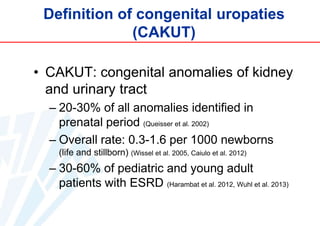
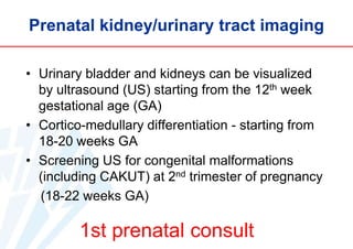
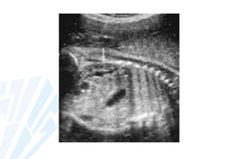
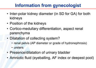
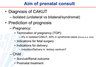
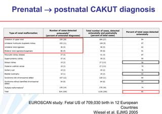
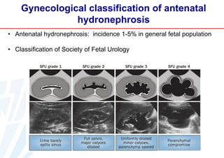
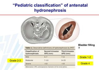
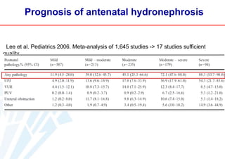
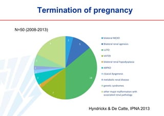
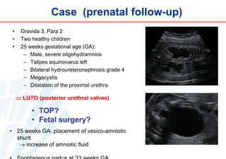
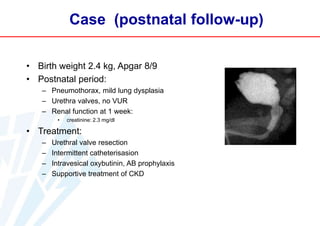
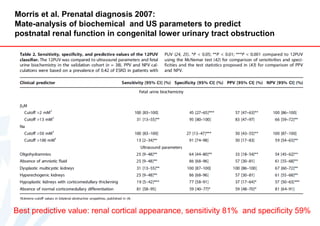
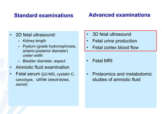
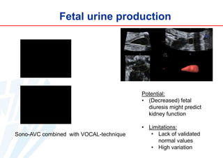
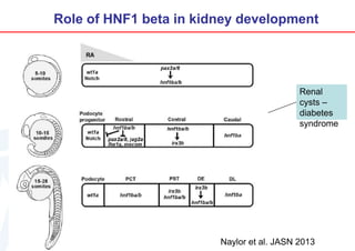
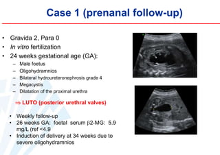
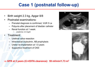
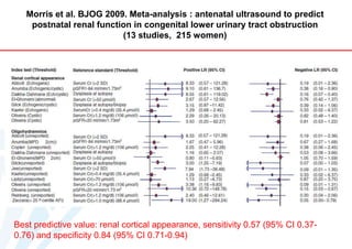
Prenatal diagnosis of congenital uropathies

Elena Levtchenko
Leuven, Belgium
Moscow, October 22, 2013
Gifted by R. Kleta

?

Pediatric
Pediatric consult consult

birth
Definition of congenital uropaties
(CAKUT)
• CAKUT: congenital anomalies of kidney
and urinary tract
– 20-30% of all anomalies identified in
prenatal period (Queisser et al. 2002)
– Overall rate: 0.3-1.6 per 1000 newborns
(life and stillborn) (Wissel et al. 2005, Caiulo et al. 2012)

– 30-60% of pediatric and young adult
patients with ESRD (Harambat et al. 2012, Wuhl et al. 2013)
Development of human pronephros

Song et al. Pediatr Nephrol
2011
Prenatal kidney/urinary tract imaging
• Urinary bladder and kidneys can be visualized
by ultrasound (US) starting from the 12th week
gestational age (GA)
• Cortico-medullary differentiation - starting from
18-20 weeks GA
• Screening US for congenital malformations
(including CAKUT) at 2nd trimester of pregnancy
(18-22 weeks GA)

1st prenatal consult
Information from gynecologist
• Inter-polar kidney diameter (in SD for GA) for both
kidneys
• Position of the kidneys
• Cortico-medullary differentiation, aspect renal
parenchyma
• Dilatation of collecting system?
• renal pelvis (AP diameter or grade of hydronephrosis)
• ureters

• Presence/dilatation of urinary bladder
• Amniotic fluid (eyeballing, AF index or deepest pool)
Aim of prenatal consult
• Diagnosis of CAKUT
– Isolated (unilateral vs bilateral/syndromal)

• Prediction of prognosis
– Pregnancy:
• Termination of pregnancy (TOP):
– 4% in isolated CAKUT, 50% in syndromal cases (Wissel et al. 2005)

• Indications for fetal surgery
• Indications for delivery:
– Induction/Delivery in tertiary centrum?

– Child
• Survival/Renal outcome
• Postnatal treatment
Prenatal  postnatal CAKUT diagnosis

EUROSCAN study: Fetal US of 709,030 birth in 12 European
Countries
Wiesel et al. EJMG 2005
Gynecological classification of antenatal
hydronephrosis
• Antenatal hydronephrosis: incidence 1-5% in general fetal population
• Classification of Society of Fetal Urology
“Pediatric classification” of antenatal
hydronephrosis

Bladder filling
?

Grade 1-2
Grade 2-3
Grade 4
Prognosis of antenatal hydronephrosis

Lee et al. Pediatrics 2006. Meta-analysis of 1,645 studies -> 17 studies sufficient
quality
Termination of pregnancy
N=50 (2008-2013)

Hyndrickx & De Catte, IPNA 2013
Case (prenatal follow-up)
•
•
•

Gravida 3, Para 2
Two healthy children
25 weeks gestational age (GA):
– Male, severe oligohydramnios
– Talipes equinovarus left
– Bilateral hydroureteronephrosis grade 4
– Megacystis
– Dilatation of the proximal urethra

 LUTO (posterior urethral valves)

• TOP?
• Fetal surgery?
• 25 weeks GA: placement of vesico-amniotic
shunt
 increase of amniotic fluid
Case (postnatal follow-up)
• Birth weight 2.4 kg, Apgar 8/9
• Postnatal period:
– Pneumothorax, mild lung dysplasia
– Urethra valves, no VUR
– Renal function at 1 week:
•

creatinine: 2.3 mg/dl

• Treatment:
–
–
–
–

Urethral valve resection
Intermittent catheterisasion
Intravesical oxybutinin, AB prophylaxis
Supportive treatment of CKD
Glomerular filtration rate at 2 years?
• < 15 ml/min/1,73 m2

• 15-30 ml/min/1,73 m2

• 30-60 ml/min/1,73 m2

• 60-90 ml/min/1,73 m2
Morris et al. Prenatal diagnosis 2007:
Mate-analysis of biochemical and US parameters to predict
postnatal renal function in congenital lower urinary tract obstruction

Best predictive value: renal cortical appearance, sensitivity 81% and specificity 59%
Klein et al. Sci Transl Med 2013: proteomics study of fetal urine to
predict postnatal renal function in congenital lower urinary tract
obstruction
Klein et al. Sci Transl Med 2013
Indications for fetal surgery
Only in Low-Urinary Tract
obstruction (LUTO)

Morris et al. Lancet 2013:
Results of PLUTO trial:
N=31 pregnancies with PUV,
16 – vesicoamniotic shunt (VAS)
15 – concervative treatment (CT)

•
•
•
•
•

VAS:
1 intrauterine death
3 TOP
4 death <28
days
8 survived
2 normal RF

•
•
•
•
•

CT:
1 intrauterine death
2 TOP
8 death <28
days
4 survived
0 normal RF
Indications for delivery induction
• Efforts to continue pregnancy to prevent
complications of premature birth:
– 34-36 weeks in case of severe
oligohydramnios?

• Indications for delivery in tertiary center:
• Anticipated requirement of renal replacement
therapy
• Anticipated requirement of complex surgery in
neonatal period
• Anticipated requirement in tertiary neonatal care
Can standard prenatal examinations
predict postnatal outcome of renal
function?

An Hindryckx

Luc De Catte

Prenatal imaging studies

Anke Raaijmakers
Postnatal follow-up
Standard examinations

• 2D fetal ultrasound:
– Kidney length
– Pyelum (grade hydronephrosis,
anterio-posterior diameter)
ureter width
– Bladder diameter, aspect

• Amniotic fluid examination
• Fetal serum (2-MG, cystatin C,
caryotype, urine (electrolytes,
osmol)

Advanced examinations

• 3D fetal ultrasound
• Fetal urine production
• Fetal cortex blood flow

• Fetal MRI
• Proteomics and metabolomic
studies of amniotic fluid
3D renal ultrasound

Kidney/pyelum volume measurement:
by Virtual Organ Computer-aided AnaLys
(VOCAL) with sono-AVC (automated volume
count) for fluid/filled spaces segmentation
3D renal ultrasound
Potential:
•

Volume is better reflecting
function compared to renal
pelvic diameter (Nam et al. 2012)

•

Distinction between pyelum and
renal parenchyma  cortex
volume

Limitations:

Inversion rendering

• Lack of standardization:
• Methodology (imaging planes,
anatomical landmarks,
repeatability)
• Lack of validated normal
values
• Data analysis and storage
(depending on producent)
Reference curve for 3D renal volume
ml

Hindryckx et al. 2013, IPNA Poster P-SUN009
Fetal urine production

Potential:
• (Decreased) fetal
diuresis might predict
kidney function

Sono-AVC combined with VOCAL-technique

• Limitations:
• Lack of validated
normal values
• High variation
Fetal kidney perfusion

• Peak systolic velocity (PSV)
• Flow velocity integral in renal vein correlated to
kidney cortex volume (FVI x HR/renal cortex
volume)
Normal fetal kidney perfusion
cm/se
c

Hindryckx et al. 2013, IPNA Poster P-SUN009
Conclusions
• Interaction between gynecologist and
pediatrician for prenatal diagnosis of
CAKUT
• Prenatal consult aimes to make diagnosis,
define severity and prognosis, pre- and
postnatal follow-up; objective information
for the parents
• Good prognostic markers for renal function
outcome are limited and are a subject of
intensive research
Pediatricians/pediatric
nephrologists should be involved
in prenatal follow-up of patients
with CAKUT!
Acknowledgments
Katholieke Universiteit Leuven
An Hindryckx
Luc De Catte
Anke Raaijmakers
Djalila Mekahli
Karel Allegaert

Inserm Toulouse
Joost Schanstra
Stéphane Decramer
Jean-Loup Bascands

UCL University College London
Paul Winyard

Universitätsklinikum Heidelberg
Franz Schaefer
Elke Wühl
Anke Doyon
THANK
YOU!

Fons Sapientiae by Jef Claerhout

Leuven, Belgium
Embriology of kidney development
Abnormal kidney development

Song et al. Pediatr Nephrol
2011
Molecular regulation of kidney development (1)

M. Little et al. Current Topics in Developmental Biology
2010
Molecular regulation of kidney development (2)

TownesBrocks
syndrome
DenisDrash
syndrome

Brachiootorenal
syndrome
Oculorenaal
syndrome

M. Little et al. Current Topics in Developmental Biology
2010
Role of HNF1 beta in kidney development

Renal
cysts –
diabetes
syndrome

Naylor et al. JASN 2013
Case 1 (prenanal follow-up)
• Gravida 2, Para 0
• In vitro fertilization
• 24 weeks gestational age (GA):
– Male foetus
– Oligohydramnios
– Bilateral hydroureteronephrosis grade 4
– Megacystis
– Dilatation of the proximal urethra

 LUTO (posterior urethral valves)
• Weekly follow-up
• 26 weeks GA: foetal serum 2-MG: 5.9
mg/L (ref <4.9
• Induction of delivery at 34 weeks due to
severe oligohydramnios
Case 1 (postnatal follow-up)
• Birth weight 2.3 kg, Apgar 8/8
• Postnatal examinations:
– Prenatal diagnosis is confirmed, VUR 5 Le
– Polyuria after placement of bladder catheter
– Renal function at 1 week:
•

creatinine: 2.3 mg/dl

• Treatment:
–
–
–
–

Urethral valve resection
Intravesical oxybutinin, AB prophylaxis
Ureter re-implantation at 1.5 years
Supportive treatment of CKD

 GFR at 2 years (Cr-EDTA clearance): 59 ml/min/1.73 m2
Morris et al. BJOG 2009. Meta-analysis : antenatal ultrasound to predict
postnatal renal function in congenital lower urinary tract obstruction
(13 studies, 215 women)

Best predictive value: renal cortical appearance, sensitivity 0.57 (95% CI 0.370.76) and specificity 0.84 (95% CI 0.71-0.94)
AF 4-quadrant index

2-2. CAKUT. Elena Levtchenko (eng)

  • 1.
    Prenatal diagnosis ofcongenital uropathies Elena Levtchenko Leuven, Belgium Moscow, October 22, 2013
  • 2.
    Gifted by R.Kleta ? Pediatric Pediatric consult consult birth
  • 3.
    Definition of congenitaluropaties (CAKUT) • CAKUT: congenital anomalies of kidney and urinary tract – 20-30% of all anomalies identified in prenatal period (Queisser et al. 2002) – Overall rate: 0.3-1.6 per 1000 newborns (life and stillborn) (Wissel et al. 2005, Caiulo et al. 2012) – 30-60% of pediatric and young adult patients with ESRD (Harambat et al. 2012, Wuhl et al. 2013)
  • 4.
    Development of humanpronephros Song et al. Pediatr Nephrol 2011
  • 5.
    Prenatal kidney/urinary tractimaging • Urinary bladder and kidneys can be visualized by ultrasound (US) starting from the 12th week gestational age (GA) • Cortico-medullary differentiation - starting from 18-20 weeks GA • Screening US for congenital malformations (including CAKUT) at 2nd trimester of pregnancy (18-22 weeks GA) 1st prenatal consult
  • 7.
    Information from gynecologist •Inter-polar kidney diameter (in SD for GA) for both kidneys • Position of the kidneys • Cortico-medullary differentiation, aspect renal parenchyma • Dilatation of collecting system? • renal pelvis (AP diameter or grade of hydronephrosis) • ureters • Presence/dilatation of urinary bladder • Amniotic fluid (eyeballing, AF index or deepest pool)
  • 8.
    Aim of prenatalconsult • Diagnosis of CAKUT – Isolated (unilateral vs bilateral/syndromal) • Prediction of prognosis – Pregnancy: • Termination of pregnancy (TOP): – 4% in isolated CAKUT, 50% in syndromal cases (Wissel et al. 2005) • Indications for fetal surgery • Indications for delivery: – Induction/Delivery in tertiary centrum? – Child • Survival/Renal outcome • Postnatal treatment
  • 9.
    Prenatal  postnatalCAKUT diagnosis EUROSCAN study: Fetal US of 709,030 birth in 12 European Countries Wiesel et al. EJMG 2005
  • 10.
    Gynecological classification ofantenatal hydronephrosis • Antenatal hydronephrosis: incidence 1-5% in general fetal population • Classification of Society of Fetal Urology
  • 11.
    “Pediatric classification” ofantenatal hydronephrosis Bladder filling ? Grade 1-2 Grade 2-3 Grade 4
  • 12.
    Prognosis of antenatalhydronephrosis Lee et al. Pediatrics 2006. Meta-analysis of 1,645 studies -> 17 studies sufficient quality
  • 13.
    Termination of pregnancy N=50(2008-2013) Hyndrickx & De Catte, IPNA 2013
  • 14.
    Case (prenatal follow-up) • • • Gravida3, Para 2 Two healthy children 25 weeks gestational age (GA): – Male, severe oligohydramnios – Talipes equinovarus left – Bilateral hydroureteronephrosis grade 4 – Megacystis – Dilatation of the proximal urethra  LUTO (posterior urethral valves) • TOP? • Fetal surgery? • 25 weeks GA: placement of vesico-amniotic shunt  increase of amniotic fluid
  • 15.
    Case (postnatal follow-up) •Birth weight 2.4 kg, Apgar 8/9 • Postnatal period: – Pneumothorax, mild lung dysplasia – Urethra valves, no VUR – Renal function at 1 week: • creatinine: 2.3 mg/dl • Treatment: – – – – Urethral valve resection Intermittent catheterisasion Intravesical oxybutinin, AB prophylaxis Supportive treatment of CKD
  • 16.
    Glomerular filtration rateat 2 years? • < 15 ml/min/1,73 m2 • 15-30 ml/min/1,73 m2 • 30-60 ml/min/1,73 m2 • 60-90 ml/min/1,73 m2
  • 17.
    Morris et al.Prenatal diagnosis 2007: Mate-analysis of biochemical and US parameters to predict postnatal renal function in congenital lower urinary tract obstruction Best predictive value: renal cortical appearance, sensitivity 81% and specificity 59%
  • 18.
    Klein et al.Sci Transl Med 2013: proteomics study of fetal urine to predict postnatal renal function in congenital lower urinary tract obstruction
  • 19.
    Klein et al.Sci Transl Med 2013
  • 20.
    Indications for fetalsurgery Only in Low-Urinary Tract obstruction (LUTO) Morris et al. Lancet 2013: Results of PLUTO trial: N=31 pregnancies with PUV, 16 – vesicoamniotic shunt (VAS) 15 – concervative treatment (CT) • • • • • VAS: 1 intrauterine death 3 TOP 4 death <28 days 8 survived 2 normal RF • • • • • CT: 1 intrauterine death 2 TOP 8 death <28 days 4 survived 0 normal RF
  • 21.
    Indications for deliveryinduction • Efforts to continue pregnancy to prevent complications of premature birth: – 34-36 weeks in case of severe oligohydramnios? • Indications for delivery in tertiary center: • Anticipated requirement of renal replacement therapy • Anticipated requirement of complex surgery in neonatal period • Anticipated requirement in tertiary neonatal care
  • 22.
    Can standard prenatalexaminations predict postnatal outcome of renal function? An Hindryckx Luc De Catte Prenatal imaging studies Anke Raaijmakers Postnatal follow-up
  • 23.
    Standard examinations • 2Dfetal ultrasound: – Kidney length – Pyelum (grade hydronephrosis, anterio-posterior diameter) ureter width – Bladder diameter, aspect • Amniotic fluid examination • Fetal serum (2-MG, cystatin C, caryotype, urine (electrolytes, osmol) Advanced examinations • 3D fetal ultrasound • Fetal urine production • Fetal cortex blood flow • Fetal MRI • Proteomics and metabolomic studies of amniotic fluid
  • 24.
    3D renal ultrasound Kidney/pyelumvolume measurement: by Virtual Organ Computer-aided AnaLys (VOCAL) with sono-AVC (automated volume count) for fluid/filled spaces segmentation
  • 25.
    3D renal ultrasound Potential: • Volumeis better reflecting function compared to renal pelvic diameter (Nam et al. 2012) • Distinction between pyelum and renal parenchyma  cortex volume Limitations: Inversion rendering • Lack of standardization: • Methodology (imaging planes, anatomical landmarks, repeatability) • Lack of validated normal values • Data analysis and storage (depending on producent)
  • 26.
    Reference curve for3D renal volume ml Hindryckx et al. 2013, IPNA Poster P-SUN009
  • 27.
    Fetal urine production Potential: •(Decreased) fetal diuresis might predict kidney function Sono-AVC combined with VOCAL-technique • Limitations: • Lack of validated normal values • High variation
  • 28.
    Fetal kidney perfusion •Peak systolic velocity (PSV) • Flow velocity integral in renal vein correlated to kidney cortex volume (FVI x HR/renal cortex volume)
  • 29.
    Normal fetal kidneyperfusion cm/se c Hindryckx et al. 2013, IPNA Poster P-SUN009
  • 30.
    Conclusions • Interaction betweengynecologist and pediatrician for prenatal diagnosis of CAKUT • Prenatal consult aimes to make diagnosis, define severity and prognosis, pre- and postnatal follow-up; objective information for the parents • Good prognostic markers for renal function outcome are limited and are a subject of intensive research
  • 31.
    Pediatricians/pediatric nephrologists should beinvolved in prenatal follow-up of patients with CAKUT!
  • 32.
    Acknowledgments Katholieke Universiteit Leuven AnHindryckx Luc De Catte Anke Raaijmakers Djalila Mekahli Karel Allegaert Inserm Toulouse Joost Schanstra Stéphane Decramer Jean-Loup Bascands UCL University College London Paul Winyard Universitätsklinikum Heidelberg Franz Schaefer Elke Wühl Anke Doyon
  • 33.
    THANK YOU! Fons Sapientiae byJef Claerhout Leuven, Belgium
  • 34.
  • 35.
    Abnormal kidney development Songet al. Pediatr Nephrol 2011
  • 36.
    Molecular regulation ofkidney development (1) M. Little et al. Current Topics in Developmental Biology 2010
  • 37.
    Molecular regulation ofkidney development (2) TownesBrocks syndrome DenisDrash syndrome Brachiootorenal syndrome Oculorenaal syndrome M. Little et al. Current Topics in Developmental Biology 2010
  • 38.
    Role of HNF1beta in kidney development Renal cysts – diabetes syndrome Naylor et al. JASN 2013
  • 39.
    Case 1 (prenanalfollow-up) • Gravida 2, Para 0 • In vitro fertilization • 24 weeks gestational age (GA): – Male foetus – Oligohydramnios – Bilateral hydroureteronephrosis grade 4 – Megacystis – Dilatation of the proximal urethra  LUTO (posterior urethral valves) • Weekly follow-up • 26 weeks GA: foetal serum 2-MG: 5.9 mg/L (ref <4.9 • Induction of delivery at 34 weeks due to severe oligohydramnios
  • 40.
    Case 1 (postnatalfollow-up) • Birth weight 2.3 kg, Apgar 8/8 • Postnatal examinations: – Prenatal diagnosis is confirmed, VUR 5 Le – Polyuria after placement of bladder catheter – Renal function at 1 week: • creatinine: 2.3 mg/dl • Treatment: – – – – Urethral valve resection Intravesical oxybutinin, AB prophylaxis Ureter re-implantation at 1.5 years Supportive treatment of CKD  GFR at 2 years (Cr-EDTA clearance): 59 ml/min/1.73 m2
  • 41.
    Morris et al.BJOG 2009. Meta-analysis : antenatal ultrasound to predict postnatal renal function in congenital lower urinary tract obstruction (13 studies, 215 women) Best predictive value: renal cortical appearance, sensitivity 0.57 (95% CI 0.370.76) and specificity 0.84 (95% CI 0.71-0.94)
  • 42.