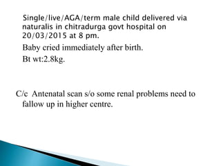
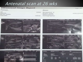
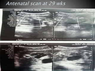
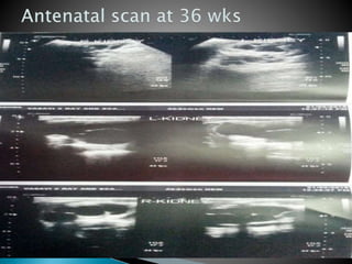
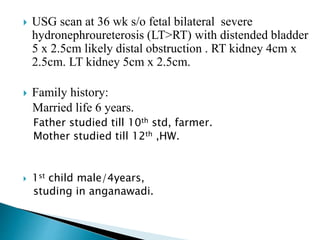
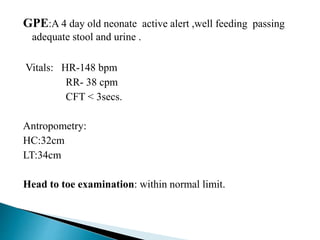
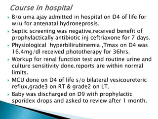
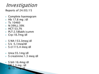
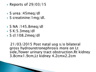
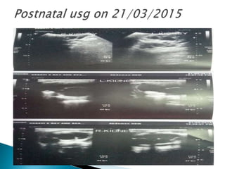
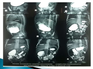
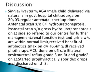
Dr Raghavendra
Fellow in neonatology.
Patient name: B/o Uma ajay
S/o Ajay
Gollaratti(P)
Chitradurga dist
DOB:20/03/2015
DOA:24/03/2015
DOD:03/04/2015
Single/live/AGA/term male child delivered via
naturalis in chitradurga govt hospital on
20/03/2015 at 8 pm.
Baby cried immediately after birth.
Bt wt:2.8kg.
C/c Antenatal scan s/o some renal problems need to
fallow up in higher centre.
 Obstretic history: ANC registered in govt hospital chitradurga.
Regular fallow up, taken iron and folic acid tablets.
Taken 2 dose of TT inj. Taken three antenatal scan.
Antenatal events un eventful.
 USG scan at 26 wks s/o fetal bilateral moderate to severe
hydroneproureterosis (LT>RT) with moderately distended
urinary bladder.?Partial posterior uretral valve. RT kidney 3 x 1.5
cm. LT kidney 3.4cm x 2.2cm.
 USG scan at 29 weeks s/o bilateral fetal moderte to severe
hydronepro ureterosis ((LT>RT) with thickened urinary mucosa
s/o bladder outlet obstruction. RT kidney 3cm x1.6cm. Lt kidney
3.8cmx 2.6cm.
 USG scan at 36 wk s/o fetal bilateral severe
hydronephroureterosis (LT>RT) with distended bladder
5 x 2.5cm likely distal obstruction . RT kidney 4cm x
2.5cm. LT kidney 5cm x 2.5cm.
 Family history:
Married life 6 years.
Father studied till 10th std, farmer.
Mother studied till 12th ,HW.
 1st child male/4years,
studing in anganawadi.
GPE:A 4 day old neonate active alert ,well feeding passing
adequate stool and urine .
Vitals: HR-148 bpm
RR- 38 cpm
CFT < 3secs.
Antropometry:
HC:32cm
LT:34cm
Head to toe examination: within normal limit.
 Systemic examination:
 CVS- S1 & S2 heard, no murmur
 R/S- B/L air entry equal, no added sounds
 P/A- soft, BS heard
 CNS- tone and reflexes normal
 B/o uma ajay admitted in hospital on D4 of life for
w/u for antenatal hydroneprosis.
 Septic screening was negative,received benefit of
prophylactically antibiotic inj ceftriaxone for 7 days.
 Physiological hyperbilirubinemia ,Tmax on D4 was
16.4mg/dl received phototherapy for 36hrs.
 Workup for renal function test and routine urine and
culture sensitivity done.reports are within normal
limits.
 MCU done on D4 of life s/o bilateral vesicoureteric
reflux,grade3 on RT & grade2 on LT.
 Baby was discharged on D9 with prophylactic
sporidex drops and asked to review after 1 month.
Reports of 24/03/15
 Complete haemogram
 Hb 17.8 mg /dl
 Tc 10460
 N:39%,L:39%
 HCT:53.7%
 PLT:2.58lakh/cumm
 Crp:18.7mg/dl
 S NA:153.3meq/dl
 S k :5.1meq?dl
 S cl:115.4 meq/dl
 Urea:55.1mg/dl
 S creatinine:1.3 meq/dl
 S bili 16.4mg/dl
 DB:1.3 mg /dl
 IB:15.1mg/dl
 Reports of 29/03/15
 S urea :45meq/dl
 S creatinine:1mg/dl.
 S NA :145.8meq/dl
 S K:5.3meq/dl
 S cl:108.2meq/dl
21/03/2015 Post natal usg s/o bilateral
gross hydrouretronephrosis more on Lt
side,?lower urinary tract obstruction.Rt kidney
3.8cmx1.9cm,Lt kidney 4.2cmx2.2cm
 Single/live/term/AGA/male child delivered via
naturalis in govt hospital chitradurga on
20/03.regular antenatal checkup done.
Antenatal scan s/o B/l hydrouretroneprosis.
Postnatal scan s/o gross hydro ureteroneprosis
on Lt side,so refered to our centre for further
management.renal function test and urine w/u
are within normal limit,received benefit of
antibiotics,tmax on d4 16.4mg/dl received
photherapy.MCU done on d5 s/o Bilateral
vesicouretral reflux grade 3 on Rt and grade2
on Lt.Started prophylactically sporidex drops
and dischared on d13.
 Antenatal hydronephrosis is the dilatation of the collecting
system of the fetal kidney.
 It is estimated that fetal urinary tract dilatation is identified in
1% of all pregnancies.
 In more than 50% cases, the antenatally detected dilatation is
transient and resolves spontaneously.
 Antenatally detected dilatation, which persists after birth is
labeled as neonatal hydronephrosis.
 Pelviureteric junction (PUJ) obstruction accounts for 50-60%
patients with neonatal hydronephrosis.
 Vesicoureteric reflux (VUR) is detected in 20-30% of such
cases.
Common
 Pelviureteric junction obstruction
 Vesicoureteric reflux
 Vesicoureteric junction obstruction
 Multicystic kidney.
Rare
 Posterior urethral valves
 Obstructive and non-obstructive megaureter
 Ureterocele
 Neurogenic bladder
 Prune-belly syndrome
 Urethral atresia
 Fetal hydronephrosis of moderate degree can be
detected as early as 15-18 weeks gestation by
ultrasonography.
 A maximum anteroposterior diameter of renal
pelvis of more than 10 mm and the ratio of
antero-posterior diameter of renal pelvis to
kidney of more than 0.5 after 30 weeks
gestation requires postnatal evaluation.
 The ultrasound study should be repeated every
6-8 weeks until delivery.
 Hydronephrosis .
 Renal pelvic anteroposterior diameter.
 Caliectasis.
 Ureteral dilatation .
 Renal echogenicity.
 Contralateral kidney: size, dilatation.
 Bladder size, thickness.
 Posterior urethral dilatation.
 Urinary flow .
 Amniotic fluid volume
At birth Clinical examination,
assess urine stream.
 Asymptomatic unilateral hydronephrosis
 0–2 weeks Ultrasonography
Blood urea, creatinine; urine culture
 4–6 weeks Micturating cystourethrogram
DTPA scan (with diuretic renography).
 Solitary kidney, suspected posterior urethral valves, bilateral
hydronephrosis or presence of symptoms
0–2 weeks Ultrasonography
Blood urea, creatinine; urine culture
Micturating cystourethrogram
 4–6 weeks DTPA scan (with diuretic
renography)
 DMSA renal scan is performed in patients with vesicoureteric reflux
 Grade 1: Slight separation of the central renal
echo complex.
 Grade 2: Renal pelvis is further dilated and a
single or a few calyces may be visualized.
 Grade 3: Renal pelvis is dilated and there are
fluid filled calyces throughout the kidney, but
renal parenchyma is of normal thickness.
 Grade 4: As grade 3, but renal parenchyma over
the calyces is thinned
 Asymptomatic unilateral hydronephrosis is most
often a benign condition.
 Hydronephrosis due to PUJ obstruction resolves
spontaneously with passage of time.
 Hydronephrosis & Hydroureteronephrosis due to
vesicoureteric junction obstruction improve.
 Kidneys with renal pelvic diameter more than 20
mm are likely to show deterioration in renal
function, which might require intervention.
 VUR is seen in 20-25% neonates with antenatally
detected hydronephrosis, more commonly in boys.
 Forty per cent neonates with VUR show features
suggestive of renal scarring on DMSA scan.
 Neonates with VUR should be managed on long-term
antibiotic prophylaxis (while awaiting spontaneous
resolution of the reflux).
 The patients are kept on close follow-up for occurrence
of break-through urinary infections.
 An ultrasound examination, and radionuclide cystogram
or MCU should be repeated at 12-15 months.
I Ureter only
II Ureter, pelvis, and calices; no dilatation; normal caliceal fornices
III Mild or moderate dilatation or tortuosity of the ureter and moderate
dilatation of the renal pelvis; no or slight blunting of the fornices
IV Moderate dilatation or tortuosity of the ureter and moderate dilatation
of the renal pelvis and calices; complete obliteration of the sharp
angle of the fornices but maintenance of the papillary impressions in
the majority of calices
V Gross dilatation and tortuosity of the ureter; gross dilatation of the
renal pelvis and calices; papillary impressions are no longer visible in
the majority of the calices
Conservative Approach
 The management of asymptomatic neonatal
hydronephrosis is essentially conservative.
 severe neonatal hydronephrosis resolve
spontaneously and deterioration of renal function, if
detected, can be reversed by prompt surgery.
Antibiotic prophylaxis.
 Cephalexin (10-15 mg/kg/day) should be used for
the initial 3 months, and cotrimoxazole (1-2
mg/kg/day) or nitro-furantoin (1 mg/kg/day).
 Urine culture should be promptly obtained if the
patient has symptoms suggestive of urinary tract
infection e.g., unexplained fever, turbid or foul
smelling urine, poor feeding and lethargy
PUJ obstruction
At initial diagnosis
 Presence of symptoms
 Solitary kidney with hydronephrosis
 Bilateral hydronephrosis
 Differential renal function of obstructed kidney <30%
 On follow-up Increasing renal pelvic dilatation >10%
decline in differential renal function
Posterior urethral valve,
ureterocele
Vesicoureteric reflux
 Grade IV-V reflux persisting beyond infancy
 New renal scars or recurrent urinary infections despite
antibiotic prophylaxis
 Behrman, R.E., Kliegman, R.M., & Jenson, H.B. (2000).
Nelson Textbook of Pediatrics (16th ed.). Page1445-1454.
 Indian Pediatrics 2001; 38: 1244-1251
Consensus Statement on Management of Antenatally Detected
Hydronephrosis.
 Indian J Nephrol. 2013 Mar-Apr; 23(2): 83–97.
 . Mallik M, Watson AR. Antenatally detected urinary tract
abnormalities: More detection but less action.Pediatr
Nephrol. 2008;23:897–904. [PubMed]
 2. Dudley JA, Haworth JM, McGraw ME, Frank JD, Tizard EJ.
Clinical relevance and implications of antenatal
hydronephrosis. Arch Dis Child Fetal Neonatal
Ed. 1997;76:F31–4. [PMC free article] [PubMed]
 Goole images
Vur reflex

Vur reflex

  • 1.
  • 2.
    Patient name: B/oUma ajay S/o Ajay Gollaratti(P) Chitradurga dist DOB:20/03/2015 DOA:24/03/2015 DOD:03/04/2015
  • 3.
    Single/live/AGA/term male childdelivered via naturalis in chitradurga govt hospital on 20/03/2015 at 8 pm. Baby cried immediately after birth. Bt wt:2.8kg. C/c Antenatal scan s/o some renal problems need to fallow up in higher centre.
  • 4.
     Obstretic history:ANC registered in govt hospital chitradurga. Regular fallow up, taken iron and folic acid tablets. Taken 2 dose of TT inj. Taken three antenatal scan. Antenatal events un eventful.  USG scan at 26 wks s/o fetal bilateral moderate to severe hydroneproureterosis (LT>RT) with moderately distended urinary bladder.?Partial posterior uretral valve. RT kidney 3 x 1.5 cm. LT kidney 3.4cm x 2.2cm.  USG scan at 29 weeks s/o bilateral fetal moderte to severe hydronepro ureterosis ((LT>RT) with thickened urinary mucosa s/o bladder outlet obstruction. RT kidney 3cm x1.6cm. Lt kidney 3.8cmx 2.6cm.
  • 8.
     USG scanat 36 wk s/o fetal bilateral severe hydronephroureterosis (LT>RT) with distended bladder 5 x 2.5cm likely distal obstruction . RT kidney 4cm x 2.5cm. LT kidney 5cm x 2.5cm.  Family history: Married life 6 years. Father studied till 10th std, farmer. Mother studied till 12th ,HW.  1st child male/4years, studing in anganawadi.
  • 9.
    GPE:A 4 dayold neonate active alert ,well feeding passing adequate stool and urine . Vitals: HR-148 bpm RR- 38 cpm CFT < 3secs. Antropometry: HC:32cm LT:34cm Head to toe examination: within normal limit.
  • 10.
     Systemic examination: CVS- S1 & S2 heard, no murmur  R/S- B/L air entry equal, no added sounds  P/A- soft, BS heard  CNS- tone and reflexes normal
  • 11.
     B/o umaajay admitted in hospital on D4 of life for w/u for antenatal hydroneprosis.  Septic screening was negative,received benefit of prophylactically antibiotic inj ceftriaxone for 7 days.  Physiological hyperbilirubinemia ,Tmax on D4 was 16.4mg/dl received phototherapy for 36hrs.  Workup for renal function test and routine urine and culture sensitivity done.reports are within normal limits.  MCU done on D4 of life s/o bilateral vesicoureteric reflux,grade3 on RT & grade2 on LT.  Baby was discharged on D9 with prophylactic sporidex drops and asked to review after 1 month.
  • 12.
    Reports of 24/03/15 Complete haemogram  Hb 17.8 mg /dl  Tc 10460  N:39%,L:39%  HCT:53.7%  PLT:2.58lakh/cumm  Crp:18.7mg/dl  S NA:153.3meq/dl  S k :5.1meq?dl  S cl:115.4 meq/dl  Urea:55.1mg/dl  S creatinine:1.3 meq/dl  S bili 16.4mg/dl  DB:1.3 mg /dl  IB:15.1mg/dl
  • 13.
     Reports of29/03/15  S urea :45meq/dl  S creatinine:1mg/dl.  S NA :145.8meq/dl  S K:5.3meq/dl  S cl:108.2meq/dl 21/03/2015 Post natal usg s/o bilateral gross hydrouretronephrosis more on Lt side,?lower urinary tract obstruction.Rt kidney 3.8cmx1.9cm,Lt kidney 4.2cmx2.2cm
  • 18.
     Single/live/term/AGA/male childdelivered via naturalis in govt hospital chitradurga on 20/03.regular antenatal checkup done. Antenatal scan s/o B/l hydrouretroneprosis. Postnatal scan s/o gross hydro ureteroneprosis on Lt side,so refered to our centre for further management.renal function test and urine w/u are within normal limit,received benefit of antibiotics,tmax on d4 16.4mg/dl received photherapy.MCU done on d5 s/o Bilateral vesicouretral reflux grade 3 on Rt and grade2 on Lt.Started prophylactically sporidex drops and dischared on d13.
  • 19.
     Antenatal hydronephrosisis the dilatation of the collecting system of the fetal kidney.  It is estimated that fetal urinary tract dilatation is identified in 1% of all pregnancies.  In more than 50% cases, the antenatally detected dilatation is transient and resolves spontaneously.  Antenatally detected dilatation, which persists after birth is labeled as neonatal hydronephrosis.  Pelviureteric junction (PUJ) obstruction accounts for 50-60% patients with neonatal hydronephrosis.  Vesicoureteric reflux (VUR) is detected in 20-30% of such cases.
  • 20.
    Common  Pelviureteric junctionobstruction  Vesicoureteric reflux  Vesicoureteric junction obstruction  Multicystic kidney. Rare  Posterior urethral valves  Obstructive and non-obstructive megaureter  Ureterocele  Neurogenic bladder  Prune-belly syndrome  Urethral atresia
  • 21.
     Fetal hydronephrosisof moderate degree can be detected as early as 15-18 weeks gestation by ultrasonography.  A maximum anteroposterior diameter of renal pelvis of more than 10 mm and the ratio of antero-posterior diameter of renal pelvis to kidney of more than 0.5 after 30 weeks gestation requires postnatal evaluation.  The ultrasound study should be repeated every 6-8 weeks until delivery.
  • 24.
     Hydronephrosis . Renal pelvic anteroposterior diameter.  Caliectasis.  Ureteral dilatation .  Renal echogenicity.  Contralateral kidney: size, dilatation.  Bladder size, thickness.  Posterior urethral dilatation.  Urinary flow .  Amniotic fluid volume
  • 25.
    At birth Clinicalexamination, assess urine stream.  Asymptomatic unilateral hydronephrosis  0–2 weeks Ultrasonography Blood urea, creatinine; urine culture  4–6 weeks Micturating cystourethrogram DTPA scan (with diuretic renography).  Solitary kidney, suspected posterior urethral valves, bilateral hydronephrosis or presence of symptoms 0–2 weeks Ultrasonography Blood urea, creatinine; urine culture Micturating cystourethrogram  4–6 weeks DTPA scan (with diuretic renography)  DMSA renal scan is performed in patients with vesicoureteric reflux
  • 26.
     Grade 1:Slight separation of the central renal echo complex.  Grade 2: Renal pelvis is further dilated and a single or a few calyces may be visualized.  Grade 3: Renal pelvis is dilated and there are fluid filled calyces throughout the kidney, but renal parenchyma is of normal thickness.  Grade 4: As grade 3, but renal parenchyma over the calyces is thinned
  • 29.
     Asymptomatic unilateralhydronephrosis is most often a benign condition.  Hydronephrosis due to PUJ obstruction resolves spontaneously with passage of time.  Hydronephrosis & Hydroureteronephrosis due to vesicoureteric junction obstruction improve.  Kidneys with renal pelvic diameter more than 20 mm are likely to show deterioration in renal function, which might require intervention.
  • 30.
     VUR isseen in 20-25% neonates with antenatally detected hydronephrosis, more commonly in boys.  Forty per cent neonates with VUR show features suggestive of renal scarring on DMSA scan.  Neonates with VUR should be managed on long-term antibiotic prophylaxis (while awaiting spontaneous resolution of the reflux).  The patients are kept on close follow-up for occurrence of break-through urinary infections.  An ultrasound examination, and radionuclide cystogram or MCU should be repeated at 12-15 months.
  • 31.
    I Ureter only IIUreter, pelvis, and calices; no dilatation; normal caliceal fornices III Mild or moderate dilatation or tortuosity of the ureter and moderate dilatation of the renal pelvis; no or slight blunting of the fornices IV Moderate dilatation or tortuosity of the ureter and moderate dilatation of the renal pelvis and calices; complete obliteration of the sharp angle of the fornices but maintenance of the papillary impressions in the majority of calices V Gross dilatation and tortuosity of the ureter; gross dilatation of the renal pelvis and calices; papillary impressions are no longer visible in the majority of the calices
  • 33.
    Conservative Approach  Themanagement of asymptomatic neonatal hydronephrosis is essentially conservative.  severe neonatal hydronephrosis resolve spontaneously and deterioration of renal function, if detected, can be reversed by prompt surgery. Antibiotic prophylaxis.  Cephalexin (10-15 mg/kg/day) should be used for the initial 3 months, and cotrimoxazole (1-2 mg/kg/day) or nitro-furantoin (1 mg/kg/day).  Urine culture should be promptly obtained if the patient has symptoms suggestive of urinary tract infection e.g., unexplained fever, turbid or foul smelling urine, poor feeding and lethargy
  • 34.
    PUJ obstruction At initialdiagnosis  Presence of symptoms  Solitary kidney with hydronephrosis  Bilateral hydronephrosis  Differential renal function of obstructed kidney <30%  On follow-up Increasing renal pelvic dilatation >10% decline in differential renal function Posterior urethral valve, ureterocele Vesicoureteric reflux  Grade IV-V reflux persisting beyond infancy  New renal scars or recurrent urinary infections despite antibiotic prophylaxis
  • 35.
     Behrman, R.E.,Kliegman, R.M., & Jenson, H.B. (2000). Nelson Textbook of Pediatrics (16th ed.). Page1445-1454.  Indian Pediatrics 2001; 38: 1244-1251 Consensus Statement on Management of Antenatally Detected Hydronephrosis.  Indian J Nephrol. 2013 Mar-Apr; 23(2): 83–97.  . Mallik M, Watson AR. Antenatally detected urinary tract abnormalities: More detection but less action.Pediatr Nephrol. 2008;23:897–904. [PubMed]  2. Dudley JA, Haworth JM, McGraw ME, Frank JD, Tizard EJ. Clinical relevance and implications of antenatal hydronephrosis. Arch Dis Child Fetal Neonatal Ed. 1997;76:F31–4. [PMC free article] [PubMed]  Goole images