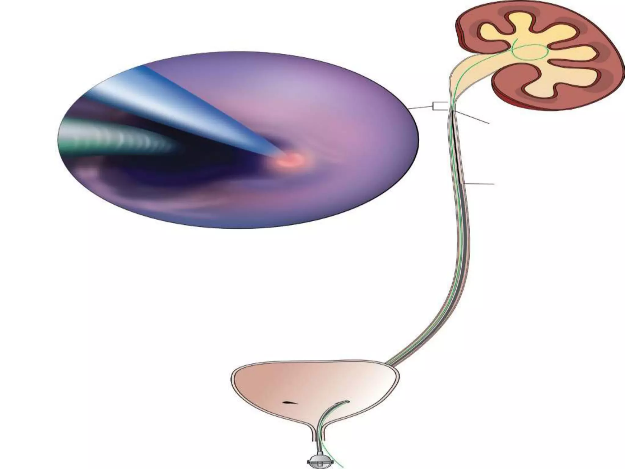
PREPARED BY :DR.TALAL BALLOUT
SUPERVISED BY:DR.WALED ZALLOUM
URETER ANATOMY
The ureters are bilateral tubular structures
responsible for transporting urine from the renal
pelvis to the bladder .
They are generally 22 to 30 cm in length with a
wall composed of multiple layers:
-transitional epithelium
-lamina propria
-smooth muscle(inner longitudinal and outer
circular)
Adventitia. -
URETER ANATOMY
Sites of ureteral narrowing:
-Ureteropelvic junction.
-Junction as the ureter crosses the iliac
vessels.
-Ureterovesical junction.
:
URETER ANATOMY
Anatomic Relationships:
-Anteriorly, the right ureter is related to the ascending
colon,cecum, colonic mesentery, and appendix.
-The left ureter is closely related to the descending and
sigmoid colon and their mesenteries.
-Approximately a third of the way to the bladder
the ureter is crossed anteriorly by the gonadal vessels.
-UPJ lies posterior to the renal artery and vein, It
then lies anterior to the psoas muscle .
-As it enters the pelvis the ureter crosses anterior to the
iliac vessel.
-In the female pelvis, the ureters are crossed anteriorly
by the uterine arteries and are closely related to the
uterine.
URETER ANATOMY
:BLOOD SUPPLY
To upper ureter branches originate from the
renal artery, gonadal artery, abdominal aorta,
and common iliac artery. After entering the
pelvis,
additional small arterial branches to
the distal ureter may arise from the internal
iliac artery or its branches, especially the vesical
and uterine arteries, but also from
the middle rectal and vaginal arteries.
URETER ANATOMY
-The venous and lymphatic drainage of the
ureter parallels the arterial supply.
-ureteral lymphatic drainage varies by ureteral level:
-In the pelvis, ureteral lymphatics drain to internal,
external, and common iliac nodes.
-In the abdomen the left para-aortic lymph nodes
are the primary drainage site for the left ureter,
whereas
the abdominal portion of the right ureter is drained
primarily to right paracaval and interaortocaval lymph
nodes.
URETER ANATOMY
 NERVE SUPPLY
 Sympathetic- T10-L1
 Parasympathetic- S2-S4
EPIDEMIOLOGY
-Most common site of urinary tract obstruction in children .
-Majority are discovered antenatally:
-It is the most common anatomical cause of
antenatal hydronephrosis
– Boys > girls
– Most cases on the left
– 10-40% bilateral
PATHOPHYSIOLOGY
-It is caused by anatomic lesions or functional
disturbances that restrict urinary flow
resulting in hydronephrosis.
- Most cases are thought to be due to partial
obstruction, because complete obstruction
results in rapid destruction of the kidney.
- In some cases, partial obstruction may also
lead to progressive deterioration of renal
function.
ETIOLOGY
-It is both congenital and acquired
conditions.
Usually caused by intrinsic stenosis of the proximal ureter, and less
commonly by extrinsic compression of the UPJ.
ETIOLOGY
congenital :
Intrinsic narrowing:
• In most cases of UPJ obstruction, the upper
segment of the ureter is narrowed or kinked,
resulting in obstruction of urinary flow.
• Although the underlying mechanism is not
proven, it is thought that there is an
embryologic disruption of the proximal ureter
that alters circular musculature development
and/or collagen fibers, and composition
between and around the muscular cells.
ETIOLOGY
Extrinsic narrowing:
In about 10 % of pediatric UPJ obstruction, an
aberrant or accessory renal artery or arterial
branch may cross the lower pole of the
kidney, resulting in compression of the UPJ
and blockage of urinary flow.
ETIOLOGY
:acquired
-vesicoureteral reflux
- Benign lesions such as fibroepithelial polyps.
- stone disease.
- Postinflammatory or postoperative scarring or
ischemia.
-
CLINICAL PRESENTATION
• Historically presented as a palpable mass.
– Newborn:
• Antenatal hydronephrosis 80%
• UTI, hematuria, failure to thrive, feeding
difficulties, sepsis,azotemia.
– Later in life:
• 30% diagnosed after UTI
• 25% diagnosed after hematuria
• Episodic abdominal pain and vomiting due
to intermittent obstruction
DIAGNOSIS
• It is generally suspected when imaging
studies,
usually ultrasonography, demonstrate
hydronephrosis.
• The diagnosis is confirmed by diuretic
renography.
DIURETIC RENOGRAPHY
• It (renal scan and the administration of a
diuretic) is used to diagnose urinary tract
obstruction.
• It measures the drainage time from the renal
pelvis (referred to as washout) and assesses
total and each individual kidney's renal
function.
•The washout measurement correlates with the
degree of obstruction.
In general, a half-life greater than 20 minutes to
clear the isotope from the kidney is
considered indicative of obstruction.
COMPUTED TOMOGRAPHIC SCAN (CT)
- It is an alternative to ultrasonography in the
symptomatic child.
-It is not the preferred modality due to its
radiation exposure.
- In UPJ obstruction, the CT scan typically
shows hydronephrosis without a dilated
ureter.
TC99M-DIETHYLENETRIAMENEPENTACETIC
ACID (99MTC-DTPA) SCAN
-help differentiate UPJ obstruction from multicystic kidney
and determine the level of obstruction.
- Multicystic kidneys rarely reveal concentration of this
isotope. When uptake is seen, the areas of functioning
tissue are initially discrete and are usually medial to the
bulk of the mass, which
itself remains a “cold” area.
In contrast, neonatal kidneys with UPJ obstruction generally
exhibit good concentration of the isotope. Furthermore,
even with severe obstruction in which only a cortical rim
remains, uptake of the isotope will be seen peripherally in
the cortex.
99MTC-MAG3
-It provides quantitative data regarding
differential renal function and obstruction,
even in hydronephrotic renal units.
-There is evidence that the diuretic
renography using MAG-3 is a most accurate
study for patients with UPJ obstruction
following therapeutic intervention
VOIDING CYSTOURETHROGRAM (VCUG)
-It is performed in patients with hydronephrosis to
confirm the presence or absence of VUR of
both the affected and contralateral kidneys.
-10% of patients with UPJ obstruction have
contralateral low-grade vesicoureteral reflux.
-Identification of VUR is important because
children with concurrent VUR and UPJ
obstruction may be at higher risk for severe
infection.
FOLLOW-UP
• U/S on day 2 - 3 of life Persistent
hydronephrosis.
• VCUG to evaluate PUV or VUR.
• Prophylactic antibiotics if VUR present.
• No PUV or VUR - repeat U/S and diuretic renal
scan at 1 month .
• Continued hydro - surgery vs. observation .
• observation - U/S and/or renal scan every 3-4
months for 1 year and then every 4-6 months.
DIFFERENTIAL DIAGNOSIS
• It includes other causes of hydronephrosis.
• Imaging studies differentiate UPJ obstruction
from the following conditions:
-Vesicoureteral reflux (VUR)
- Other urological anomalies including
posterior urethral valves, congenital
megaureter, ureterocele,,,,.
MANAGEMENT
Conservative:
• Principles:
– 50% of antenatal hydro resolved postpartum .
– observations that asymptomatic hydronephrosis can resolve
spontaneously.
• Studies with infants with renal function >35-40% in
the affected kidney and variable washout patterns:
– “Rule of 1/3” - 1/3 stay the same, 1/3 improve, 1/3
worsen.
SURGICAL
Indications for Surgical Intervention:
• Presence of symptoms associated with the
obstruction.
• Impairment of overall renal function.
• Progressive impairment of ipsilateral
function.
• Development of stones or infection .
• Hypertension.
OPEN PYELOPLASTY
– Gold Standard.
– Dismembered pyeloplasty is the most common
(Anderson Hynes ).
Advantages:
-This approach can be used regardless of whether the ureteral insertion is
high on
the pelvis or already dependent.
-It also permits reduction of a redundant pelvis or straightening of a tortuous
proximal ureter.
-anterior or posterior transposition of the UPJ can be achieved when the
obstruction is due to accessory or aberrant lower pole vessels.
-only a dismembered pyeloplasty allows complete excision of the
anatomically or functionally abnormal UPJ itself.
Disadvantages:
Dismembered pyeloplasty is not well suited to UPJ obstruction associated
with lengthy or multiple proximal ureteral strictures or to patients in whom
the UPJ obstruction is associated with a small intrarenal
pelvis.
• FOLEY V-Y-PLASTY
Best for high inserting ureter-
Best with relatively small pelvis-
--contraindicated :
a-when transposition of lower pole vessels is
necessary.
b-redundant renal pelvis
• SPIRAL FLAP
-Best for large, readily accessible
extrarenal pelves in which the ureteral
insertion is
already in a dependent position.
Best for long segment of ureteral narrowing or
stricture.
URETEROCALYCOSTOMY
-Used when small intrarenal pelvis
-When the UPJ is associated with rotational
anomalies such as horseshoe kidney .
-ureterocalycostomy is a well-accepted
salvage technique for the failed
pyeloplasty.
• ENDOPYELOTOMY
– Antegrade or retrograde endopyelotomy
-Direct vision antegrade approach is most
common
-dilation balloon with hot wire
– 86% success in adults
– Slightly less effective in children
Contraindications include relatively long
areas of
obstruction
• LAPAROSCOPIC PYELOPLASTY
– Same indications as open or endourologic
procedures
– Dismembered pyeloplasty is most common
procedure performed :
• Without crossing vessels, may do any
number of flap procedures
• Up to 94% success rate, similar to open
pyeloplasty
THANK YOU!!!

Puj obstruction

  • 1.
    PREPARED BY :DR.TALALBALLOUT SUPERVISED BY:DR.WALED ZALLOUM
  • 2.
    URETER ANATOMY The uretersare bilateral tubular structures responsible for transporting urine from the renal pelvis to the bladder . They are generally 22 to 30 cm in length with a wall composed of multiple layers: -transitional epithelium -lamina propria -smooth muscle(inner longitudinal and outer circular) Adventitia. -
  • 3.
    URETER ANATOMY Sites ofureteral narrowing: -Ureteropelvic junction. -Junction as the ureter crosses the iliac vessels. -Ureterovesical junction. :
  • 4.
    URETER ANATOMY Anatomic Relationships: -Anteriorly,the right ureter is related to the ascending colon,cecum, colonic mesentery, and appendix. -The left ureter is closely related to the descending and sigmoid colon and their mesenteries. -Approximately a third of the way to the bladder the ureter is crossed anteriorly by the gonadal vessels. -UPJ lies posterior to the renal artery and vein, It then lies anterior to the psoas muscle . -As it enters the pelvis the ureter crosses anterior to the iliac vessel. -In the female pelvis, the ureters are crossed anteriorly by the uterine arteries and are closely related to the uterine.
  • 6.
    URETER ANATOMY :BLOOD SUPPLY Toupper ureter branches originate from the renal artery, gonadal artery, abdominal aorta, and common iliac artery. After entering the pelvis, additional small arterial branches to the distal ureter may arise from the internal iliac artery or its branches, especially the vesical and uterine arteries, but also from the middle rectal and vaginal arteries.
  • 7.
    URETER ANATOMY -The venousand lymphatic drainage of the ureter parallels the arterial supply. -ureteral lymphatic drainage varies by ureteral level: -In the pelvis, ureteral lymphatics drain to internal, external, and common iliac nodes. -In the abdomen the left para-aortic lymph nodes are the primary drainage site for the left ureter, whereas the abdominal portion of the right ureter is drained primarily to right paracaval and interaortocaval lymph nodes.
  • 9.
    URETER ANATOMY  NERVESUPPLY  Sympathetic- T10-L1  Parasympathetic- S2-S4
  • 10.
    EPIDEMIOLOGY -Most common siteof urinary tract obstruction in children . -Majority are discovered antenatally: -It is the most common anatomical cause of antenatal hydronephrosis – Boys > girls – Most cases on the left – 10-40% bilateral
  • 11.
    PATHOPHYSIOLOGY -It is causedby anatomic lesions or functional disturbances that restrict urinary flow resulting in hydronephrosis. - Most cases are thought to be due to partial obstruction, because complete obstruction results in rapid destruction of the kidney. - In some cases, partial obstruction may also lead to progressive deterioration of renal function.
  • 12.
    ETIOLOGY -It is bothcongenital and acquired conditions. Usually caused by intrinsic stenosis of the proximal ureter, and less commonly by extrinsic compression of the UPJ.
  • 13.
    ETIOLOGY congenital : Intrinsic narrowing: •In most cases of UPJ obstruction, the upper segment of the ureter is narrowed or kinked, resulting in obstruction of urinary flow. • Although the underlying mechanism is not proven, it is thought that there is an embryologic disruption of the proximal ureter that alters circular musculature development and/or collagen fibers, and composition between and around the muscular cells.
  • 14.
    ETIOLOGY Extrinsic narrowing: In about10 % of pediatric UPJ obstruction, an aberrant or accessory renal artery or arterial branch may cross the lower pole of the kidney, resulting in compression of the UPJ and blockage of urinary flow.
  • 15.
    ETIOLOGY :acquired -vesicoureteral reflux - Benignlesions such as fibroepithelial polyps. - stone disease. - Postinflammatory or postoperative scarring or ischemia. -
  • 16.
    CLINICAL PRESENTATION • Historicallypresented as a palpable mass. – Newborn: • Antenatal hydronephrosis 80% • UTI, hematuria, failure to thrive, feeding difficulties, sepsis,azotemia. – Later in life: • 30% diagnosed after UTI • 25% diagnosed after hematuria • Episodic abdominal pain and vomiting due to intermittent obstruction
  • 17.
    DIAGNOSIS • It isgenerally suspected when imaging studies, usually ultrasonography, demonstrate hydronephrosis. • The diagnosis is confirmed by diuretic renography.
  • 18.
    DIURETIC RENOGRAPHY • It(renal scan and the administration of a diuretic) is used to diagnose urinary tract obstruction. • It measures the drainage time from the renal pelvis (referred to as washout) and assesses total and each individual kidney's renal function. •The washout measurement correlates with the degree of obstruction. In general, a half-life greater than 20 minutes to clear the isotope from the kidney is considered indicative of obstruction.
  • 20.
    COMPUTED TOMOGRAPHIC SCAN(CT) - It is an alternative to ultrasonography in the symptomatic child. -It is not the preferred modality due to its radiation exposure. - In UPJ obstruction, the CT scan typically shows hydronephrosis without a dilated ureter.
  • 23.
    TC99M-DIETHYLENETRIAMENEPENTACETIC ACID (99MTC-DTPA) SCAN -helpdifferentiate UPJ obstruction from multicystic kidney and determine the level of obstruction. - Multicystic kidneys rarely reveal concentration of this isotope. When uptake is seen, the areas of functioning tissue are initially discrete and are usually medial to the bulk of the mass, which itself remains a “cold” area. In contrast, neonatal kidneys with UPJ obstruction generally exhibit good concentration of the isotope. Furthermore, even with severe obstruction in which only a cortical rim remains, uptake of the isotope will be seen peripherally in the cortex.
  • 24.
    99MTC-MAG3 -It provides quantitativedata regarding differential renal function and obstruction, even in hydronephrotic renal units. -There is evidence that the diuretic renography using MAG-3 is a most accurate study for patients with UPJ obstruction following therapeutic intervention
  • 26.
    VOIDING CYSTOURETHROGRAM (VCUG) -Itis performed in patients with hydronephrosis to confirm the presence or absence of VUR of both the affected and contralateral kidneys. -10% of patients with UPJ obstruction have contralateral low-grade vesicoureteral reflux. -Identification of VUR is important because children with concurrent VUR and UPJ obstruction may be at higher risk for severe infection.
  • 27.
    FOLLOW-UP • U/S onday 2 - 3 of life Persistent hydronephrosis. • VCUG to evaluate PUV or VUR. • Prophylactic antibiotics if VUR present. • No PUV or VUR - repeat U/S and diuretic renal scan at 1 month . • Continued hydro - surgery vs. observation . • observation - U/S and/or renal scan every 3-4 months for 1 year and then every 4-6 months.
  • 28.
    DIFFERENTIAL DIAGNOSIS • Itincludes other causes of hydronephrosis. • Imaging studies differentiate UPJ obstruction from the following conditions: -Vesicoureteral reflux (VUR) - Other urological anomalies including posterior urethral valves, congenital megaureter, ureterocele,,,,.
  • 29.
    MANAGEMENT Conservative: • Principles: – 50%of antenatal hydro resolved postpartum . – observations that asymptomatic hydronephrosis can resolve spontaneously. • Studies with infants with renal function >35-40% in the affected kidney and variable washout patterns: – “Rule of 1/3” - 1/3 stay the same, 1/3 improve, 1/3 worsen.
  • 30.
    SURGICAL Indications for SurgicalIntervention: • Presence of symptoms associated with the obstruction. • Impairment of overall renal function. • Progressive impairment of ipsilateral function. • Development of stones or infection . • Hypertension.
  • 31.
    OPEN PYELOPLASTY – GoldStandard. – Dismembered pyeloplasty is the most common (Anderson Hynes ). Advantages: -This approach can be used regardless of whether the ureteral insertion is high on the pelvis or already dependent. -It also permits reduction of a redundant pelvis or straightening of a tortuous proximal ureter. -anterior or posterior transposition of the UPJ can be achieved when the obstruction is due to accessory or aberrant lower pole vessels. -only a dismembered pyeloplasty allows complete excision of the anatomically or functionally abnormal UPJ itself. Disadvantages: Dismembered pyeloplasty is not well suited to UPJ obstruction associated with lengthy or multiple proximal ureteral strictures or to patients in whom the UPJ obstruction is associated with a small intrarenal pelvis.
  • 35.
    • FOLEY V-Y-PLASTY Bestfor high inserting ureter- Best with relatively small pelvis- --contraindicated : a-when transposition of lower pole vessels is necessary. b-redundant renal pelvis
  • 37.
    • SPIRAL FLAP -Bestfor large, readily accessible extrarenal pelves in which the ureteral insertion is already in a dependent position. Best for long segment of ureteral narrowing or stricture.
  • 39.
    URETEROCALYCOSTOMY -Used when smallintrarenal pelvis -When the UPJ is associated with rotational anomalies such as horseshoe kidney . -ureterocalycostomy is a well-accepted salvage technique for the failed pyeloplasty.
  • 41.
    • ENDOPYELOTOMY – Antegradeor retrograde endopyelotomy -Direct vision antegrade approach is most common -dilation balloon with hot wire – 86% success in adults – Slightly less effective in children Contraindications include relatively long areas of obstruction
  • 43.
    • LAPAROSCOPIC PYELOPLASTY –Same indications as open or endourologic procedures – Dismembered pyeloplasty is most common procedure performed : • Without crossing vessels, may do any number of flap procedures • Up to 94% success rate, similar to open pyeloplasty
  • 44.