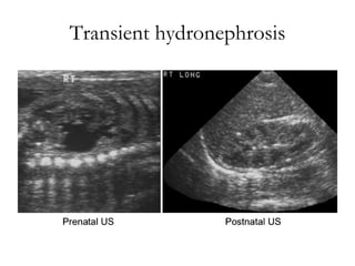
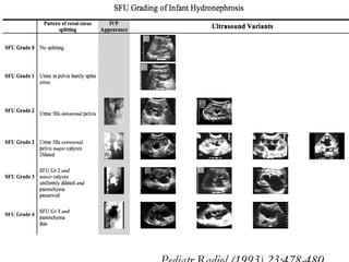
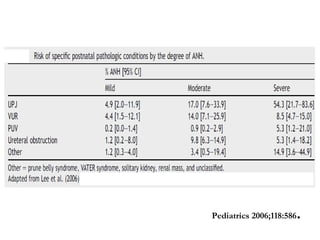
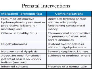
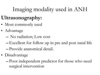
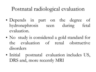
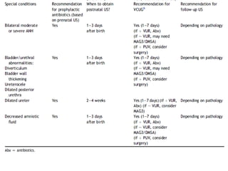
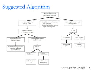
Antenataly detected  hydronephrosis
Introduction One of the most common abnormalities detected on prenatal ultrasonography Routine prenatal USG:  Detects urinary tract obstruction / reflux  prior to the development of complications
Fetal Renal Sonography Fetal kidney visualized (USG) 12-13th wk 12-40 weeks Length increases from 1.0 to 2.7 cm* APD from 0.8 to 2.6 cm Transverse diameter from 0.9 to 2.6 cm. AJR Am J Roentgenol 1991;157:545 .
 
Renal Physiology Urine formation- first seen at the 5-8th week. Urine output  5 cc/h at the 20th week  50 cc/h by the 40th week. Bladder : visualized by  10-14th week  Bladder capacity  10 cc at the 30th week to 50 cc at term Br J Obstet Gynaecol 1977;84:205 .
Renal Physiology Urine formation- first seen at the 5-8th week. Urine output  5 cc/h at the 20th week  50 cc/h by the 40th week. Bladder : visualized by  10-14th week  Bladder capacity  10 cc at the 30th week to 50 cc at term Br J Obstet Gynaecol 1977;84:205.
Amniotic fluid and indices  Volume dependent on urine production since 16 th  wk Amount  of fluid (USG)  amniotic fluid index (AFI).  Polyhydramnios  amniotic  fluid  volume > 1500 cc or AFI > 20-24 Oligohydramnios amniotic fluid  volume < 500 cc or  AFI < 5-6.
Defining ANH  Anteroposterior diameter (APD) of the renal pelvis in the transverse plane  most studied parameter for assessing ANH in utero. Threshold APD value  which separates normal from abnormal does not exist.  No consensus on the optimal APD threshold for need of postnatal follow up.
APN Guidelines 2001 e = APPD
AP Diameter  Factors affecting APD  gestational age  hydration status of the mother degree of bladder distension.  Lower cut offs  more sensitive in detecting postnatal pathology  but has higher false positive rates.
 
90 th   10 th   50 th   2 nd   3 rd
APD>4-5 mm  Threshold for abnormal . APD>15mm Severe hydronephrosis Journal of Paediatric Urology (2010) 6, 212-231
Disadvantages and limitations: APD  May not accurately reflect the degree of hydronephrosis Does not consider calyceal dilation / parenchymal changes  may reflect more severe obstruction.
Epidemiology  1-5 % of all pregnancy Male :female = 2:1 Bilateral 20-40%
Indian studies:  0.2 % among live born babies
Etiology: Antenatal Hydronephrosis
Transient hydronephrosis Incidence: ranges from 41 to 88% Etiology :  may be a narrowing of the ureteropelvic junction (UPJ) natural kinks and folds that occur early in development that resolve as the patient matures.  Transient  hydronephrosis versus clinically significant UPJ obstruction:  controversial challenges in modern pediatric urology.  Most children with a APPD  <6 mm diagnosed during the 2nd trimester or < 8 mm diagnosed during the 3rd trimester have transient hydronephrosis.
Transient hydronephrosis
Posterior Urethral Valves Sonography Dilated ureter and renal pelvis  and  dilated bladder with a dilated posterior urethra
Posterior Urethral Valves Sonography
UPJ obstruction
Multicystic dysplastic kidney
Grading of ANH Using APD thresholds ANH classified in the 2nd & 3rd trimester  Provides  prognostic information.
Estimate of distribution by severity  J obstet gynecol 2005 ;25:119
Grading of Hydronephrosis  Society for Fetal Urology (SFU)  proposed a 5-point numerical grading system  Based on the postnatal appearance of the renal pelvis, calyces, and renal parenchyma. Pediatr Radiol (1993) 23:478-480
Pediatr Radiol (1993) 23:478-480
Other sonographic parameters Poor corticomedullary differentiation  (lack of US visualization of the renal pyramids) Increased echogenicity  Presence of renal cysts  Perinephric urinoma
Post Natal Pathology likely Oligohydramnios Parenchymal thinning Calyceal dilatation Ureteral dilatation Chromosomal anomalies or multiple system malformations.
Severe bladder outlet obstruction Oligohydramnios, dilated posterior urethra (keyhole sign), ANH, thick-walled bladder, and increased renal echogenicity  Therapy Counselling &  Possible fetal intervention such as  early delivery or  vesicoamniotic shunting
Predictive value of APD-defined ANH for pathology The risk of postnatal pathology per degree of antenatal hydronephrosis  11.9% for mild 45.1% for moderate  88.3% for severe Risk of vesicoureteral reflux was similar for all degrees of antenatal hydronephrosis. Pediatrics 2006;118:586
Pediatrics 2006;118:586 .
Natural history of ANH M ajority diagnosed during the second trimester  resolve during follow-up in the third trimester. not associated with clinically significant postnatal pathology. Hydronephrosis  which is  stable/persistent or worsened during pregnancy  More variable course
Timing of diagnosis and  prognostic value First trimester :- poor outcome. Second trimester :-  overall favourable prognosis.  Resolve  or improve in the majority (approximately 80%) . Prognosis better: milder hydronephrosis. 3rd trimester :  higher rates of postnatally confirmed pathology that may require operative intervention   Ultrasound Obstet Gynecol 2005;25:483.   Ultrasound Obstet Gynecol 2001;17:191.
Natural History of ANH 30-40% of ANH persists postnatally  Of these 30-40% resolve spontaneously . The timing of resolution  Variable Occurring during the first few years of life. Earlier with milder grades of hydronephrosis Majority  of SFU Gd 1-2 resolve by 18 mths If increasing hydronephrosis occurs it does so early in life,  often during the first year.
Recommendation for prenatal evaluation of ANH Journal of Pediatric Urology (2010) 6, 212-231
Suspected Bladder Outflow Obstruction Suspected PUV (oligohydramnios, dilated bladder, bilateral hydroureteronephrosis, male gender)  Warrants monitoring throughout pregnancy. Level 3 US to exclude other organ system abnormalities. Fetal imaging every 4 weeks depending on the severity of oligohydramnios  In increasing  oligohydramnios, fetal intervention such as vesicoamniotic shunting may be offered Ideal  time period to intervention for suspected bladder outlet obstruction is  mid-second trimester.  Allows return of amniotic fluid & promotes lung development.
Predictor of renal function in fetus  By analysis of fetal urine biochemistries and electrolytes  If favorable urine electrolytes -> intervention may be offered as  option.
 
Prenatal Interventions
Imaging modality used in ANH Ultrasonography:   Most commonly used Advantage  No radiation; Low cost  Excellent for follow up in pre and post natal life Provide anatomical detail.  Disadvantage Poor independent predictor for those who need surgical intervention
Doppler Ultrasonography  Based  on the fact that obstruction causes an increase in intrarenal arterial resistance resulting in a relative reduction in diastolic flow compared to systolic flow. Use for the evaluation of renal obstructive disorders  currently controversial and not widely utilized.
Postnatal radiological evaluation Depends in part on the degree of hydronephrosis seen during fetal evaluation. No  study is considered a gold standard for the evaluation of renal obstructive disorders  Initial  postnatal  evaluation includes US, DRS and, more recently MRI
Principle of Management Follow hydronephrotic kidneys with serial radiological exams and  use decreasing differential renal function or  worsening hydronephrosis Indicators for surgery
Modalities used for Postnatal  evaluation Journal of Pediatric Urology (2010) 6, 212-231
Dynamic renal Scintigraphy adjunct test  estimate differential renal function and  the severity of obstruction. Performed after 6 weeks . Useful for serial follow up and postoperative assessment of patients with UPJ obstruction and megaureter.
DYNAMIC RENAL SCINTIGRAPHY Tc-MAG3 90% bound to plasma proteins  Principally cleared by tubular secretion. Demonstrates parenchymal and collecting system definition. Also provides functional quantification Lower radiation dose Preferred over DTPA Tc- DTPA Little plasma protein binding  Cleared exclusively by glomerular filtration. Provides  excellent visualization of the pelvicalyceal system, ureter and bladder Not good for visualization of parenchymal abnormalities. Results  suboptimal in infants with immature kidneys and a low GFR or in patients  with compromised renal function.
Who requires postnatal evaluation? Degree  of hydronephrosis useful in decision  diagnostic imaging  treatment. SFU grades I and II  resolve with time and  usually only require US surveillance.
Who requires postnatal evaluation? In moderate hydronephrosis (SFU grade III), DRS may be helpful in determining the timing and role of further studies a normal DRS followed by US,  while an indeterminate DRS may require additional DRS or MRU.  the criteria for surgical intervention variable  confounded by the surgeon’s and parents’ wishes.
Who requires postnatal evaluation? Severe hydronephrosis (SFU IV),  a functional evaluation is recommended More likely to have significant urologic pathology  require surgical intervention. SFU IV hydronephrosis should prompt either DRS or MRU. For solitary kidneys or bilateral renal involvement, MRU is superior as individual kidney function (GFR) may be assessed.
The timing of postnatal evaluation Unilateral ANH with a normal contralateral kidney : evaluation within the first week of life with a renal US  In an increased risk of UTI (e.g. girls, uncircumcised boys, moderate to severe antenatal hydronephrosis, familial VUR, etc.)  Prophylactic antibiotics until the evaluation Management discuss with the family
The timing of postnatal evaluation Bilateral  hydronephrosis and hydronephrosis in solitary kidneys or in patients with suspected bladder outlet obstruction early postnatal imaging is suggested.
Indian Pediatrics 2001; 38: 1244-1251    .  Early evaluation is necessary in neonates with solitary kidney, bilateral hydronephrosis or suspected bladder outlet obstruction.
 
 
Suggested Algorithm Curr Opn Ped 2009;207-13
Role  of VCUG Performed in conjunction with renal studies to rule out VUR Most patients with VUR and low-grade hydronephrosis can be followed without surgical intervention.
VUR as a cause of ANH No clear evidence to support or to avoid postnatal imaging for VUR. Incidence of VUR  Up to 30% in children with ANH, including those with resolved hydronephrosis. Remains unproven whether the identification and treatment of children with VUR confers any clinical benefit.
Follow-up evaluation A single normal US within the first week of life not adequate to verify absence of obstruction. A second US is recommended at 1 month of age as initial follow-up testing.
Late Worsening/ Recurrence 1-5%, with all grades of initial hydronephrosis . The severity of hydronephrosis is quite significant, being of grade III-IV Majority are symptomatic . Timing ranges from a few months to 5-6 years .
Follow Up Long term follow up recommended Length of surveillance yet to be determined. Discharge children with mild or grade I-III hydronephrosis on the 1-month US from further surveillance Others recommend serial US and UTI surveillance every 6 or 12 months or in 2-3 years.
Role of antibiotic Prophylaxis Infants with antenatal hydronephrosis are nearly 12 times more likely to have pyelonephritis-related hospitalizations in the first year of life.  Association is stronger in girls. Urology 2007 ;69:970-4 . Risk of UTI increases with grade of hydronephrosis .  Pediatr Nephrol 2007;22:1727-34 J Urol 2008;179:1524
Antibiotic Prophylaxis No statistical difference in the incidence of UTI in children with ANH on or off prophylactic antibiotics.  Pediatr Nephrol 2004;19:819. Children with prenatal hydronephrosis with persistent grade II hydronephrosis secondary to VUR, use of prophylactic antibiotics significantly reduces the risk of febrile UTIs. J Urol 2008;181:801.
Antibiotic Prophylaxis Prophylactic antibiotic in an effort to prevent infant UTIs in high-risk populations higher grades of hydronephrosis,  hydroureteronephrosis ,  VUR,  or obstructive drainage patterns.
Surgical Intervention Recommendation for surgical intervention obstructive wash-out curve ( T½ exceeds 20 min)  & significant discrepancy in split renal function (<40%).  Exception to split renal function- severe bilateral hydronephrosis or obstruction. As the absence of a normal contralateral kidney with which to compare the hydronephrotic kidney.  Renal unit that demonstrates the least function should be repaired first .
Indication for chromosomal evaluation . ANH common in fetuses with serious chromosomal anomalies Do  not recommend routine karyotyping for all cases of isolated hydronephrosis. Consider in the presence of multiple system anomalies.
 

Antenatal Hydronephrosis

  • 1.
    Antenataly detected hydronephrosis
  • 2.
    Introduction One ofthe most common abnormalities detected on prenatal ultrasonography Routine prenatal USG: Detects urinary tract obstruction / reflux prior to the development of complications
  • 3.
    Fetal Renal SonographyFetal kidney visualized (USG) 12-13th wk 12-40 weeks Length increases from 1.0 to 2.7 cm* APD from 0.8 to 2.6 cm Transverse diameter from 0.9 to 2.6 cm. AJR Am J Roentgenol 1991;157:545 .
  • 4.
  • 5.
    Renal Physiology Urineformation- first seen at the 5-8th week. Urine output 5 cc/h at the 20th week 50 cc/h by the 40th week. Bladder : visualized by 10-14th week Bladder capacity 10 cc at the 30th week to 50 cc at term Br J Obstet Gynaecol 1977;84:205 .
  • 6.
    Renal Physiology Urineformation- first seen at the 5-8th week. Urine output 5 cc/h at the 20th week 50 cc/h by the 40th week. Bladder : visualized by 10-14th week Bladder capacity 10 cc at the 30th week to 50 cc at term Br J Obstet Gynaecol 1977;84:205.
  • 7.
    Amniotic fluid andindices Volume dependent on urine production since 16 th wk Amount of fluid (USG) amniotic fluid index (AFI). Polyhydramnios amniotic fluid volume > 1500 cc or AFI > 20-24 Oligohydramnios amniotic fluid volume < 500 cc or AFI < 5-6.
  • 8.
    Defining ANH Anteroposterior diameter (APD) of the renal pelvis in the transverse plane most studied parameter for assessing ANH in utero. Threshold APD value which separates normal from abnormal does not exist. No consensus on the optimal APD threshold for need of postnatal follow up.
  • 9.
  • 10.
    AP Diameter Factors affecting APD gestational age hydration status of the mother degree of bladder distension. Lower cut offs more sensitive in detecting postnatal pathology but has higher false positive rates.
  • 11.
  • 12.
    90 th 10 th 50 th 2 nd 3 rd
  • 13.
    APD>4-5 mm Threshold for abnormal . APD>15mm Severe hydronephrosis Journal of Paediatric Urology (2010) 6, 212-231
  • 14.
    Disadvantages and limitations:APD May not accurately reflect the degree of hydronephrosis Does not consider calyceal dilation / parenchymal changes may reflect more severe obstruction.
  • 15.
    Epidemiology 1-5% of all pregnancy Male :female = 2:1 Bilateral 20-40%
  • 16.
    Indian studies: 0.2 % among live born babies
  • 17.
  • 18.
    Transient hydronephrosis Incidence:ranges from 41 to 88% Etiology : may be a narrowing of the ureteropelvic junction (UPJ) natural kinks and folds that occur early in development that resolve as the patient matures. Transient hydronephrosis versus clinically significant UPJ obstruction: controversial challenges in modern pediatric urology. Most children with a APPD <6 mm diagnosed during the 2nd trimester or < 8 mm diagnosed during the 3rd trimester have transient hydronephrosis.
  • 19.
  • 20.
    Posterior Urethral ValvesSonography Dilated ureter and renal pelvis and dilated bladder with a dilated posterior urethra
  • 21.
  • 22.
  • 23.
  • 24.
    Grading of ANHUsing APD thresholds ANH classified in the 2nd & 3rd trimester Provides prognostic information.
  • 25.
    Estimate of distributionby severity J obstet gynecol 2005 ;25:119
  • 26.
    Grading of Hydronephrosis Society for Fetal Urology (SFU) proposed a 5-point numerical grading system Based on the postnatal appearance of the renal pelvis, calyces, and renal parenchyma. Pediatr Radiol (1993) 23:478-480
  • 27.
  • 28.
    Other sonographic parametersPoor corticomedullary differentiation (lack of US visualization of the renal pyramids) Increased echogenicity Presence of renal cysts Perinephric urinoma
  • 29.
    Post Natal Pathologylikely Oligohydramnios Parenchymal thinning Calyceal dilatation Ureteral dilatation Chromosomal anomalies or multiple system malformations.
  • 30.
    Severe bladder outletobstruction Oligohydramnios, dilated posterior urethra (keyhole sign), ANH, thick-walled bladder, and increased renal echogenicity Therapy Counselling & Possible fetal intervention such as early delivery or vesicoamniotic shunting
  • 31.
    Predictive value ofAPD-defined ANH for pathology The risk of postnatal pathology per degree of antenatal hydronephrosis 11.9% for mild 45.1% for moderate 88.3% for severe Risk of vesicoureteral reflux was similar for all degrees of antenatal hydronephrosis. Pediatrics 2006;118:586
  • 32.
  • 33.
    Natural history ofANH M ajority diagnosed during the second trimester resolve during follow-up in the third trimester. not associated with clinically significant postnatal pathology. Hydronephrosis which is stable/persistent or worsened during pregnancy More variable course
  • 34.
    Timing of diagnosisand prognostic value First trimester :- poor outcome. Second trimester :- overall favourable prognosis. Resolve or improve in the majority (approximately 80%) . Prognosis better: milder hydronephrosis. 3rd trimester : higher rates of postnatally confirmed pathology that may require operative intervention Ultrasound Obstet Gynecol 2005;25:483. Ultrasound Obstet Gynecol 2001;17:191.
  • 35.
    Natural History ofANH 30-40% of ANH persists postnatally Of these 30-40% resolve spontaneously . The timing of resolution Variable Occurring during the first few years of life. Earlier with milder grades of hydronephrosis Majority of SFU Gd 1-2 resolve by 18 mths If increasing hydronephrosis occurs it does so early in life, often during the first year.
  • 36.
    Recommendation for prenatalevaluation of ANH Journal of Pediatric Urology (2010) 6, 212-231
  • 37.
    Suspected Bladder OutflowObstruction Suspected PUV (oligohydramnios, dilated bladder, bilateral hydroureteronephrosis, male gender) Warrants monitoring throughout pregnancy. Level 3 US to exclude other organ system abnormalities. Fetal imaging every 4 weeks depending on the severity of oligohydramnios In increasing oligohydramnios, fetal intervention such as vesicoamniotic shunting may be offered Ideal time period to intervention for suspected bladder outlet obstruction is mid-second trimester. Allows return of amniotic fluid & promotes lung development.
  • 38.
    Predictor of renalfunction in fetus By analysis of fetal urine biochemistries and electrolytes If favorable urine electrolytes -> intervention may be offered as option.
  • 39.
  • 40.
  • 41.
    Imaging modality usedin ANH Ultrasonography: Most commonly used Advantage No radiation; Low cost Excellent for follow up in pre and post natal life Provide anatomical detail. Disadvantage Poor independent predictor for those who need surgical intervention
  • 42.
    Doppler Ultrasonography Based on the fact that obstruction causes an increase in intrarenal arterial resistance resulting in a relative reduction in diastolic flow compared to systolic flow. Use for the evaluation of renal obstructive disorders currently controversial and not widely utilized.
  • 43.
    Postnatal radiological evaluationDepends in part on the degree of hydronephrosis seen during fetal evaluation. No study is considered a gold standard for the evaluation of renal obstructive disorders Initial postnatal evaluation includes US, DRS and, more recently MRI
  • 44.
    Principle of ManagementFollow hydronephrotic kidneys with serial radiological exams and use decreasing differential renal function or worsening hydronephrosis Indicators for surgery
  • 45.
    Modalities used forPostnatal evaluation Journal of Pediatric Urology (2010) 6, 212-231
  • 46.
    Dynamic renal Scintigraphyadjunct test estimate differential renal function and the severity of obstruction. Performed after 6 weeks . Useful for serial follow up and postoperative assessment of patients with UPJ obstruction and megaureter.
  • 47.
    DYNAMIC RENAL SCINTIGRAPHYTc-MAG3 90% bound to plasma proteins Principally cleared by tubular secretion. Demonstrates parenchymal and collecting system definition. Also provides functional quantification Lower radiation dose Preferred over DTPA Tc- DTPA Little plasma protein binding Cleared exclusively by glomerular filtration. Provides excellent visualization of the pelvicalyceal system, ureter and bladder Not good for visualization of parenchymal abnormalities. Results suboptimal in infants with immature kidneys and a low GFR or in patients with compromised renal function.
  • 48.
    Who requires postnatalevaluation? Degree of hydronephrosis useful in decision diagnostic imaging treatment. SFU grades I and II resolve with time and usually only require US surveillance.
  • 49.
    Who requires postnatalevaluation? In moderate hydronephrosis (SFU grade III), DRS may be helpful in determining the timing and role of further studies a normal DRS followed by US, while an indeterminate DRS may require additional DRS or MRU. the criteria for surgical intervention variable confounded by the surgeon’s and parents’ wishes.
  • 50.
    Who requires postnatalevaluation? Severe hydronephrosis (SFU IV), a functional evaluation is recommended More likely to have significant urologic pathology require surgical intervention. SFU IV hydronephrosis should prompt either DRS or MRU. For solitary kidneys or bilateral renal involvement, MRU is superior as individual kidney function (GFR) may be assessed.
  • 51.
    The timing ofpostnatal evaluation Unilateral ANH with a normal contralateral kidney : evaluation within the first week of life with a renal US In an increased risk of UTI (e.g. girls, uncircumcised boys, moderate to severe antenatal hydronephrosis, familial VUR, etc.) Prophylactic antibiotics until the evaluation Management discuss with the family
  • 52.
    The timing ofpostnatal evaluation Bilateral hydronephrosis and hydronephrosis in solitary kidneys or in patients with suspected bladder outlet obstruction early postnatal imaging is suggested.
  • 53.
    Indian Pediatrics 2001;38: 1244-1251   . Early evaluation is necessary in neonates with solitary kidney, bilateral hydronephrosis or suspected bladder outlet obstruction.
  • 54.
  • 55.
  • 56.
    Suggested Algorithm CurrOpn Ped 2009;207-13
  • 57.
    Role ofVCUG Performed in conjunction with renal studies to rule out VUR Most patients with VUR and low-grade hydronephrosis can be followed without surgical intervention.
  • 58.
    VUR as acause of ANH No clear evidence to support or to avoid postnatal imaging for VUR. Incidence of VUR Up to 30% in children with ANH, including those with resolved hydronephrosis. Remains unproven whether the identification and treatment of children with VUR confers any clinical benefit.
  • 59.
    Follow-up evaluation Asingle normal US within the first week of life not adequate to verify absence of obstruction. A second US is recommended at 1 month of age as initial follow-up testing.
  • 60.
    Late Worsening/ Recurrence1-5%, with all grades of initial hydronephrosis . The severity of hydronephrosis is quite significant, being of grade III-IV Majority are symptomatic . Timing ranges from a few months to 5-6 years .
  • 61.
    Follow Up Longterm follow up recommended Length of surveillance yet to be determined. Discharge children with mild or grade I-III hydronephrosis on the 1-month US from further surveillance Others recommend serial US and UTI surveillance every 6 or 12 months or in 2-3 years.
  • 62.
    Role of antibioticProphylaxis Infants with antenatal hydronephrosis are nearly 12 times more likely to have pyelonephritis-related hospitalizations in the first year of life. Association is stronger in girls. Urology 2007 ;69:970-4 . Risk of UTI increases with grade of hydronephrosis . Pediatr Nephrol 2007;22:1727-34 J Urol 2008;179:1524
  • 63.
    Antibiotic Prophylaxis Nostatistical difference in the incidence of UTI in children with ANH on or off prophylactic antibiotics. Pediatr Nephrol 2004;19:819. Children with prenatal hydronephrosis with persistent grade II hydronephrosis secondary to VUR, use of prophylactic antibiotics significantly reduces the risk of febrile UTIs. J Urol 2008;181:801.
  • 64.
    Antibiotic Prophylaxis Prophylacticantibiotic in an effort to prevent infant UTIs in high-risk populations higher grades of hydronephrosis, hydroureteronephrosis , VUR, or obstructive drainage patterns.
  • 65.
    Surgical Intervention Recommendationfor surgical intervention obstructive wash-out curve ( T½ exceeds 20 min) & significant discrepancy in split renal function (<40%). Exception to split renal function- severe bilateral hydronephrosis or obstruction. As the absence of a normal contralateral kidney with which to compare the hydronephrotic kidney. Renal unit that demonstrates the least function should be repaired first .
  • 66.
    Indication for chromosomalevaluation . ANH common in fetuses with serious chromosomal anomalies Do not recommend routine karyotyping for all cases of isolated hydronephrosis. Consider in the presence of multiple system anomalies.
  • 67.

Editor's Notes

  • #8 The AFI is the score obtained by adding centimeters of depth of four pockets of fluid.
  • #40 How to explain the table