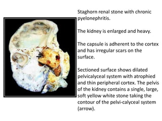
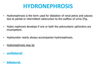
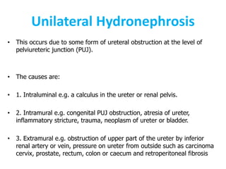
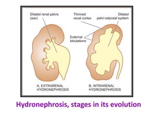
The KIDNEY
OBSTRUCTIVE UROPATHY
DR. ROOPAM JAIN
PROFESSOR & HEAD, PATHOLOGY
OBSTRUCTIVE
UROPATHY
Causes of obstructive uropathy
Causes of
obstructive
uropathy
NEPHROLITHIASIS
• Nephrolithiasis or urolithiasis is formation of urinary calculi at any level
of the urinary tract.
• Urinary calculi are worldwide in distribution but are particularly common
in some geographic locations such as in parts of the United States,
South Africa, India and South-East Asia.
• 2% of the population experiences renal stone disease
• male-female ratio of 2:1.
• peak incidence is observed in 2nd to 3rd decades of life.
• Renal calculi are characterised clinically by colicky pain (renal colic) as
they pass down along the ureter and manifest by haematuria.
NEPHROLITHIASIS
Salient features of urinary calculi
Staghorn renal stone with chronic
pyelonephritis.
The kidney is enlarged and heavy.
The capsule is adherent to the cortex
and has irregular scars on the
surface.
Sectioned surface shows dilated
pelvicalyceal system with atrophied
and thin peripheral cortex. The pelvis
of the kidney contains a single, large,
soft yellow white stone taking the
contour of the pelvi-calyceal system
(arrow).
HYDRONEPHROSIS
• Hydronephrosis is the term used for dilatation of renal pelvis and calyces
due to partial or intermittent obstruction to the outflow of urine (Fig.
• Hydro nephrosis develops if one or both the pelviureteric sphincters are
incompetent.
• Hydroureter nearly always accompanies hydronephrosis.
• Hydronephrosis may be
• unilateral
• bilateral.
Unilateral Hydronephrosis
• This occurs due to some form of ureteral obstruction at the level of
pelviureteric junction (PUJ).
• The causes are:
• 1. Intraluminal e.g. a calculus in the ureter or renal pelvis.
• 2. Intramural e.g. congenital PUJ obstruction, atresia of ureter,
inflammatory stricture, trauma, neoplasm of ureter or bladder.
• 3. Extramural e.g. obstruction of upper part of the ureter by inferior
renal artery or vein, pressure on ureter from outside such as carcinoma
cervix, prostate, rectum, colon or caecum and retroperitoneal fibrosis
Bilateral Hydronephrosis
• 1. Congenital
• e.g. atresia of the urethral meatus,
• congenital posterior urethral valve.
• 2. Acquired
• e.g. bladder tumour involving both ureteric orifices,
• prostatic enlargement,
• prostatic carcinoma
• prostatitis,
• bladder neck stenosis,
• inflammatory or traumatic urethral stricture
• phimosis.
Hydronephrosis, stages in its evolution
Hydronephrosis with
nephrolithiasis.
The kidney is enlarged and heavy.
On cut section, the renal pelvis
and calyces are dilated and cystic
and contain a large stone in the
pelvis of the kidney (arrow).
The cystic change is seen to
extend into renal parenchyma,
compressing the cortex as a thin
rim at the periphery. Unlike
polycystic kidney, however, these
cysts are communicating with the
pelvi-calyceal system.
The Kidney: OBSTRUCTIVE UROPATHY

The Kidney: OBSTRUCTIVE UROPATHY

  • 1.
    The KIDNEY OBSTRUCTIVE UROPATHY DR.ROOPAM JAIN PROFESSOR & HEAD, PATHOLOGY
  • 2.
  • 3.
  • 4.
  • 5.
    NEPHROLITHIASIS • Nephrolithiasis orurolithiasis is formation of urinary calculi at any level of the urinary tract. • Urinary calculi are worldwide in distribution but are particularly common in some geographic locations such as in parts of the United States, South Africa, India and South-East Asia. • 2% of the population experiences renal stone disease • male-female ratio of 2:1. • peak incidence is observed in 2nd to 3rd decades of life. • Renal calculi are characterised clinically by colicky pain (renal colic) as they pass down along the ureter and manifest by haematuria.
  • 6.
  • 8.
    Staghorn renal stonewith chronic pyelonephritis. The kidney is enlarged and heavy. The capsule is adherent to the cortex and has irregular scars on the surface. Sectioned surface shows dilated pelvicalyceal system with atrophied and thin peripheral cortex. The pelvis of the kidney contains a single, large, soft yellow white stone taking the contour of the pelvi-calyceal system (arrow).
  • 9.
    HYDRONEPHROSIS • Hydronephrosis isthe term used for dilatation of renal pelvis and calyces due to partial or intermittent obstruction to the outflow of urine (Fig. • Hydro nephrosis develops if one or both the pelviureteric sphincters are incompetent. • Hydroureter nearly always accompanies hydronephrosis. • Hydronephrosis may be • unilateral • bilateral.
  • 10.
    Unilateral Hydronephrosis • Thisoccurs due to some form of ureteral obstruction at the level of pelviureteric junction (PUJ). • The causes are: • 1. Intraluminal e.g. a calculus in the ureter or renal pelvis. • 2. Intramural e.g. congenital PUJ obstruction, atresia of ureter, inflammatory stricture, trauma, neoplasm of ureter or bladder. • 3. Extramural e.g. obstruction of upper part of the ureter by inferior renal artery or vein, pressure on ureter from outside such as carcinoma cervix, prostate, rectum, colon or caecum and retroperitoneal fibrosis
  • 11.
    Bilateral Hydronephrosis • 1.Congenital • e.g. atresia of the urethral meatus, • congenital posterior urethral valve. • 2. Acquired • e.g. bladder tumour involving both ureteric orifices, • prostatic enlargement, • prostatic carcinoma • prostatitis, • bladder neck stenosis, • inflammatory or traumatic urethral stricture • phimosis.
  • 12.
  • 13.
    Hydronephrosis with nephrolithiasis. The kidneyis enlarged and heavy. On cut section, the renal pelvis and calyces are dilated and cystic and contain a large stone in the pelvis of the kidney (arrow). The cystic change is seen to extend into renal parenchyma, compressing the cortex as a thin rim at the periphery. Unlike polycystic kidney, however, these cysts are communicating with the pelvi-calyceal system.