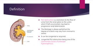
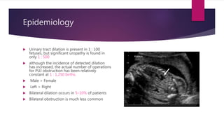
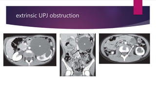
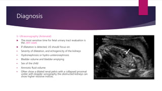
PUJ Obstruction
HASAN MH MATLOOB
RESIDENT DOCTOR IN PAEDIATRIC SURGERY
2ND YEAR ARABIC BOARD
Definition
 PUJ obstruction is a restriction to the flow of
urine from the renal pelvis to the ureter,
which if left uncorrected, may lead to
progressive renal deterioration.
 The blockage is always partial but the
degree of a block may vary from minimal to
severe.
 It can be congenital or acquired.
 congenital PUJ obstruction being one of the
commonest causes of antenatal
hydronephrosis.
Epidemiology
 Urinary tract dilation is present in 1 : 100
fetuses, but significant uropathy is found in
only 1 : 500
 although the incidence of detected dilation
has increased, the actual number of operations
for PUJ obstruction has been relatively
constant at 1 : 1,250 births.
 Male > Female
 Left > Right
 Bilateral dilation occurs in 5–10% of patients
 Bilateral obstruction is much less common
Etiology
 Failure to recanalize adequately is thought to be
the cause of most intrinsic PUJ obstructions.
 Other causes of intrinsic PUJ obstruction include
ureteral valves, polyps, and leiomyomas.
 In 20–30% of patients, the ureter is draped over a
lower-pole vessel, producing an extrinsic PUJ
obstruction.
extrinsic UPJ obstruction
Clinical Presentation
 Most renal dilation and obstruction are detected
prenatally.
 Less frequently, it is detected because of an
abdominal mass, urinary tract infection (UTI), or
associated with other congenital anomalies
(VACTERL syndrome).
 Hematuria after minor trauma or vigorous exercise
may be a presenting feature, most likely secondary
to rupture of mucosal vessels in the dilated
collecting system
 Can be found in conjunction with high-grade
vesicoureteral reflux (VUR).
Diagnosis
1- Ultrasonography (Antenatal)
 The most sensitive time for fetal urinary tract evaluation is
the 28th week
 If dilatation is detected, US should focus on:
 Severity of dilatation, and echogenicity of the kidneys
 Hydronephrosis or hydro-ureteronephrosis
 Bladder volume and bladder emptying
 Sex of the child
 Amniotic fluid volume
 Often show a dilated renal pelvis with a collapsed proximal
ureter with doppler sonography the obstructed kidneys can
show higher resistive indices.
Diagnosis
 Pelvic AP diameter
 PAPD > 7 mm at 18-23 weeks gestation
 PAPD > 10 mm during last trimester
 PAPD > 12 mm at birth
should be considered as abnormal
When the antenatal diagnosis of PUJ obstruction is made, the initial postpartum evaluation
should be performed at 10 to 14 days of life.
In older children with abdominal pain and suspected PUJ obstruction, an ultrasound
examination should be performed during the acute painful episode (Dietl's crisis) to
demonstrate hydronephrosis
Diagnosis
2- Voiding Cystourethrogram (VCUG)
 In newborns with identified UUT dilatation, the primary or important associated factors
that must be detected include:
 vesicoureteral reflux (15-25% of affected children)
 urethral valves
 Ureteroceles
 Diverticula
 neurogenic bladder
 Conventional VCUG is the method of choice for primary diagnostic procedures
Diagnosis
3- Diuretic Renography:
In general, diuretic renography can be performed after six weeks of life because
immediate surgical intervention is rarely required.
It is performed to differentiate between obstructive vs nonobstructive hydronephrosis.
 PUJ "obstruction" will demonstrate excretion (downward slope on renogram) after
administration of diuretic from the collecting system.
 Mechanical obstructive hydronephrosis will demonstrate no downward slope on renogram,
with retained tracer in collecting system.
 Good drainage on renography is surely a define sign of the absence of obstruction
 The two most common radiolabelled pharmaceutical agents used are:
Tc99m-MAG3 and Tc99m-DTPA (diethylenetriaminepentacetate)
Diagnosis
4- CT
May show evidence of hydronephrosis +/- calyectasis with collapsed ureters. Useful
for assessing crossing vessels at the PUJ especially when surgical intervention is
planned.
5- FLUOROSCOPY - IVU
Dilatation of the renal pelvis and caliceal system with a stenotic ureteropelvic
segment.
Intravenous urography is often not used in children, since better alternatives (e.g. MR
urography) are available
Diagnosis
6- MR urography
 MRU It is used in assessing PUJ obstruction. The study also provides details of
renal vasculature, renal pelvis anatomy, location of crossing vessels, renal cortical
scarring, and ureteral fetal folds in the proximal ureter.
Treatment
MEDICAL THERAPY
 Currently, no available medical therapy is capable of reversing UPJ obstruction in
either adults or children. In children initially conservative treatment with
monitoring. Intervention is indicated in the event of significantly impaired renal
drainage or poor renal growth.
Treatment
SUGRICAL THERAPY
 The accepted criteria for intervention in infants and children including:
1- clearance half-time (T 1/2) greater than 20 minutes
2- Differential function less than 40%.
3- Ongoing parenchymal thinning with or without contralateral compensatory
hypertrophy.
4- Associated symptoms like: pain, hypertension, hematuria, secondary renal calculi,
and recurrent urinary tract infections.
Treatment
OPEN PYELOPLASTY
 The obstructed segment is completely resected,
with re-anastomosis of the renal pelvis and
ureter in a dependent funneled fashion.
 The success rate exceeds 95%.
 Gold Standard
 Anderson-Hynes dismembered pyeloplasty is the
most common.
Treatment
ENDOPYELOTOMY
 Incision of the area with a balloon catheter to
help ensure a complete incision followed by
prolonged ureteral stenting, for a period of 4-8
weeks. Success rates are 80-90%.
 Anterograde or retrograde
 Slightly less effective in children
Treatment
FLAP PYELOPLASTY
 Better in adults
 Useful when a long-strictured segment of
diseased ureter is encountered.
 The proximal ureter is re-created with
redundant renal pelvis that is tubularized.
Treatment
LAPAROSCOPIC PYELOPLASTY
 Laparoscopic pyeloplasty offers a minimally invasive
 Used in patients with either primary or secondary
UPJ obstruction
 Offers the advantages of decreased morbidity,
shorter hospital stay, and quicker recovery.
Video
Follow up
TIPS
 Prophylactic antibiotic therapy should be given postoperatively.
 Remove the endopyelotomy stent after 4-8 weeks.
 Follow up with renal ultrasonography 1-3 months after surgery.
 Follow up with IVP or nuclear medicine renal scan 3-6 months after surgery.
 Serial renal imaging is recommended for the first year after surgery.
THANK YOU FOR
LISTENING

PUJ obstruction.pptx

  • 1.
    PUJ Obstruction HASAN MHMATLOOB RESIDENT DOCTOR IN PAEDIATRIC SURGERY 2ND YEAR ARABIC BOARD
  • 2.
    Definition  PUJ obstructionis a restriction to the flow of urine from the renal pelvis to the ureter, which if left uncorrected, may lead to progressive renal deterioration.  The blockage is always partial but the degree of a block may vary from minimal to severe.  It can be congenital or acquired.  congenital PUJ obstruction being one of the commonest causes of antenatal hydronephrosis.
  • 3.
    Epidemiology  Urinary tractdilation is present in 1 : 100 fetuses, but significant uropathy is found in only 1 : 500  although the incidence of detected dilation has increased, the actual number of operations for PUJ obstruction has been relatively constant at 1 : 1,250 births.  Male > Female  Left > Right  Bilateral dilation occurs in 5–10% of patients  Bilateral obstruction is much less common
  • 4.
    Etiology  Failure torecanalize adequately is thought to be the cause of most intrinsic PUJ obstructions.  Other causes of intrinsic PUJ obstruction include ureteral valves, polyps, and leiomyomas.  In 20–30% of patients, the ureter is draped over a lower-pole vessel, producing an extrinsic PUJ obstruction.
  • 5.
  • 6.
    Clinical Presentation  Mostrenal dilation and obstruction are detected prenatally.  Less frequently, it is detected because of an abdominal mass, urinary tract infection (UTI), or associated with other congenital anomalies (VACTERL syndrome).  Hematuria after minor trauma or vigorous exercise may be a presenting feature, most likely secondary to rupture of mucosal vessels in the dilated collecting system  Can be found in conjunction with high-grade vesicoureteral reflux (VUR).
  • 7.
    Diagnosis 1- Ultrasonography (Antenatal) The most sensitive time for fetal urinary tract evaluation is the 28th week  If dilatation is detected, US should focus on:  Severity of dilatation, and echogenicity of the kidneys  Hydronephrosis or hydro-ureteronephrosis  Bladder volume and bladder emptying  Sex of the child  Amniotic fluid volume  Often show a dilated renal pelvis with a collapsed proximal ureter with doppler sonography the obstructed kidneys can show higher resistive indices.
  • 8.
    Diagnosis  Pelvic APdiameter  PAPD > 7 mm at 18-23 weeks gestation  PAPD > 10 mm during last trimester  PAPD > 12 mm at birth should be considered as abnormal When the antenatal diagnosis of PUJ obstruction is made, the initial postpartum evaluation should be performed at 10 to 14 days of life. In older children with abdominal pain and suspected PUJ obstruction, an ultrasound examination should be performed during the acute painful episode (Dietl's crisis) to demonstrate hydronephrosis
  • 9.
    Diagnosis 2- Voiding Cystourethrogram(VCUG)  In newborns with identified UUT dilatation, the primary or important associated factors that must be detected include:  vesicoureteral reflux (15-25% of affected children)  urethral valves  Ureteroceles  Diverticula  neurogenic bladder  Conventional VCUG is the method of choice for primary diagnostic procedures
  • 10.
    Diagnosis 3- Diuretic Renography: Ingeneral, diuretic renography can be performed after six weeks of life because immediate surgical intervention is rarely required. It is performed to differentiate between obstructive vs nonobstructive hydronephrosis.  PUJ "obstruction" will demonstrate excretion (downward slope on renogram) after administration of diuretic from the collecting system.  Mechanical obstructive hydronephrosis will demonstrate no downward slope on renogram, with retained tracer in collecting system.  Good drainage on renography is surely a define sign of the absence of obstruction  The two most common radiolabelled pharmaceutical agents used are: Tc99m-MAG3 and Tc99m-DTPA (diethylenetriaminepentacetate)
  • 11.
    Diagnosis 4- CT May showevidence of hydronephrosis +/- calyectasis with collapsed ureters. Useful for assessing crossing vessels at the PUJ especially when surgical intervention is planned. 5- FLUOROSCOPY - IVU Dilatation of the renal pelvis and caliceal system with a stenotic ureteropelvic segment. Intravenous urography is often not used in children, since better alternatives (e.g. MR urography) are available
  • 12.
    Diagnosis 6- MR urography MRU It is used in assessing PUJ obstruction. The study also provides details of renal vasculature, renal pelvis anatomy, location of crossing vessels, renal cortical scarring, and ureteral fetal folds in the proximal ureter.
  • 13.
    Treatment MEDICAL THERAPY  Currently,no available medical therapy is capable of reversing UPJ obstruction in either adults or children. In children initially conservative treatment with monitoring. Intervention is indicated in the event of significantly impaired renal drainage or poor renal growth.
  • 14.
    Treatment SUGRICAL THERAPY  Theaccepted criteria for intervention in infants and children including: 1- clearance half-time (T 1/2) greater than 20 minutes 2- Differential function less than 40%. 3- Ongoing parenchymal thinning with or without contralateral compensatory hypertrophy. 4- Associated symptoms like: pain, hypertension, hematuria, secondary renal calculi, and recurrent urinary tract infections.
  • 15.
    Treatment OPEN PYELOPLASTY  Theobstructed segment is completely resected, with re-anastomosis of the renal pelvis and ureter in a dependent funneled fashion.  The success rate exceeds 95%.  Gold Standard  Anderson-Hynes dismembered pyeloplasty is the most common.
  • 16.
    Treatment ENDOPYELOTOMY  Incision ofthe area with a balloon catheter to help ensure a complete incision followed by prolonged ureteral stenting, for a period of 4-8 weeks. Success rates are 80-90%.  Anterograde or retrograde  Slightly less effective in children
  • 17.
    Treatment FLAP PYELOPLASTY  Betterin adults  Useful when a long-strictured segment of diseased ureter is encountered.  The proximal ureter is re-created with redundant renal pelvis that is tubularized.
  • 18.
    Treatment LAPAROSCOPIC PYELOPLASTY  Laparoscopicpyeloplasty offers a minimally invasive  Used in patients with either primary or secondary UPJ obstruction  Offers the advantages of decreased morbidity, shorter hospital stay, and quicker recovery. Video
  • 19.
    Follow up TIPS  Prophylacticantibiotic therapy should be given postoperatively.  Remove the endopyelotomy stent after 4-8 weeks.  Follow up with renal ultrasonography 1-3 months after surgery.  Follow up with IVP or nuclear medicine renal scan 3-6 months after surgery.  Serial renal imaging is recommended for the first year after surgery.
  • 20.