Downloaded 2,741 times
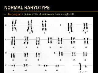


This document discusses chromosomal abnormalities, including both numerical and structural abnormalities. It provides examples of various chromosomal abnormalities such as trisomy 21 (Down syndrome), trisomy 18, trisomy 13 (Patau syndrome), Turner syndrome, and Klinefelter syndrome. It also discusses methods used in cytogenetic analysis such as karyotyping, G-banding, fluorescent in situ hybridization (FISH), and spectral karyotyping. Overall, the document provides an overview of common chromosomal abnormalities and the techniques used to identify them.