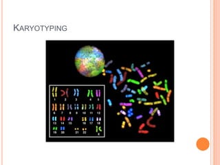
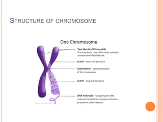
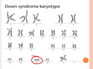
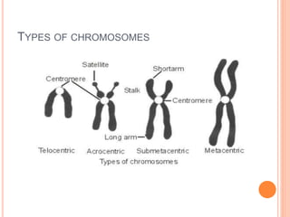
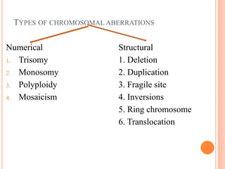
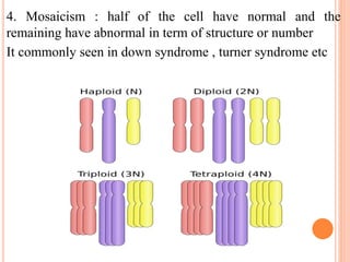
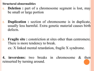
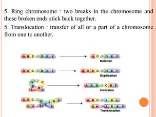
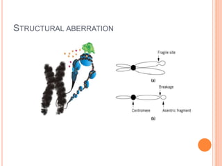
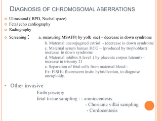
CHROMOSOMAL ABERRATION
Prepared by:
Vijayakumar . M . B
Lecturer, charuast university
 MEANING OF ABERAATION
 Any departure or deviation from normal, usually
expected.
DEFINITION : any aberration in the shape, size or
structure of a chromosome is called chromosomal
aberration.
It reflects an atypical number or a structure in one or
more chromosomes.
BASIC TERMS TO BE REMEMBERED
 Karyotyping : refers to a full set of chromosome from an
individual, or a photographic arrangement of a set of
chromosomes of an individual.
 Chromatid : either of the two daughter strands of
replicated chromosome that are joined by centromere and
separate during cell division.
 Constituent of chromosome: DNA, RNA , HISTONES (5
kinds)
 No of chromosomes: 22 pairs of autosomes& a pair of sex
chromosomes.
 Diploid: 46. haploid : 23
KARYOTYPING
STRUCTURE OF CHROMOSOME
TYPES OF CHROMOSOMES
TYPES OF CHROMOSOMAL ABERRATIONS
Numerical Structural
1. Trisomy 1. Deletion
2. Monosomy 2. Duplication
3. Polyploidy 3. Fragile site
4. Mosaicism 4. Inversions
5. Ring chromosome
6. Translocation
Numerical aberrations :
ANEUPLOIDY : having too many or too few chromosomes.
1. TRISOMY : 3 copies of particular chromosome.
Common trisomy is down syndrome, trisomy 21.
Reason : fails to go to opposite poles of the dividing cells.
2. MONOSOMY: missing of a copy of chromosome.
Ex: 45x, and monosomy are always lethal, the only
compatible condition is turner syndrome( only one x chromosome)
3. POLYPLOIDY : more than normal diploid number of chromosome.
Ex: triploidy – 6pxxx
Tertroploidy – 92XXXX
Reason : Polyspermy, failed meiosis( diploid cell) , self fertilize (
tetraploid zygote)
4. Mosaicism : half of the cell have normal and the
remaining have abnormal in term of structure or number
It commonly seen in down syndrome , turner syndrome etc
Structural abnormalities
1. Deletion : part of a chromosome segment is lost, may
be small or large portion
2. Duplication : section of chromosome is in duplicate,
usually less harmful. Extra genetic material causes birth
defects.
3. Fragile site : constriction at sites other than centromere.
There is more tendency to break.
ex: X linked mental retardation, fragile X syndrome.
4. inversions: two breaks in chromosome & then
reinserted by turning around.
5. Ring chromosome : two breaks in the chromosome and
these broken ends stick back together.
5. Translocation : transfer of all or a part of a chromosome
from one to another.
STRUCTURAL ABERRATION
DIAGNOSIS OF CHROMOSOMAL ABERRATIONS
 Ultrasound ( BPD, Nuchal space)
 Fetal echo cardiography
 Radiography
 Screening : a. measuring MSAFP( by yolk sac) – decrease in down syndrome
b. Maternal unconjugated estriol - (decrease in down syndrome
c. Maternal serum human HCG – (produced by trophoblast)
increase in down syndrome
d. Maternal inhibin A level ( by placenta corpus luteum) –
increase in trisomy 21
e. Separation of fetal cells from maternal blood :
Ex: FISH:- fluorescent insitu hybridization, to diagnose
uneuploidy.
• Other invasive
Embryoscopy
fetal tissue sampling : - amniocentesis
- Chorianic villai sampling
- Cordocentesis
THANKS

Chromosomal aberration

  • 1.
    CHROMOSOMAL ABERRATION Prepared by: Vijayakumar. M . B Lecturer, charuast university
  • 2.
     MEANING OFABERAATION  Any departure or deviation from normal, usually expected. DEFINITION : any aberration in the shape, size or structure of a chromosome is called chromosomal aberration. It reflects an atypical number or a structure in one or more chromosomes.
  • 3.
    BASIC TERMS TOBE REMEMBERED  Karyotyping : refers to a full set of chromosome from an individual, or a photographic arrangement of a set of chromosomes of an individual.  Chromatid : either of the two daughter strands of replicated chromosome that are joined by centromere and separate during cell division.  Constituent of chromosome: DNA, RNA , HISTONES (5 kinds)  No of chromosomes: 22 pairs of autosomes& a pair of sex chromosomes.  Diploid: 46. haploid : 23
  • 4.
  • 5.
  • 7.
  • 8.
    TYPES OF CHROMOSOMALABERRATIONS Numerical Structural 1. Trisomy 1. Deletion 2. Monosomy 2. Duplication 3. Polyploidy 3. Fragile site 4. Mosaicism 4. Inversions 5. Ring chromosome 6. Translocation
  • 9.
    Numerical aberrations : ANEUPLOIDY: having too many or too few chromosomes. 1. TRISOMY : 3 copies of particular chromosome. Common trisomy is down syndrome, trisomy 21. Reason : fails to go to opposite poles of the dividing cells. 2. MONOSOMY: missing of a copy of chromosome. Ex: 45x, and monosomy are always lethal, the only compatible condition is turner syndrome( only one x chromosome) 3. POLYPLOIDY : more than normal diploid number of chromosome. Ex: triploidy – 6pxxx Tertroploidy – 92XXXX Reason : Polyspermy, failed meiosis( diploid cell) , self fertilize ( tetraploid zygote)
  • 10.
    4. Mosaicism :half of the cell have normal and the remaining have abnormal in term of structure or number It commonly seen in down syndrome , turner syndrome etc
  • 11.
    Structural abnormalities 1. Deletion: part of a chromosome segment is lost, may be small or large portion 2. Duplication : section of chromosome is in duplicate, usually less harmful. Extra genetic material causes birth defects. 3. Fragile site : constriction at sites other than centromere. There is more tendency to break. ex: X linked mental retardation, fragile X syndrome. 4. inversions: two breaks in chromosome & then reinserted by turning around.
  • 12.
    5. Ring chromosome: two breaks in the chromosome and these broken ends stick back together. 5. Translocation : transfer of all or a part of a chromosome from one to another.
  • 13.
  • 14.
    DIAGNOSIS OF CHROMOSOMALABERRATIONS  Ultrasound ( BPD, Nuchal space)  Fetal echo cardiography  Radiography  Screening : a. measuring MSAFP( by yolk sac) – decrease in down syndrome b. Maternal unconjugated estriol - (decrease in down syndrome c. Maternal serum human HCG – (produced by trophoblast) increase in down syndrome d. Maternal inhibin A level ( by placenta corpus luteum) – increase in trisomy 21 e. Separation of fetal cells from maternal blood : Ex: FISH:- fluorescent insitu hybridization, to diagnose uneuploidy. • Other invasive Embryoscopy fetal tissue sampling : - amniocentesis - Chorianic villai sampling - Cordocentesis
  • 15.