Downloaded 154 times
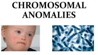


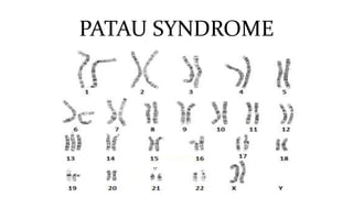
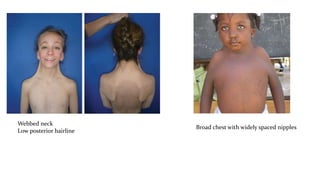
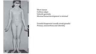


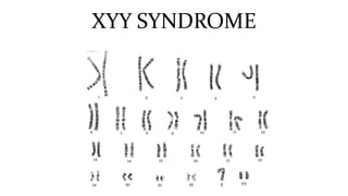

The document covers various chromosomal anomalies, detailing their classifications into numeric and structural abnormalities and emphasizing the incidence and clinical features of conditions like Down syndrome, Patau syndrome, and Turner syndrome. It explains the impact of maternal age on the occurrence of these conditions and provides insight into prenatal screening methods. Additionally, it outlines specific syndromes, their genetic underpinnings, and associated physical and mental health issues.


