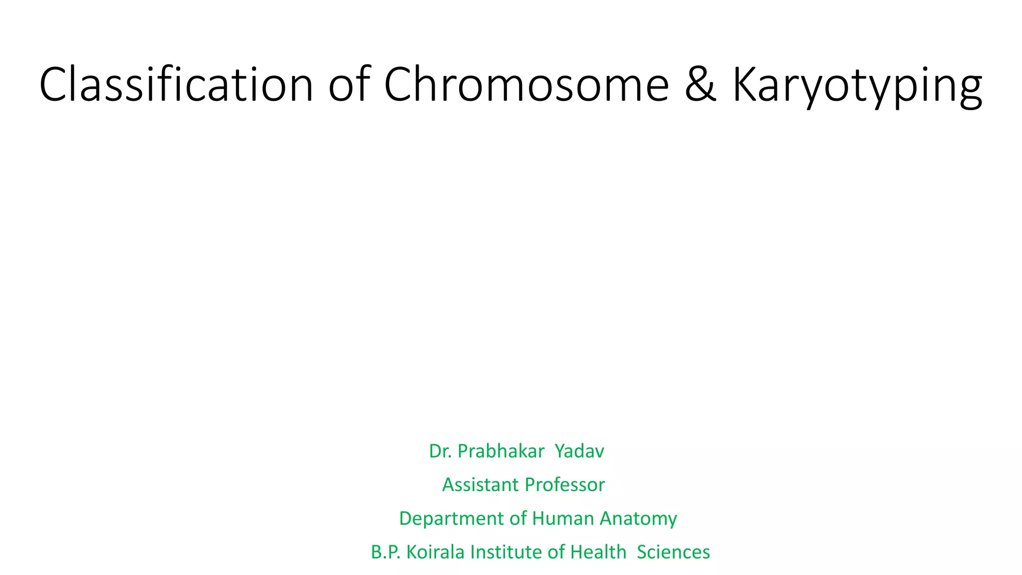
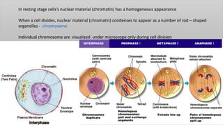
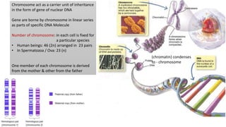
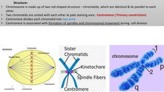
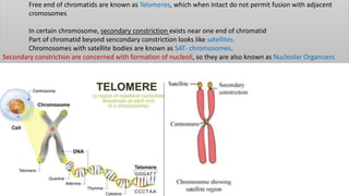
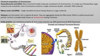
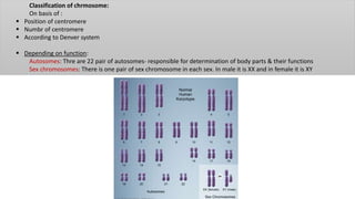
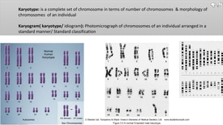
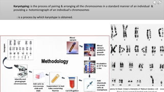
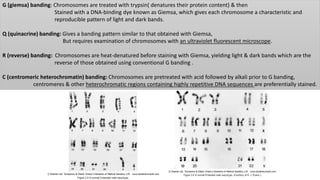
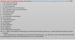
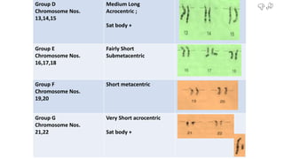
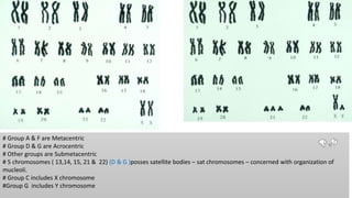
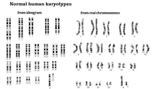
Classification of Chromosome & Karyotyping
Dr. Prabhakar Yadav
Assistant Professor
Department of Human Anatomy
B.P. Koirala Institute of Health Sciences
In resting stage cells’s nuclear material (chromatin) has a homogeneous appearance
When a cell divides, nuclear material (chromatin) condenses to appear as a number of rod – shaped
organelles - chromosome
Individual chromosome are visualized under microscope only during cell division
(chromatin) condenses
to - chromosome
Chromosome act as a carrier unit of inheritance
in the form of gene of nuclear DNA
Gene are borne by chromosome in linear series
as parts of specific DNA Molecule
Number of chromosome: in each cell is fixed for
a particular species
• Human beings: 46 (2n) arranged in 23 pairs
• In Spermatozoa / Ova: 23 (n)
One member of each chromosome is derived
from the mother & other from the father
Types:
22 out of 23 pairs are identical and are known as autosomes
Chromosome in the 23rd pair is known as sex chromosome
In female both sex chromosome are identical and are called X- chromosome
In Male 2 sex chromosome are not identical and are called X & Y- chromome
• Female germ cell always has X chromosome where as Male germ cell may either have X or Y chromosome
Structure:
• Chromosome is made up of two rod shaped structure – chromatids, which are identical & lie parallel to each
other.
• Two chromatids are united with each other at pale staining area : Centromere ( Primary constriction)
• Centromere divides each chromatid into two arms
• Centromere is associated with formation of spindles and chromosomal movement during cell division
Free end of chromatids are known as Telomeres, which when intact do not permit fusion with adjacent
cromosomes
In certain chromosome, secondary constriction exists near one end of chromatid
Part of chromatid beyond sencondary constriction looks like satellites.
Chromosomes with satellite bodies are known as SAT- chromosomes.
Secondary constriction are concerned with formation of nucleoli, so they are also known as Nucleolar Organizers
Chemical composition of chromosome:
Deoxyribonuclic acid (DNA): Most essential & stable molecular constituent of chromosomes. It is made up of deoxyribose sugar
molecule and nucleotides. Each chromosome contains a single continuous double –stranded DNA molecule
Ribose nuclic acid (RNA) : Single stranded structure having ribose as a sugar molecule
Histones: are the protein rich in arginine & lysine. They are aggregated along the DNA strand, Which is coiled around each
particle to form a complex body known as nucleosomes having 4 histones
Acidic proteins: are nonhistone proteins & form many enzymes e.g. DNA polymerase & RNA Polymerase
Classification of chrmosome:
On basis of :
 Position of centromere
 Numbr of centromere
 According to Denver system
 Depending on function:
Autosomes: Thre are 22 pair of autosomes- responsible for determination of body parts & their functions
Sex chromosomes: There is one pair of sex chromosome in each sex. In male it is XX and in female it is XY
According to position of centromere:
1. Metacentric: Chromosome with a centromere located in middle of chromosome ; Two arm are almost equal
2. Submetacentric: Chromosome with a centromere located slightly away form midpoint; Two arm are unequal
3. Acrocentric: centromere occupying subterminal position; one are is very long & other is short
4. Telocentric: centromere at its Terminal position; Each chromatid therefore has only one arm
According to number of centromere:
1. Monocentric: Having one centromere only, which is usual and normal
2. Dicentric: Having two centromere, which is found in some species of wheat
3. Polycentric: Having more than two centromere seen in some forms of roundworm
4. A-centric: Represents only a fragment of chromosome having no centromere. It is not visible
Karyotype: is a complete set of chromosome in terms of number of chromosomes & morphology of
chromosomes of an individual
Karyogram( karyotype/ idiogram): Photomicrograph of chromosomes of an individual arranged in a
standard manner/ Standard classification
Karyotyping: is the process of pairing & arranging all the chromosomes in a standard manner of an individual &
providing a hotomicrograph of an individual's chromosomes
: is a process by which karyotype is obtained.
Indications
1. Women with amenorrhoea & couples with Infertility or habitual abortion
2. Pregnancy in a elderly woman (fetal chromosome analysis)
3. Still births & Neonatal deaths
4. Problems in early growth- Developmental delay, Dysmorphic facies,
Multiple malformations,
Short stature,
Ambiguous genitalia,
Mental retardation.
5. First degree relatives of a known or suspected case of chromosome abnormality
6. Neoplasia
Selection of tissue for Karyotyping:
Any cell capable of growth & rapid cell division in a tissue culture medium is suitable for karyotyping.
A. T-lymphpcytes in peripheral blood- most commonly used
B. Skin Fibroblasts (from skin biopsy)
C. Bone marrow cells
D. Fetal blood
E. Amniotic fluid cells For Prenatal diagnosis
F. chorionic villi
Procedure
1. Collection of peripheral venous blood in strictly aseptic condition in heparinized vaccute
2. Planting: should be done within 4-5 hours of collection
Blood sample is transferred to the culture tube containing:
a. Culture Media: RPMI( Roswell Park Memorial Institute), TC 199 etc.
b. Neonatal calf serum/ fetal bovine serum: to nourish the culture cells
c. Phytoheamaglutinin : Mitotic aget – to increase the rate of mitosis of cultured cells
d. Antibiotics: Penicilllin & streptomycin combination – to prevent bacterial growth
3. Culture tube is Incubated at 37 degree C for 3 days – during this period lymphocytes undergo mitosis
4. Harvasting: 69-72 hours after planting, Colchicin is added to culture tube to arrest mitosis at metaphase by
preventing the formation of spindle tubules.
5. After 2 hours cells are collected by Centrifugation (for separating the cells)
6. After discarding the supernatant fluid, Cell pellate are treated with Hypotonic solution {sodium citrate /( 0.56% KCL)}
and incubated for 20 min so that the cells swells & chromosomes are dispersed
7. Hypotonic solution is discarded by centrifugation.
7. Cell pellate is fixed with methanol and acetic acid mixture and then the test tube is shaken
9. Cell suspension is dropped form a height on a clean slide, dried at room temperatue or spirit lamp flame & then stained
10. Staining: For chromosome analysis various banding technique are vailabe:
G(giemsa)-banding, Q (quinacrine)-banding, R(reverse)-banding, C(centromeric heterochromatin) -banding & NOR Banding
G- banding:
slides with chromosome prepration are first treated with solution of trypsin (Trypsin denatures the chromosome protein)
Slides are then treated with Giemsa solution
Chromosome shows dark and light band that can be observed under microscope
 Banding pattern helps to identify individual chromosomes
11. Micoscopy: Stained slide are examined under the oil immersion lens and Photomicrograph is taken
13. From the Enlarged photographs individual members of each homologous pair are matched and placed side by side.
Prepration of Karyotype:
Individual chromosome are cut manually from the photograph
22 autosomes & a pair of sex chromosome are identified and arranged in pairs using following parameters:
• Shape of chromosome
• Length of chromosome
• Centromeric Index =
𝑆ℎ𝑜𝑟𝑡 𝑎𝑟𝑚 𝑙𝑒𝑛𝑡ℎ
𝑇𝑜𝑡𝑎𝑙 𝑐ℎ𝑟𝑜𝑚𝑜𝑠𝑜𝑚𝑒 𝑙𝑒𝑛𝑔𝑡ℎ
G (giemsa) banding: Chromosomes are treated with trypsin( denatures their protein content) & then
Stained with a DNA-binding dye known as Giemsa, which gives each chromosome a characteristic and
reproducible pattern of light and dark bands.
Q (quinacrine) banding: Gives a banding pattern similar to that obtained with Giemsa,
But requires examination of chromosomes with an ultraviolet fluorescent microscope.
R (reverse) banding: Chromosomes are heat-denatured before staining with Giemsa, yielding light & dark bands which are the
reverse of those obtained using conventional G banding .
C (centromeric heterochromatin) banding: Chromosomes are pretreated with acid followed by alkali prior to G banding,
centromeres & other heterochromatic regions containing highly repetitive DNA sequences are preferentially stained.
Symboles used in describing karyotype: {According to International system of Human cytogenetic Nomenclature (ISCN) 1985}
1. A-G: Chromosome groups
2. 1-22: Autosome numbers
3. X,Y: Sex Chromosome
4. p(= petite): Short arm of chromosome
5. Q ('g' = grande): Long arm of chromosome
6. mat: Maternal origin
7. pat: paternal origin
8. t: translocation
9. rob: robertsonian translocation
10. rep: reciprocal
11. r: ring chromomsome
12. i: Iso chromosoe
13. del: Deletion
14. dup: Duplication
15. inv: Inversion
16. fra: Fragile site
17. ter: terminal end of chromome
18. + or - : Befor a chromosome: indicate gain or loss of that chromosome. Eg:47, xx+21; means female with trisomy 21;Down’s
syndrome
After a chromosome: indicate gain or loss of part of that chromosome , e.g. 46, XY, 5p-; means male with 46 chromosome
but having deletion of short arm of chromosome 5 ( cri-du – chat syndrome
According to Denver System:
In 1960 at conference of genetics at Denver, human chromosome including sex chromosome were arranged into 7 groups
form I to VII depending upon size of chromosome – Denver System
According to modern convention, chromosome are subdivided into Group A to Group G depending upon position of centromere-
patau’s modification of Denver method
`
Group A
Chromosome Nos. 1,2,3
Long metacentric
ChromosomeNo 2 is
submetacentric
Group B
Chromosome Nos. 4,5
Fairly long
Submetacentric
Group C
Chromosome Nos. 6 to
12 & X
Medium Long
Submetacentric
`Group D
Chromosome Nos.
13,14,15
Medium Long
Acrocentric ;
Sat body +
Group E
Chromosome Nos.
16,17,18
Fairly Short
Submetacentric
Group F
Chromosome Nos.
19,20
Short metacentric
Group G
Chromosome Nos.
21,22
Very Short acrocentric
Sat body +
# Group A & F are Metacentric
# Group D & G are Acrocentric
# Other groups are Submetacentric
# 5 chromosomes ( 13,14, 15, 21 & 22) (D & G )posses satellite bodies – sat chromosomes – concerned with organization of
mucleoli.
# Group C includes X chromosome
#Group G includes Y chromosome
HAPPY NEW YEAR 2018

Classification of chromosome & karyotyping

  • 1.
    Classification of Chromosome& Karyotyping Dr. Prabhakar Yadav Assistant Professor Department of Human Anatomy B.P. Koirala Institute of Health Sciences
  • 2.
    In resting stagecells’s nuclear material (chromatin) has a homogeneous appearance When a cell divides, nuclear material (chromatin) condenses to appear as a number of rod – shaped organelles - chromosome Individual chromosome are visualized under microscope only during cell division
  • 3.
    (chromatin) condenses to -chromosome Chromosome act as a carrier unit of inheritance in the form of gene of nuclear DNA Gene are borne by chromosome in linear series as parts of specific DNA Molecule Number of chromosome: in each cell is fixed for a particular species • Human beings: 46 (2n) arranged in 23 pairs • In Spermatozoa / Ova: 23 (n) One member of each chromosome is derived from the mother & other from the father
  • 4.
    Types: 22 out of23 pairs are identical and are known as autosomes Chromosome in the 23rd pair is known as sex chromosome In female both sex chromosome are identical and are called X- chromosome In Male 2 sex chromosome are not identical and are called X & Y- chromome • Female germ cell always has X chromosome where as Male germ cell may either have X or Y chromosome
  • 5.
    Structure: • Chromosome ismade up of two rod shaped structure – chromatids, which are identical & lie parallel to each other. • Two chromatids are united with each other at pale staining area : Centromere ( Primary constriction) • Centromere divides each chromatid into two arms • Centromere is associated with formation of spindles and chromosomal movement during cell division
  • 6.
    Free end ofchromatids are known as Telomeres, which when intact do not permit fusion with adjacent cromosomes In certain chromosome, secondary constriction exists near one end of chromatid Part of chromatid beyond sencondary constriction looks like satellites. Chromosomes with satellite bodies are known as SAT- chromosomes. Secondary constriction are concerned with formation of nucleoli, so they are also known as Nucleolar Organizers
  • 7.
    Chemical composition ofchromosome: Deoxyribonuclic acid (DNA): Most essential & stable molecular constituent of chromosomes. It is made up of deoxyribose sugar molecule and nucleotides. Each chromosome contains a single continuous double –stranded DNA molecule Ribose nuclic acid (RNA) : Single stranded structure having ribose as a sugar molecule Histones: are the protein rich in arginine & lysine. They are aggregated along the DNA strand, Which is coiled around each particle to form a complex body known as nucleosomes having 4 histones Acidic proteins: are nonhistone proteins & form many enzymes e.g. DNA polymerase & RNA Polymerase
  • 8.
    Classification of chrmosome: Onbasis of :  Position of centromere  Numbr of centromere  According to Denver system  Depending on function: Autosomes: Thre are 22 pair of autosomes- responsible for determination of body parts & their functions Sex chromosomes: There is one pair of sex chromosome in each sex. In male it is XX and in female it is XY
  • 9.
    According to positionof centromere: 1. Metacentric: Chromosome with a centromere located in middle of chromosome ; Two arm are almost equal 2. Submetacentric: Chromosome with a centromere located slightly away form midpoint; Two arm are unequal 3. Acrocentric: centromere occupying subterminal position; one are is very long & other is short 4. Telocentric: centromere at its Terminal position; Each chromatid therefore has only one arm
  • 10.
    According to numberof centromere: 1. Monocentric: Having one centromere only, which is usual and normal 2. Dicentric: Having two centromere, which is found in some species of wheat 3. Polycentric: Having more than two centromere seen in some forms of roundworm 4. A-centric: Represents only a fragment of chromosome having no centromere. It is not visible
  • 11.
    Karyotype: is acomplete set of chromosome in terms of number of chromosomes & morphology of chromosomes of an individual Karyogram( karyotype/ idiogram): Photomicrograph of chromosomes of an individual arranged in a standard manner/ Standard classification
  • 12.
    Karyotyping: is theprocess of pairing & arranging all the chromosomes in a standard manner of an individual & providing a hotomicrograph of an individual's chromosomes : is a process by which karyotype is obtained.
  • 13.
    Indications 1. Women withamenorrhoea & couples with Infertility or habitual abortion 2. Pregnancy in a elderly woman (fetal chromosome analysis) 3. Still births & Neonatal deaths 4. Problems in early growth- Developmental delay, Dysmorphic facies, Multiple malformations, Short stature, Ambiguous genitalia, Mental retardation. 5. First degree relatives of a known or suspected case of chromosome abnormality 6. Neoplasia
  • 14.
    Selection of tissuefor Karyotyping: Any cell capable of growth & rapid cell division in a tissue culture medium is suitable for karyotyping. A. T-lymphpcytes in peripheral blood- most commonly used B. Skin Fibroblasts (from skin biopsy) C. Bone marrow cells D. Fetal blood E. Amniotic fluid cells For Prenatal diagnosis F. chorionic villi
  • 16.
    Procedure 1. Collection ofperipheral venous blood in strictly aseptic condition in heparinized vaccute 2. Planting: should be done within 4-5 hours of collection Blood sample is transferred to the culture tube containing: a. Culture Media: RPMI( Roswell Park Memorial Institute), TC 199 etc. b. Neonatal calf serum/ fetal bovine serum: to nourish the culture cells c. Phytoheamaglutinin : Mitotic aget – to increase the rate of mitosis of cultured cells d. Antibiotics: Penicilllin & streptomycin combination – to prevent bacterial growth
  • 17.
    3. Culture tubeis Incubated at 37 degree C for 3 days – during this period lymphocytes undergo mitosis 4. Harvasting: 69-72 hours after planting, Colchicin is added to culture tube to arrest mitosis at metaphase by preventing the formation of spindle tubules.
  • 18.
    5. After 2hours cells are collected by Centrifugation (for separating the cells) 6. After discarding the supernatant fluid, Cell pellate are treated with Hypotonic solution {sodium citrate /( 0.56% KCL)} and incubated for 20 min so that the cells swells & chromosomes are dispersed 7. Hypotonic solution is discarded by centrifugation.
  • 19.
    7. Cell pellateis fixed with methanol and acetic acid mixture and then the test tube is shaken 9. Cell suspension is dropped form a height on a clean slide, dried at room temperatue or spirit lamp flame & then stained 10. Staining: For chromosome analysis various banding technique are vailabe: G(giemsa)-banding, Q (quinacrine)-banding, R(reverse)-banding, C(centromeric heterochromatin) -banding & NOR Banding G- banding: slides with chromosome prepration are first treated with solution of trypsin (Trypsin denatures the chromosome protein) Slides are then treated with Giemsa solution Chromosome shows dark and light band that can be observed under microscope  Banding pattern helps to identify individual chromosomes 11. Micoscopy: Stained slide are examined under the oil immersion lens and Photomicrograph is taken 13. From the Enlarged photographs individual members of each homologous pair are matched and placed side by side.
  • 20.
    Prepration of Karyotype: Individualchromosome are cut manually from the photograph 22 autosomes & a pair of sex chromosome are identified and arranged in pairs using following parameters: • Shape of chromosome • Length of chromosome • Centromeric Index = 𝑆ℎ𝑜𝑟𝑡 𝑎𝑟𝑚 𝑙𝑒𝑛𝑡ℎ 𝑇𝑜𝑡𝑎𝑙 𝑐ℎ𝑟𝑜𝑚𝑜𝑠𝑜𝑚𝑒 𝑙𝑒𝑛𝑔𝑡ℎ
  • 21.
    G (giemsa) banding:Chromosomes are treated with trypsin( denatures their protein content) & then Stained with a DNA-binding dye known as Giemsa, which gives each chromosome a characteristic and reproducible pattern of light and dark bands. Q (quinacrine) banding: Gives a banding pattern similar to that obtained with Giemsa, But requires examination of chromosomes with an ultraviolet fluorescent microscope. R (reverse) banding: Chromosomes are heat-denatured before staining with Giemsa, yielding light & dark bands which are the reverse of those obtained using conventional G banding . C (centromeric heterochromatin) banding: Chromosomes are pretreated with acid followed by alkali prior to G banding, centromeres & other heterochromatic regions containing highly repetitive DNA sequences are preferentially stained.
  • 22.
    Symboles used indescribing karyotype: {According to International system of Human cytogenetic Nomenclature (ISCN) 1985} 1. A-G: Chromosome groups 2. 1-22: Autosome numbers 3. X,Y: Sex Chromosome 4. p(= petite): Short arm of chromosome 5. Q ('g' = grande): Long arm of chromosome 6. mat: Maternal origin 7. pat: paternal origin 8. t: translocation 9. rob: robertsonian translocation 10. rep: reciprocal 11. r: ring chromomsome 12. i: Iso chromosoe 13. del: Deletion 14. dup: Duplication 15. inv: Inversion 16. fra: Fragile site 17. ter: terminal end of chromome 18. + or - : Befor a chromosome: indicate gain or loss of that chromosome. Eg:47, xx+21; means female with trisomy 21;Down’s syndrome After a chromosome: indicate gain or loss of part of that chromosome , e.g. 46, XY, 5p-; means male with 46 chromosome but having deletion of short arm of chromosome 5 ( cri-du – chat syndrome
  • 23.
    According to DenverSystem: In 1960 at conference of genetics at Denver, human chromosome including sex chromosome were arranged into 7 groups form I to VII depending upon size of chromosome – Denver System According to modern convention, chromosome are subdivided into Group A to Group G depending upon position of centromere- patau’s modification of Denver method
  • 24.
    ` Group A Chromosome Nos.1,2,3 Long metacentric ChromosomeNo 2 is submetacentric Group B Chromosome Nos. 4,5 Fairly long Submetacentric Group C Chromosome Nos. 6 to 12 & X Medium Long Submetacentric
  • 25.
    `Group D Chromosome Nos. 13,14,15 MediumLong Acrocentric ; Sat body + Group E Chromosome Nos. 16,17,18 Fairly Short Submetacentric Group F Chromosome Nos. 19,20 Short metacentric Group G Chromosome Nos. 21,22 Very Short acrocentric Sat body +
  • 26.
    # Group A& F are Metacentric # Group D & G are Acrocentric # Other groups are Submetacentric # 5 chromosomes ( 13,14, 15, 21 & 22) (D & G )posses satellite bodies – sat chromosomes – concerned with organization of mucleoli. # Group C includes X chromosome #Group G includes Y chromosome
  • 28.