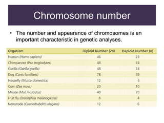
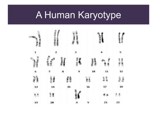
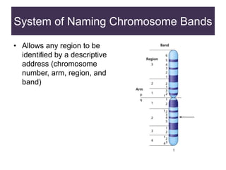
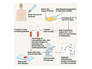
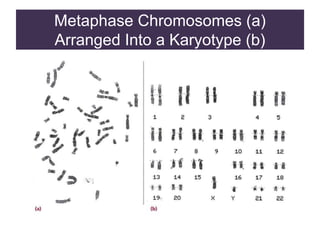
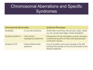
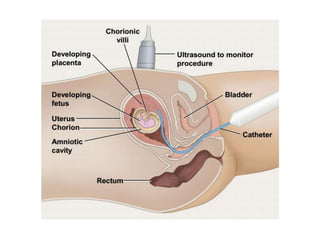
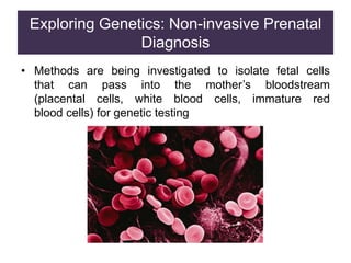
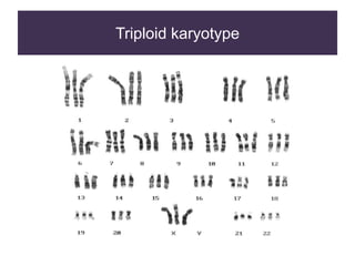
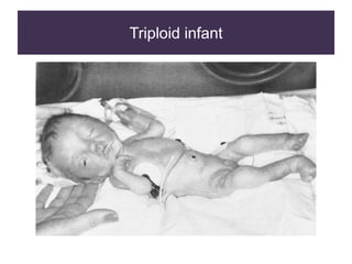
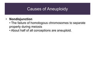
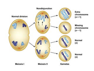
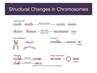
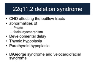
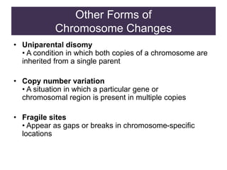
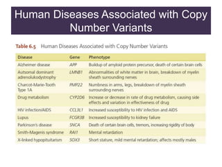
Normal karyotype and
cytogenetic disorders
SLOs
• What is karyotype?
• Structure of chromosome
• How to do cytogenetics?
• Common variations in cytogenetics
Chromosome number
• The number and appearance of chromosomes is an
important characteristic in genetic analyses.
Chromosome shape
• As chromosomes condense and become visible during
cell division, certain structural features can be
recognized
Centromere
• A region of a chromosome to which microtubule fibers
attach during cell division
• The location of a centromere gives a chromosome its
characteristic shape
Human chromosomes
• Replicated chromosomes at metaphase consist of sister
chromatids joined by a single centromere
Centromere Location
Metacentric
• A chromosome that has a centrally placed
centromere
Submetacentric
• A chromosome whose centromere is placed closer
to one end than the other
Acrocentric
• A chromosome whose centromere is placed very
close to, but not at, one end
Metaphase Chromosomes
• Chromosomes are identified by size, centromere
location, banding pattern
A Set of Human Chromosomes
• Human chromosomes are analyzed by construction
of karyotypes
Karyotype
• A complete set of chromosomes from a cell that has
been photographed during cell division and arranged
in a standard sequence
A Human Karyotype
System of Naming Chromosome Bands
• Allows any region to be
identified by a descriptive
address (chromosome
number, arm, region, and
band)
Metaphase Chromosomes (a)
Arranged Into a Karyotype (b)
Constructing and
Analyzing Karyotypes
• Different stains and dyes produce banding patterns
specific to each chromosome
• Karyotypes reveal variations in chromosomal
structure and number
– 1959: Discovery that Down syndrome is caused by
an extra copy of chromosome 21
• Chromosome banding and other techniques can
identify small changes in chromosomal structure
Information Obtained from a Karyotype
• Number of chromosomes
• Sex chromosome content
• Presence or absence of individual chromosomes
• Nature and extent of large structural abnormalities
Four Common Chromosome Staining
Procedures
Chromosome Painting
• New techniques using fluorescent dyes generate unique
patterns for each chromosome
Chromosomal Aberrations and Specific
Syndromes
Obtaining Cells for Chromosome Studies
• Any nucleus can be used to make karyotype
– Lymphocytes, skin cells, cells from biopsies, tumor
cells
• Sampling cells before birth
– Amniocentesis
– Chorionic villus sampling (CVS)
Amniocentesis
• A method of sampling the fluid surrounding the
developing fetus by inserting a hollow needle and
withdrawing suspended fetal cells and fluid
– Used in diagnosing fetal genetic and developmental
disorders
– Usually performed in the sixteenth week of
pregnancy
Chorionic Villus Sampling (CVS)
• A method of sampling the fluid surrounding the
developing fetus by inserting a hollow needle and
withdrawing suspended fetal cells and fluid
– Used in diagnosing fetal genetic and developmental
disorders
– Usually performed in the sixteenth week of
pregnancy
Exploring Genetics: Non-invasive Prenatal
Diagnosis
• Methods are being investigated to isolate fetal cells
that can pass into the mother’s bloodstream
(placental cells, white blood cells, immature red
blood cells) for genetic testing
Variations in Chromosome Number
• Changes in chromosome number or chromosome
structure can cause genetic disorders
• Two major types of chromosomal changes can be
detected in a karyotype
• A change in chromosomal number
• A change in chromosomal arrangement
Changes in Chromosome Number
• Polyploidy
• Duplication of an entire set of chromosomes
• 3N = triploid
• 4N = tetraploid
• Aneuploidy
• Refers to a single chromosome
• Trisomy = one extra chromosome (three copies)
• Monosomy = missing one chromosome of a pair
Triploid karyotype
Triploid infant
Causes of Aneuploidy
• Nondisjunction
• The failure of homologous chromosomes to separate
properly during meiosis
• About half of all conceptions are aneuploid.
Effects of Monosomy and Trisomy
• Autosomal monosomy is a lethal condition
• Eliminated early in development (spontaneous
abortion)
• Some autosomal trisomies are relatively common
• Most result in spontaneous abortion
• Three types can result in live births (13, 18, 21)
Trisomies in Spontaneous Abortions
Trisomy 13: Patau Syndrome (47,+13)
• A lethal condition
• 1 in 10,000 births
Trisomy 18: Edwards Syndrome (47,+18)
• A lethal condition
• 1 in 11,000 births
• 80% are females
Trisomy 21: Down Syndrome (47, +21)
• Trisomy 21 is the only autosomal trisomy that allows
survival into adulthood
What Are the Risks
for Autosomal Trisomy?
• The causes of autosomal trisomy are unknown
• Factors that have been proposed include:
• Genetic predisposition
• Exposure to radiation
• Viral infection
• Abnormal hormone levels
• Maternal age is the leading risk factor for trisomy
• 94% of nondisjunctions occur in the mother
• Meiosis is not completed until ovulation
• Embryo-uterine interactions that normally abort
abnormal embryos become less effective
Maternal Age and Trisomy 21
Aneuploidy of the Sex
Chromosomes
• More common than autosomal aneuploidy
• Can involve both X and Y chromosomes
• A balance is needed for normal development
• At least one copy of the X chromosome is required for
development
• Increasing numbers of X or Y chromosomes causes
progressively greater disturbances in phenotype and
behavior
Turner Syndrome (45,X)
• Monosomy of the X chromosome that results in
female sterility. Other phenotypic characteristics
but otherwise normal.
Klinefelter Syndrome (47, XXY)
• Individuals (males) have some fertility
problems but few additional symptoms
XYY Syndrome (47,XYY)
• Affected individuals are usually taller than normal and
some, but not all, have personality disorders
Structural Alterations
Within Chromosomes
• Changes in the structure of chromosomes
• Deletion—loss of DNA
• Duplication—extra DNA
• Translocation—DNA that changes location
• Inversion—order of DNA changes
Structural Changes in Chromosomes
22q11.2 deletion syndrome
• CHD affecting the outflow tracts
• abnormalities of
– Palate
– facial dysmorphism
• Developmental delay
• Thymic hypoplasia
• Parathyroid hypoplasia
• DiGeorge syndrome and velocardiofacial
syndrome
Other Forms of
Chromosome Changes
• Uniparental disomy
• A condition in which both copies of a chromosome are
inherited from a single parent
• Copy number variation
• A situation in which a particular gene or
chromosomal region is present in multiple copies
• Fragile sites
• Appear as gaps or breaks in chromosome-specific
locations
Human Diseases Associated with Copy
Number Variants
Fragile Sites
• Appear as gaps or breaks in chromosomes
• One fragile site on the X chromosome is associated with a
common form of mental retardation in males know as
Fragile X Syndrome
• Females can also have this, but the phenotypes are
much more mild
Fragile Sites on the X Chromosome
Summarise
Questions ?

Normal karyotype and cytogenetics disorders

  • 1.
  • 2.
    SLOs • What iskaryotype? • Structure of chromosome • How to do cytogenetics? • Common variations in cytogenetics
  • 3.
    Chromosome number • Thenumber and appearance of chromosomes is an important characteristic in genetic analyses.
  • 4.
    Chromosome shape • Aschromosomes condense and become visible during cell division, certain structural features can be recognized Centromere • A region of a chromosome to which microtubule fibers attach during cell division • The location of a centromere gives a chromosome its characteristic shape
  • 5.
    Human chromosomes • Replicatedchromosomes at metaphase consist of sister chromatids joined by a single centromere
  • 6.
    Centromere Location Metacentric • Achromosome that has a centrally placed centromere Submetacentric • A chromosome whose centromere is placed closer to one end than the other Acrocentric • A chromosome whose centromere is placed very close to, but not at, one end
  • 7.
    Metaphase Chromosomes • Chromosomesare identified by size, centromere location, banding pattern
  • 8.
    A Set ofHuman Chromosomes • Human chromosomes are analyzed by construction of karyotypes Karyotype • A complete set of chromosomes from a cell that has been photographed during cell division and arranged in a standard sequence
  • 9.
  • 10.
    System of NamingChromosome Bands • Allows any region to be identified by a descriptive address (chromosome number, arm, region, and band)
  • 12.
  • 13.
    Constructing and Analyzing Karyotypes •Different stains and dyes produce banding patterns specific to each chromosome • Karyotypes reveal variations in chromosomal structure and number – 1959: Discovery that Down syndrome is caused by an extra copy of chromosome 21 • Chromosome banding and other techniques can identify small changes in chromosomal structure
  • 14.
    Information Obtained froma Karyotype • Number of chromosomes • Sex chromosome content • Presence or absence of individual chromosomes • Nature and extent of large structural abnormalities
  • 15.
    Four Common ChromosomeStaining Procedures
  • 16.
    Chromosome Painting • Newtechniques using fluorescent dyes generate unique patterns for each chromosome
  • 17.
    Chromosomal Aberrations andSpecific Syndromes
  • 18.
    Obtaining Cells forChromosome Studies • Any nucleus can be used to make karyotype – Lymphocytes, skin cells, cells from biopsies, tumor cells • Sampling cells before birth – Amniocentesis – Chorionic villus sampling (CVS)
  • 19.
    Amniocentesis • A methodof sampling the fluid surrounding the developing fetus by inserting a hollow needle and withdrawing suspended fetal cells and fluid – Used in diagnosing fetal genetic and developmental disorders – Usually performed in the sixteenth week of pregnancy
  • 21.
    Chorionic Villus Sampling(CVS) • A method of sampling the fluid surrounding the developing fetus by inserting a hollow needle and withdrawing suspended fetal cells and fluid – Used in diagnosing fetal genetic and developmental disorders – Usually performed in the sixteenth week of pregnancy
  • 23.
    Exploring Genetics: Non-invasivePrenatal Diagnosis • Methods are being investigated to isolate fetal cells that can pass into the mother’s bloodstream (placental cells, white blood cells, immature red blood cells) for genetic testing
  • 24.
    Variations in ChromosomeNumber • Changes in chromosome number or chromosome structure can cause genetic disorders • Two major types of chromosomal changes can be detected in a karyotype • A change in chromosomal number • A change in chromosomal arrangement
  • 25.
    Changes in ChromosomeNumber • Polyploidy • Duplication of an entire set of chromosomes • 3N = triploid • 4N = tetraploid • Aneuploidy • Refers to a single chromosome • Trisomy = one extra chromosome (three copies) • Monosomy = missing one chromosome of a pair
  • 26.
  • 27.
  • 28.
    Causes of Aneuploidy •Nondisjunction • The failure of homologous chromosomes to separate properly during meiosis • About half of all conceptions are aneuploid.
  • 31.
    Effects of Monosomyand Trisomy • Autosomal monosomy is a lethal condition • Eliminated early in development (spontaneous abortion) • Some autosomal trisomies are relatively common • Most result in spontaneous abortion • Three types can result in live births (13, 18, 21)
  • 32.
  • 33.
    Trisomy 13: PatauSyndrome (47,+13) • A lethal condition • 1 in 10,000 births
  • 34.
    Trisomy 18: EdwardsSyndrome (47,+18) • A lethal condition • 1 in 11,000 births • 80% are females
  • 35.
    Trisomy 21: DownSyndrome (47, +21) • Trisomy 21 is the only autosomal trisomy that allows survival into adulthood
  • 36.
    What Are theRisks for Autosomal Trisomy? • The causes of autosomal trisomy are unknown • Factors that have been proposed include: • Genetic predisposition • Exposure to radiation • Viral infection • Abnormal hormone levels • Maternal age is the leading risk factor for trisomy • 94% of nondisjunctions occur in the mother • Meiosis is not completed until ovulation • Embryo-uterine interactions that normally abort abnormal embryos become less effective
  • 37.
    Maternal Age andTrisomy 21
  • 38.
    Aneuploidy of theSex Chromosomes • More common than autosomal aneuploidy • Can involve both X and Y chromosomes • A balance is needed for normal development • At least one copy of the X chromosome is required for development • Increasing numbers of X or Y chromosomes causes progressively greater disturbances in phenotype and behavior
  • 39.
    Turner Syndrome (45,X) •Monosomy of the X chromosome that results in female sterility. Other phenotypic characteristics but otherwise normal.
  • 40.
    Klinefelter Syndrome (47,XXY) • Individuals (males) have some fertility problems but few additional symptoms
  • 41.
    XYY Syndrome (47,XYY) •Affected individuals are usually taller than normal and some, but not all, have personality disorders
  • 42.
    Structural Alterations Within Chromosomes •Changes in the structure of chromosomes • Deletion—loss of DNA • Duplication—extra DNA • Translocation—DNA that changes location • Inversion—order of DNA changes
  • 43.
  • 44.
    22q11.2 deletion syndrome •CHD affecting the outflow tracts • abnormalities of – Palate – facial dysmorphism • Developmental delay • Thymic hypoplasia • Parathyroid hypoplasia • DiGeorge syndrome and velocardiofacial syndrome
  • 45.
    Other Forms of ChromosomeChanges • Uniparental disomy • A condition in which both copies of a chromosome are inherited from a single parent • Copy number variation • A situation in which a particular gene or chromosomal region is present in multiple copies • Fragile sites • Appear as gaps or breaks in chromosome-specific locations
  • 46.
    Human Diseases Associatedwith Copy Number Variants
  • 47.
    Fragile Sites • Appearas gaps or breaks in chromosomes • One fragile site on the X chromosome is associated with a common form of mental retardation in males know as Fragile X Syndrome • Females can also have this, but the phenotypes are much more mild
  • 48.
    Fragile Sites onthe X Chromosome
  • 49.
  • 50.