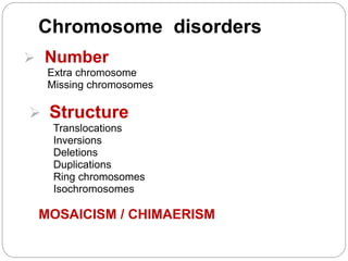
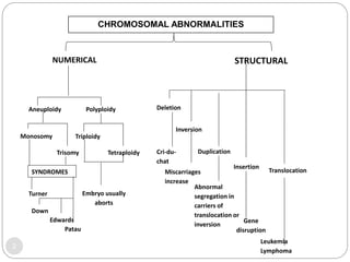
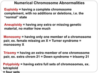
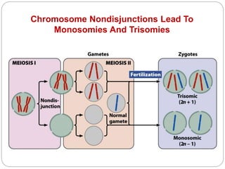
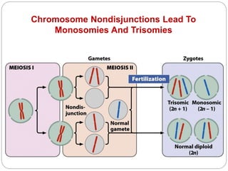
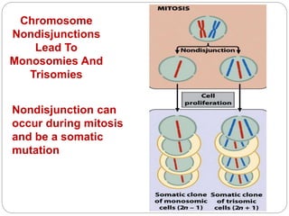
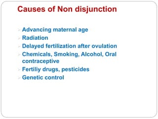
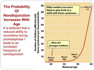
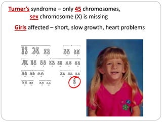
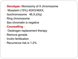
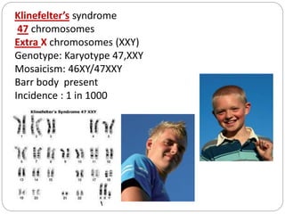
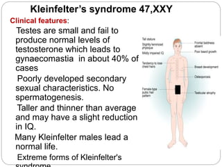
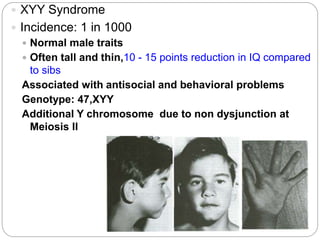
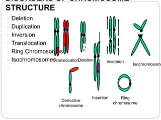
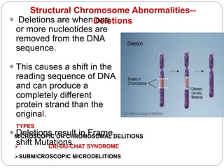
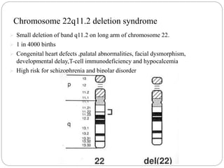
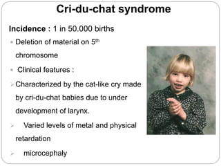
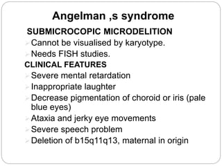
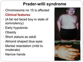
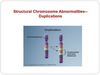
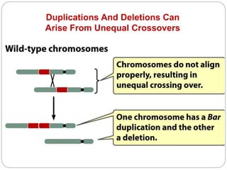
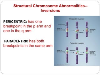
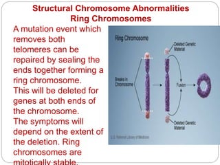
Chromosome disorders
 Number
Extra chromosome
Missing chromosomes
 Structure
Translocations
Inversions
Deletions
Duplications
Ring chromosomes
Isochromosomes
MOSAICISM / CHIMAERISM
CHROMOSOMAL ABNORMALITIES
NUMERICAL STRUCTURAL
Aneuploidy Polyploidy
Monosomy
Trisomy
SYNDROMES
Turner
Down
Edwards
Patau
Triploidy
Tetraploidy
Embryo usually
aborts
Deletion
Cri-du-
chat
Inversion
Miscarriages
increase
Duplication
Insertion
Translocation
Abnormal
segregation in
carriers of
translocation or
inversion
Gene
disruption
Leukemia
Lymphoma
2
Numerical Chromosome Abnormalities
Euploidy = having a complete chromosome
complement, with no additions or deletions, i.e. the
“normal” state
Polyploidy = having extra full sets of chromosomes, ex.
tetraploid
Aneuploidy = having any extra or missing genetic
material, no matter how much
Trisomy = having an extra member of one chromosome
pair, ex. extra chrom 21 = Down syndrome = trisomy 21
Monosomy = having only one member of a chromosome
pair, ex. female missing an X = Turner syndrome =
monosomy X
Chromosome Nondisjunctions Lead To
Monosomies And Trisomies
Chromosome Nondisjunctions Lead To
Monosomies And Trisomies
Chromosome
Nondisjunctions
Lead To
Monosomies And
Trisomies
Nondisjunction can
occur during mitosis
and be a somatic
mutation
Causes of Non disjunction
 Advancing maternal age
 Radiation
 Delayed fertilization after ovulation
 Chemicals, Smoking, Alcohol, Oral
contraceptive
 Fertiliy drugs, pesticides
 Genetic control
The Probability
Of
Nondisjunction
Increases With
Age
It is believed that a
reduced ability to
recombine during
prometaphase I
leads to an
increased
frequency of
nondisjunction
In Humans, Only Trisomy 21
Produces Viable Offspring
DISORDERS OF CHROMOSOME
NUMBER
Down’s syndrome (MONGOLISM)
(Trisomy 21) 47 chromosomes
Clinical features:
Incidence 1/700
2/3 of fetus spontaneously abort
Trisomy 21 in 94% of cases with extra
chromosome from mother
mostly(95%)
Risk correlate with maternal age
<25 y/o 1/1600
>40 y/o 1/80
2% are mosaics
Clinical features
 Mentally retarded IQ.25-75
 Poor growth, short stature
 May have heart and stomach illnesses
 Small head circumference
(brachycephaly)
 Hypotonia without weakness
 Protruded tongue, epicanthic fold
 Small ears, slopping palpebral fissures
 Small up turned nose, depressed
nasal bridge.
 Hands are short and broad. Simian
Genotype: 47+XY or 47+XX
 Non disjunction in the first meiotic division, the
father
contributing the extra chromosome in 15% of
cases.
 Mosaic due to non disjunction in an early
zygotic division.
 Translocation in 4% of cases.
Recurrence risk : 1-3% if father carrier
15% if mother is a carrier .Increases with
mothers age
Life Span: Mean age:16 yrs.
Develop Alzheimers disease.
Counselling:
Amniocentesis
13
EDWARD,S SYNDROME
 Trisomy of chromosome 18
 Incidence 1/8000
 Spontaneous abortion, >90% dead in 1st year
 CLINICAL FEATURES
 Retarded growth & development
 Small face with prominent occiput
 Small sternum and pelvis
 Flexion deformity of the finger
 VSD and horseshoe kidney
 Hypertonia, Small chin
Low set malformed ears
 Child holds fingers in typical way
 Heart defects; VSD, PDA
 Genotype:Trisomy 18
 Counselling: Most babies die in the first year and many within the first
15
Trisomy 13: PATAU,S SYNDROME
Incidence:1 in 5000 live births.
Most spontaneous abortion
50% die within first month, very
few survive beyond first year.
There are multiple dysmorphic
features.
Genotype: Trisomy of
chromosome 13
Mosaicism & Robertsonian
translocation
in rare cases.
Counselling;
Small risk of recurrence
Increases with advanced maternal
Clinical features
Severe physical & mental
retardation
Small skull & eyes.
Cleft lip & cleft palate
Extra finger or malformed
thumb
Malformations of CVS,
CNS and Excretory systems
Incidence: 1 in 5000 female births
99% of Turner syndrome embryos are
spontaneously aborted.
Clinical features:
Individuals are very short, infertile. webbed neck
Broad chest, widely spaced nipples and
underdeveloped
breasts
Lack of expected secondary sexual
characteristics
Coarctation of the aorta, VSD
Abnormalities of urinary system
IQ and lifespan are unaffected
Turner’s syndrome – only 45 chromosomes,
sex chromosome (X) is missing
Girls affected – short, slow growth, heart problems
 Genotype: Monosomy of X chromosome
 Mosaisim (15%) 45XO/46XX,
 Isochromosome 46,X,i(Xq)
 Ring chromosome
 Sex chromatin is negative
 Counselling :
 Oestrogen replacement therapy
 Remove gonads
 Invitro fertilization
 Recurrence risk is 1-2%
XXX Syndrome
 0.1% of all females
 Trisomy X
 Female
 Little or no visible differences
 Tall stature .Normal reproductive life
Reduction in IQ ,Learning disabilities
 Genotype:
XXX chromosomes
2 Barr bodies
Females with > 3X chromosomes can also
survive
Sterile, severe mental retardation, many
phenotypic effects
Klinefelter’s syndrome
47 chromosomes
Extra X chromosomes (XXY)
Genotype: Karyotype 47,XXY
Mosaicism: 46XY/47XXY
Barr body present
Incidence : 1 in 1000
Kleinfelter’s syndrome 47,XXY
Clinical features:
Testes are small and fail to
produce normal levels of
testosterone which leads to
gynaecomastia in about 40% of
cases
Poorly developed secondary
sexual characteristics. No
spermatogenesis.
Taller and thinner than average
and may have a slight reduction
in IQ.
Many Kleinfelter males lead a
normal life.
Extreme forms of Kleinfelter's
 XYY Syndrome
 Incidence: 1 in 1000
 Normal male traits
 Often tall and thin,10 - 15 points reduction in IQ compared
to sibs
Associated with antisocial and behavioral problems
Genotype: 47,XYY
Additional Y chromosome due to non dysjunction at
Meiosis II
DISORDERS OF CHROMOSOME
STRUCTURE
 Deletion
 Duplication
 Inversion
 Translocation
 Ring Chromosomes
 Isochromosomes

Translocation
Deletion
Insertion
Inversion
Isochromosome
Ring
chromosome
Derivative
chromosome
Structural Chromosome Abnormalities--
Deletions
 Deletions are when one
or more nucleotides are
removed from the DNA
sequence.
 This causes a shift in the
reading sequence of DNA
and can produce a
completely different
protein strand than the
original.
 Deletions result in Frame
shift Mutations
TYPES
MICROSCOPIC OR CHROMOSOMAL DELITIONS
 CRI-DU-CHAT SYNDROME
SUBMICROSCOPIC MICRODELITIONS
Chromosome 22q11.2 deletion syndrome
 Small deletion of band q11.2 on long arm of chromosome 22.
 1 in 4000 births
 Congenital heart defects ,palatal abnormalities, facial dysmorphism,
developmental delay,T-cell immunodeficiency and hypocalcemia
 High risk for schizophrenia and bipolar disorder
Cri-du-chat syndrome
Incidence : 1 in 50.000 births
 Deletion of material on 5th
chromosome
 Clinical features :
Characterized by the cat-like cry made
by cri-du-chat babies due to under
development of larynx.
 Varied levels of metal and physical
retardation
 microcephaly
Angelman ,s syndrome
SUBMICROCOPIC MICRODELITION
Cannot be visualised by karyotype.
Needs FISH studies.
CLINICAL FEATURES
Severe mental retardation
Inappropriate laughter
Decrease pigmentation of choroid or iris (pale
blue eyes)
Ataxia and jerky eye movements
Severe speech problem
Deletion of b15q11q13, maternal in origin
Prader-willi syndrome
 Chromosome no 15 is affected
 Clinical features
 (A fat red faced boy in state of
somnolency)
 Early hypotonia
 Obesity
 Short stature as adult
 Almond shaped blue eyes
 Mental retardation (mild to
moderate)
 Narrow hands
Structural Chromosome Abnormalities--
Duplications
Duplications And Deletions Can
Arise From Unequal Crossovers
Repeated sequences can cause uneven alignments
PERICENTRIC: has one
breakpoint in the p arm and
one in the q arm
Structural Chromosome Abnormalities--
Inversions
PARACENTRIC has both
breakpoints in the same arm
Structural Chromosome Abnormalities
Ring Chromosomes
Ex. Female with ring chrom 4, breakpoints p23.2 and q 12.4 =
46,XX,r(4)(p23.2q12.4)
A mutation event which
removes both
telomeres can be
repaired by sealing the
ends together forming a
ring chromosome.
This will be deleted for
genes at both ends of
the chromosome.
The symptoms will
depend on the extent of
the deletion. Ring
chromosomes are
A chromosome can split "the
wrong way" in mitosis (or
meiosis II) so that both long
arms remain attached and
move to one pole, and both
short arms to the other
pole.
The consequence is the
formation of an
isochromosome. These are
simultaneously duplicated
for the genes in the
retained arm and deleted
for the genes in the other.
Isochromosomes
Structural Chromosome Abnormalities—
Reciprocal Translocations
Structural
Chromosome
Abnormalities
Reciprocal
Translocations
46,XX, t(1;18)
Note that the two
chromosomes 1
are of unequal
size, as are the
two chromosomes
18
Structural Chromosome Abnormalities
Robertsonian Translocations
p arm of acrocentric chromosome
contains hundreds of copies of
rRNA gene, so losing two p arms
does not cause an abnormal
phenotype.
A Robertsonian translocation is
considered a single chromosome.
Acrocentric chromosomes 13, 14,
15, 21 and 22 form Robertsonian
translocations
If you have 5 Robertsonian
translocations, and don’t lose all
of the p arm material from all
Robertsonian Translocations Can Result In
Trisomies
The Robertsonian translocation is actually two fully
functioning chromosomes
It also has two centromeres,
one dominates over the other.
when it comes time of pulling
one set of chromosomes into
each daughter cell during
anaphase of meiosis I
When the cell divides during
meiosis I, the Robertsonian
translocation is treated as if it
was a single chromosome,
and the daughter cell does not
realize it has obtained a copy
Important Issues Pertinent
To Structural Rearrangements
Balanced = No DNA was lost
when the chromosomes
broke
The individual has all
his/her genes
Rarely causes a genetic
disorder
Will only cause a genetic
disorder if
one of the breakpoints
interrupts a gene
Unbalanced = DNA was
lost
when the chromosomes
broke
The individual is missing
one or
more of his/her genes
Often causes a genetic
disorder
Severity of effect is often
proportional to the amount
of
Important Issues Pertinent
To Structural Rearrangements
Are The Individual's Children At Risk?
 A balanced rearrangement that does not cause a
genetic disorder in the individual can still pose a risk
for the individual's offspring
The chromosomes cannot line up evenly during
meiosis
This may result in the egg or sperm having an
unbalanced genetic complement, i.e. missing
material, extra material, often a combination of both

Chromosome disorders.pptx

  • 1.
    Chromosome disorders  Number Extrachromosome Missing chromosomes  Structure Translocations Inversions Deletions Duplications Ring chromosomes Isochromosomes MOSAICISM / CHIMAERISM
  • 2.
    CHROMOSOMAL ABNORMALITIES NUMERICAL STRUCTURAL AneuploidyPolyploidy Monosomy Trisomy SYNDROMES Turner Down Edwards Patau Triploidy Tetraploidy Embryo usually aborts Deletion Cri-du- chat Inversion Miscarriages increase Duplication Insertion Translocation Abnormal segregation in carriers of translocation or inversion Gene disruption Leukemia Lymphoma 2
  • 3.
    Numerical Chromosome Abnormalities Euploidy= having a complete chromosome complement, with no additions or deletions, i.e. the “normal” state Polyploidy = having extra full sets of chromosomes, ex. tetraploid Aneuploidy = having any extra or missing genetic material, no matter how much Trisomy = having an extra member of one chromosome pair, ex. extra chrom 21 = Down syndrome = trisomy 21 Monosomy = having only one member of a chromosome pair, ex. female missing an X = Turner syndrome = monosomy X
  • 4.
    Chromosome Nondisjunctions LeadTo Monosomies And Trisomies
  • 5.
    Chromosome Nondisjunctions LeadTo Monosomies And Trisomies
  • 6.
    Chromosome Nondisjunctions Lead To Monosomies And Trisomies Nondisjunctioncan occur during mitosis and be a somatic mutation
  • 7.
    Causes of Nondisjunction  Advancing maternal age  Radiation  Delayed fertilization after ovulation  Chemicals, Smoking, Alcohol, Oral contraceptive  Fertiliy drugs, pesticides  Genetic control
  • 8.
    The Probability Of Nondisjunction Increases With Age Itis believed that a reduced ability to recombine during prometaphase I leads to an increased frequency of nondisjunction
  • 9.
    In Humans, OnlyTrisomy 21 Produces Viable Offspring
  • 10.
    DISORDERS OF CHROMOSOME NUMBER Down’ssyndrome (MONGOLISM) (Trisomy 21) 47 chromosomes Clinical features: Incidence 1/700 2/3 of fetus spontaneously abort Trisomy 21 in 94% of cases with extra chromosome from mother mostly(95%) Risk correlate with maternal age <25 y/o 1/1600 >40 y/o 1/80 2% are mosaics
  • 11.
    Clinical features  Mentallyretarded IQ.25-75  Poor growth, short stature  May have heart and stomach illnesses  Small head circumference (brachycephaly)  Hypotonia without weakness  Protruded tongue, epicanthic fold  Small ears, slopping palpebral fissures  Small up turned nose, depressed nasal bridge.  Hands are short and broad. Simian
  • 12.
    Genotype: 47+XY or47+XX  Non disjunction in the first meiotic division, the father contributing the extra chromosome in 15% of cases.  Mosaic due to non disjunction in an early zygotic division.  Translocation in 4% of cases. Recurrence risk : 1-3% if father carrier 15% if mother is a carrier .Increases with mothers age Life Span: Mean age:16 yrs. Develop Alzheimers disease. Counselling: Amniocentesis
  • 13.
  • 14.
    EDWARD,S SYNDROME  Trisomyof chromosome 18  Incidence 1/8000  Spontaneous abortion, >90% dead in 1st year  CLINICAL FEATURES  Retarded growth & development  Small face with prominent occiput  Small sternum and pelvis  Flexion deformity of the finger  VSD and horseshoe kidney  Hypertonia, Small chin Low set malformed ears  Child holds fingers in typical way  Heart defects; VSD, PDA  Genotype:Trisomy 18  Counselling: Most babies die in the first year and many within the first
  • 15.
  • 16.
    Trisomy 13: PATAU,SSYNDROME Incidence:1 in 5000 live births. Most spontaneous abortion 50% die within first month, very few survive beyond first year. There are multiple dysmorphic features. Genotype: Trisomy of chromosome 13 Mosaicism & Robertsonian translocation in rare cases. Counselling; Small risk of recurrence Increases with advanced maternal
  • 17.
    Clinical features Severe physical& mental retardation Small skull & eyes. Cleft lip & cleft palate Extra finger or malformed thumb Malformations of CVS, CNS and Excretory systems
  • 18.
    Incidence: 1 in5000 female births 99% of Turner syndrome embryos are spontaneously aborted. Clinical features: Individuals are very short, infertile. webbed neck Broad chest, widely spaced nipples and underdeveloped breasts Lack of expected secondary sexual characteristics Coarctation of the aorta, VSD Abnormalities of urinary system IQ and lifespan are unaffected
  • 19.
    Turner’s syndrome –only 45 chromosomes, sex chromosome (X) is missing Girls affected – short, slow growth, heart problems
  • 20.
     Genotype: Monosomyof X chromosome  Mosaisim (15%) 45XO/46XX,  Isochromosome 46,X,i(Xq)  Ring chromosome  Sex chromatin is negative  Counselling :  Oestrogen replacement therapy  Remove gonads  Invitro fertilization  Recurrence risk is 1-2%
  • 21.
    XXX Syndrome  0.1%of all females  Trisomy X  Female  Little or no visible differences  Tall stature .Normal reproductive life Reduction in IQ ,Learning disabilities  Genotype: XXX chromosomes 2 Barr bodies Females with > 3X chromosomes can also survive Sterile, severe mental retardation, many phenotypic effects
  • 22.
    Klinefelter’s syndrome 47 chromosomes ExtraX chromosomes (XXY) Genotype: Karyotype 47,XXY Mosaicism: 46XY/47XXY Barr body present Incidence : 1 in 1000
  • 23.
    Kleinfelter’s syndrome 47,XXY Clinicalfeatures: Testes are small and fail to produce normal levels of testosterone which leads to gynaecomastia in about 40% of cases Poorly developed secondary sexual characteristics. No spermatogenesis. Taller and thinner than average and may have a slight reduction in IQ. Many Kleinfelter males lead a normal life. Extreme forms of Kleinfelter's
  • 24.
     XYY Syndrome Incidence: 1 in 1000  Normal male traits  Often tall and thin,10 - 15 points reduction in IQ compared to sibs Associated with antisocial and behavioral problems Genotype: 47,XYY Additional Y chromosome due to non dysjunction at Meiosis II
  • 25.
    DISORDERS OF CHROMOSOME STRUCTURE Deletion  Duplication  Inversion  Translocation  Ring Chromosomes  Isochromosomes  Translocation Deletion Insertion Inversion Isochromosome Ring chromosome Derivative chromosome
  • 26.
    Structural Chromosome Abnormalities-- Deletions Deletions are when one or more nucleotides are removed from the DNA sequence.  This causes a shift in the reading sequence of DNA and can produce a completely different protein strand than the original.  Deletions result in Frame shift Mutations TYPES MICROSCOPIC OR CHROMOSOMAL DELITIONS  CRI-DU-CHAT SYNDROME SUBMICROSCOPIC MICRODELITIONS
  • 27.
    Chromosome 22q11.2 deletionsyndrome  Small deletion of band q11.2 on long arm of chromosome 22.  1 in 4000 births  Congenital heart defects ,palatal abnormalities, facial dysmorphism, developmental delay,T-cell immunodeficiency and hypocalcemia  High risk for schizophrenia and bipolar disorder
  • 28.
    Cri-du-chat syndrome Incidence :1 in 50.000 births  Deletion of material on 5th chromosome  Clinical features : Characterized by the cat-like cry made by cri-du-chat babies due to under development of larynx.  Varied levels of metal and physical retardation  microcephaly
  • 29.
    Angelman ,s syndrome SUBMICROCOPICMICRODELITION Cannot be visualised by karyotype. Needs FISH studies. CLINICAL FEATURES Severe mental retardation Inappropriate laughter Decrease pigmentation of choroid or iris (pale blue eyes) Ataxia and jerky eye movements Severe speech problem Deletion of b15q11q13, maternal in origin
  • 30.
    Prader-willi syndrome  Chromosomeno 15 is affected  Clinical features  (A fat red faced boy in state of somnolency)  Early hypotonia  Obesity  Short stature as adult  Almond shaped blue eyes  Mental retardation (mild to moderate)  Narrow hands
  • 31.
  • 32.
    Duplications And DeletionsCan Arise From Unequal Crossovers Repeated sequences can cause uneven alignments
  • 33.
    PERICENTRIC: has one breakpointin the p arm and one in the q arm Structural Chromosome Abnormalities-- Inversions PARACENTRIC has both breakpoints in the same arm
  • 34.
    Structural Chromosome Abnormalities RingChromosomes Ex. Female with ring chrom 4, breakpoints p23.2 and q 12.4 = 46,XX,r(4)(p23.2q12.4) A mutation event which removes both telomeres can be repaired by sealing the ends together forming a ring chromosome. This will be deleted for genes at both ends of the chromosome. The symptoms will depend on the extent of the deletion. Ring chromosomes are
  • 35.
    A chromosome cansplit "the wrong way" in mitosis (or meiosis II) so that both long arms remain attached and move to one pole, and both short arms to the other pole. The consequence is the formation of an isochromosome. These are simultaneously duplicated for the genes in the retained arm and deleted for the genes in the other. Isochromosomes
  • 36.
  • 37.
    Structural Chromosome Abnormalities Reciprocal Translocations 46,XX, t(1;18) Note thatthe two chromosomes 1 are of unequal size, as are the two chromosomes 18
  • 38.
    Structural Chromosome Abnormalities RobertsonianTranslocations p arm of acrocentric chromosome contains hundreds of copies of rRNA gene, so losing two p arms does not cause an abnormal phenotype. A Robertsonian translocation is considered a single chromosome. Acrocentric chromosomes 13, 14, 15, 21 and 22 form Robertsonian translocations If you have 5 Robertsonian translocations, and don’t lose all of the p arm material from all
  • 40.
    Robertsonian Translocations CanResult In Trisomies The Robertsonian translocation is actually two fully functioning chromosomes It also has two centromeres, one dominates over the other. when it comes time of pulling one set of chromosomes into each daughter cell during anaphase of meiosis I When the cell divides during meiosis I, the Robertsonian translocation is treated as if it was a single chromosome, and the daughter cell does not realize it has obtained a copy
  • 41.
    Important Issues Pertinent ToStructural Rearrangements Balanced = No DNA was lost when the chromosomes broke The individual has all his/her genes Rarely causes a genetic disorder Will only cause a genetic disorder if one of the breakpoints interrupts a gene
  • 42.
    Unbalanced = DNAwas lost when the chromosomes broke The individual is missing one or more of his/her genes Often causes a genetic disorder Severity of effect is often proportional to the amount of
  • 43.
    Important Issues Pertinent ToStructural Rearrangements Are The Individual's Children At Risk?  A balanced rearrangement that does not cause a genetic disorder in the individual can still pose a risk for the individual's offspring The chromosomes cannot line up evenly during meiosis This may result in the egg or sperm having an unbalanced genetic complement, i.e. missing material, extra material, often a combination of both