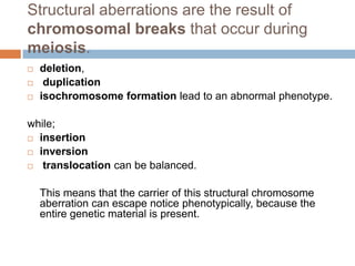
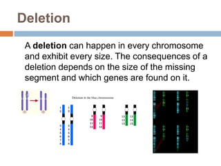
CHROMOSOMAL
    AND
GENE ABERRATION
(Duplication, Deletion, Inversion, Translocation)


     ENDAYA, DANICA FAYE M.
     MANGOSING VIDA FAYE S.
Introduction


Congenital abnormalities can be caused by an
anomaly in the number or structure of the
chromosomes. Disorders in the
gametogenesis, that is, in the first and second
meiotic divisions in the formation of oocytes
and sperm cells, lead to such defects.
Staining method
Chromosomal mutations can be
made visible with staining methods
(banding techniques). Following
the staining the chromosomes are
analyzed using a 1000x
enlargement. With this one can
recognize the chromosomes as
striped cords and display them in a
karyogram, after they have been
ordered according to their size and
the positions of the centromeres.
Various groups of similar
chromosomes are created in this
Today, small aberrations are examined using a
molecular cytogenetic (FISH) approach.
Among other things, the FISH (fluorescence-in-
situ-hybridizing) method makes possible the
targeted identification of partial aneuploidies
such as deletions, duplications and unbalanced
translocations (see below) that cannot be
resolved with a light microscope.
 A comprehensive array of DNA probes and
techniques are today available from which one
can choose for utilization, depending on the
diagnostic problem being worked on. With the
FISH method chromosomal alterations can be
analyzed down to a size of ca. 5 million
nucleotides
Origin of the
deviating
chromosome
structure
Structural aberrations are the result of
chromosomal breaks that occur during
meiosis.
   deletion,
    duplication
   isochromosome formation lead to an abnormal phenotype.

while;
 insertion

 inversion

 translocation can be balanced.


    This means that the carrier of this structural chromosome
    aberration can escape notice phenotypically, because the
    entire genetic material is present.
Deletion
 A deletion can happen in every chromosome
 and exhibit every size. The consequences of a
 deletion depends on the size of the missing
 segment and which genes are found on it.
Cri du Chat syndrome
   A partial deletion on the short
    arm (p) of chromosome 5 is
    responsible for the
    “cri du chat" syndrome
   The "cri du chat" syndrome
    manifests itself through cat-like
    crying of the newborn. This
    disorder is accompanied by
    microcephaly, severe
    psychosomatic and mental
    retardation and cardiac defects.
Duplication
   A chromosome duplication is the
    doubling of a chromosome piece.
    A duplication is sometimes termed a
    "partial trisomy".
   If, therefore, a duplication is
    present, the person is equipped with
    3 copies of the genes in the
    associated chromosome segment.
    This means that extra directions
    (genes) are present, leading to
    congenital abnormalities or
    developmental problems.
Fragile X syndrome

   The fragile X syndrome results
    from multiple duplications of a
    CGG segment (trinucleotide) in
    the 5' untranslated region of the
    FMR1 gene on the X chromosome
    (Xq27).
   Since it is an X linked mutation
    men are more affected than
    women. The fragile X syndrome
    leads to one of the most frequent
    forms of inherited mental
    retardation. In addition, the
    syndrome causes a
    prognathism, the face is long and
    small and the patient has large
Inversion
   If chromosome pieces that have been broken
    out become inserted again, but reversed, an
    inversion has occurred.

   The phenotype of this disorder is usually
    unobtrusive, since the entire chromosomal
    information is still present.
   When the interchanged region includes the
    centromere, one refers to it as a pericentric
    inversion,
   otherwise to a paracentric inversion
Insertion
   If chromosome pieces are reinserted
    somewhere else, this is referred to as an
    insertion. Carriers of such insertions can be
    phenotypically inconspicuous because no
    information has been lost.
Isochromosomiebildung
   Isochromosome formation is a relatively frequent
    chromosomal aberration, mainly in X
    chromosomes. Here the chromosomes are not
    divided along their length (see the normal division
    of the chromosomes figure) but transversely.
   The resulting isochromosomes (karyogram) either
    have two short or two long arms. Persons with this
    X chromosome anomaly have the same
    phenotype as patients suffering from Turner's
    syndrome (45, X0). This is explained by the fact
    that a X chromosome arm is missing.
Normal division
                  Isochromosome formation
of the
chromosome
Reciprocal translocation


   In a reciprocal translocation two broken
    off chromosome pieces of non-
    homologous chromosomes are
    exchanged. This is a relatively frequent
    anomaly. One finds it with an incidence
    of 1:500 newborns.
   Reciprocal translocations are frequently
    balanced because the entire genetic material
    is present. Problems occur, though, in gamete
    formation.
Cancer and Tumors
Today it is known that balanced
translocations can also lead to
pathogenic disorders in that proto-
oncogenes, which as normal
genes in their customary
environment are frequently
responsible for the controlling cell
proliferation, can be transformed
into oncogenes through
translocation events. They are
the cause for the origin of many
tumors and types of cancer
because in other environments they
achieve totally different effects.
Robertsonian translocation
   Another frequently observed
    anomaly (1:1'000 newborns) is
    the robertsonian
    translocation, which occurs
    between two acrocentric
    chromosomes of groups G
    and D. It is also referred to as
    the centric fusion of two
    acrocentric chromosomes.
   Carriers of such robertsonian translocations
    are phenotypically inconspicuous. Also
    here, though, problems arise when it comes to
    gamete formation because, normally, the
    diploid chromosome set is halved thereby.
END
 

Genetics

  • 1.
    CHROMOSOMAL AND GENE ABERRATION (Duplication, Deletion, Inversion, Translocation) ENDAYA, DANICA FAYE M. MANGOSING VIDA FAYE S.
  • 2.
    Introduction Congenital abnormalities canbe caused by an anomaly in the number or structure of the chromosomes. Disorders in the gametogenesis, that is, in the first and second meiotic divisions in the formation of oocytes and sperm cells, lead to such defects.
  • 3.
    Staining method Chromosomal mutationscan be made visible with staining methods (banding techniques). Following the staining the chromosomes are analyzed using a 1000x enlargement. With this one can recognize the chromosomes as striped cords and display them in a karyogram, after they have been ordered according to their size and the positions of the centromeres. Various groups of similar chromosomes are created in this
  • 4.
    Today, small aberrationsare examined using a molecular cytogenetic (FISH) approach. Among other things, the FISH (fluorescence-in- situ-hybridizing) method makes possible the targeted identification of partial aneuploidies such as deletions, duplications and unbalanced translocations (see below) that cannot be resolved with a light microscope. A comprehensive array of DNA probes and techniques are today available from which one can choose for utilization, depending on the diagnostic problem being worked on. With the FISH method chromosomal alterations can be analyzed down to a size of ca. 5 million nucleotides
  • 5.
  • 6.
    Structural aberrations arethe result of chromosomal breaks that occur during meiosis.  deletion,  duplication  isochromosome formation lead to an abnormal phenotype. while;  insertion  inversion  translocation can be balanced. This means that the carrier of this structural chromosome aberration can escape notice phenotypically, because the entire genetic material is present.
  • 7.
    Deletion A deletioncan happen in every chromosome and exhibit every size. The consequences of a deletion depends on the size of the missing segment and which genes are found on it.
  • 8.
    Cri du Chatsyndrome  A partial deletion on the short arm (p) of chromosome 5 is responsible for the “cri du chat" syndrome  The "cri du chat" syndrome manifests itself through cat-like crying of the newborn. This disorder is accompanied by microcephaly, severe psychosomatic and mental retardation and cardiac defects.
  • 9.
    Duplication  A chromosome duplication is the doubling of a chromosome piece.  A duplication is sometimes termed a "partial trisomy".  If, therefore, a duplication is present, the person is equipped with 3 copies of the genes in the associated chromosome segment. This means that extra directions (genes) are present, leading to congenital abnormalities or developmental problems.
  • 10.
    Fragile X syndrome  The fragile X syndrome results from multiple duplications of a CGG segment (trinucleotide) in the 5' untranslated region of the FMR1 gene on the X chromosome (Xq27).  Since it is an X linked mutation men are more affected than women. The fragile X syndrome leads to one of the most frequent forms of inherited mental retardation. In addition, the syndrome causes a prognathism, the face is long and small and the patient has large
  • 11.
    Inversion  If chromosome pieces that have been broken out become inserted again, but reversed, an inversion has occurred.  The phenotype of this disorder is usually unobtrusive, since the entire chromosomal information is still present.
  • 12.
    When the interchanged region includes the centromere, one refers to it as a pericentric inversion,
  • 13.
    otherwise to a paracentric inversion
  • 14.
    Insertion  If chromosome pieces are reinserted somewhere else, this is referred to as an insertion. Carriers of such insertions can be phenotypically inconspicuous because no information has been lost.
  • 15.
    Isochromosomiebildung  Isochromosome formation is a relatively frequent chromosomal aberration, mainly in X chromosomes. Here the chromosomes are not divided along their length (see the normal division of the chromosomes figure) but transversely.  The resulting isochromosomes (karyogram) either have two short or two long arms. Persons with this X chromosome anomaly have the same phenotype as patients suffering from Turner's syndrome (45, X0). This is explained by the fact that a X chromosome arm is missing.
  • 16.
    Normal division Isochromosome formation of the chromosome
  • 17.
    Reciprocal translocation  In a reciprocal translocation two broken off chromosome pieces of non- homologous chromosomes are exchanged. This is a relatively frequent anomaly. One finds it with an incidence of 1:500 newborns.
  • 18.
    Reciprocal translocations are frequently balanced because the entire genetic material is present. Problems occur, though, in gamete formation.
  • 19.
    Cancer and Tumors Todayit is known that balanced translocations can also lead to pathogenic disorders in that proto- oncogenes, which as normal genes in their customary environment are frequently responsible for the controlling cell proliferation, can be transformed into oncogenes through translocation events. They are the cause for the origin of many tumors and types of cancer because in other environments they achieve totally different effects.
  • 20.
    Robertsonian translocation  Another frequently observed anomaly (1:1'000 newborns) is the robertsonian translocation, which occurs between two acrocentric chromosomes of groups G and D. It is also referred to as the centric fusion of two acrocentric chromosomes.
  • 21.
    Carriers of such robertsonian translocations are phenotypically inconspicuous. Also here, though, problems arise when it comes to gamete formation because, normally, the diploid chromosome set is halved thereby.
  • 22.